Hematologic and Oncologic Disorders
Blood is the only organ that travels. When it fails, it fails everywhere at once — the brain gets confused, the heart races, the wound will not close, the fever that should have come never arrives. This chapter walks the three jobs blood does and what happens when each one breaks: carrying oxygen (the anemias), stopping and starting clot (thrombocytopenia, disseminated intravascular coagulation, anticoagulation), and defending the body (neutropenia and the marrow malignancies). Then it takes you into transfusion therapy, where the nurse is the last safety check between a labelled bag and a fatal mismatch, and into oncology, where the emergencies are quiet at the start and lethal by the end of the shift. Throughout, the same discipline applies: understand the mechanism, read the actual number, and know which finding makes you pick up the phone before you finish charting.
By the end of this chapter you can
- Interpret a complete blood count — hemoglobin, hematocrit, MCV, reticulocyte count and ANC — and classify an anemia as loss, destruction or failure of production.
- Differentiate iron deficiency, vitamin B12 deficiency, aplastic anemia and sickle cell disease by pathophysiology, laboratory pattern and nursing management.
- Distinguish thrombocytopenia, immune thrombocytopenia, heparin-induced thrombocytopenia and disseminated intravascular coagulation, and state the first nursing action for each.
- Monitor anticoagulant and antiplatelet therapy using the correct laboratory test, therapeutic range and reversal agent.
- Perform a safe blood transfusion and recognise acute hemolytic, febrile nonhemolytic, TACO and TRALI reactions within minutes of onset.
- Implement neutropenic precautions and manage febrile neutropenia as a time-critical emergency.
- Describe nursing care for leukemia, lymphoma and multiple myeloma, including nadir timing and bleeding precautions.
- Recognise and prioritise the oncologic emergencies — tumour lysis syndrome, spinal cord compression, superior vena cava syndrome, hypercalcemia of malignancy and extravasation.
- Deliver an SBAR report for a hematologic or oncologic red flag and delegate appropriately to LPN and UAP.
Section 1Anemia: When the Blood Stops Carrying Oxygen
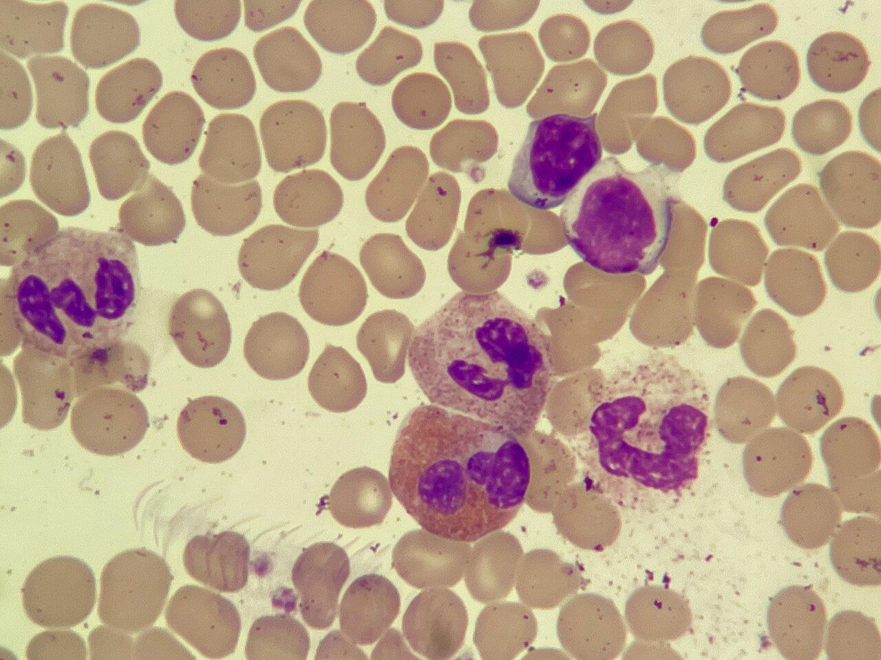
The mechanism, and why every anemia looks the same at first
Anemia is not a diagnosis. It is a finding — a reduction in circulating hemoglobin, hematocrit or red cell mass — and it always has a cause underneath it. Normal hemoglobin runs about 14 to 18 g/dL in men and 12 to 16 g/dL in women, with hematocrit roughly three times the hemoglobin (42 to 52 percent in men, 37 to 47 percent in women). When hemoglobin falls, oxygen delivery falls with it, and the body compensates in a predictable order: the heart speeds up and stroke volume rises, blood is shunted from skin and gut to brain and myocardium, and the respiratory rate climbs. That compensation is exactly why the early manifestations are so uniform — fatigue, pallor, tachycardia, exertional dyspnea, dizziness, cold intolerance — regardless of whether the patient is bleeding from a duodenal ulcer or missing intrinsic factor.
The manifestations also track how fast the anemia developed, not just how low the number is. A patient who bleeds two units in an hour is tachycardic, hypotensive and diaphoretic at a hemoglobin of 9 g/dL. A patient who has been oozing slowly from a colonic lesion for six months may walk into clinic at a hemoglobin of 6 g/dL with nothing but fatigue, because plasma volume expanded and cardiac output adapted. Do not let a stable-looking patient with a terrible number lull you, and do not dismiss a symptomatic patient because the hemoglobin is only mildly low.
Every anemia belongs to one of three buckets, and sorting the patient into a bucket is the single most useful thing a nurse can do at the bedside. Blood loss — obvious trauma or occult gastrointestinal bleeding. Increased destruction — hemolysis, whether inherited (sickle cell, G6PD deficiency, thalassemia) or acquired (autoimmune, mechanical valve, transfusion reaction). Decreased production — missing raw material (iron, B12, folate), missing signal (erythropoietin in chronic kidney disease), or a marrow that has stopped working (aplastic anemia, marrow infiltration by leukemia or metastasis).
Reading the numbers: MCV and reticulocytes do the sorting
Two indices narrow the field faster than anything else. Mean corpuscular volume (MCV, normal 80 to 100 fL) tells you cell size. Microcytic anemia (MCV under 80 fL) is iron deficiency until proven otherwise, with thalassemia and anemia of chronic disease as the other contenders. Macrocytic anemia (MCV over 100 fL) points to B12 or folate deficiency, alcohol use, liver disease or myelodysplasia. Normocytic anemia (MCV 80 to 100 fL) is acute blood loss, hemolysis, chronic kidney disease or marrow failure.
The reticulocyte count (normal 0.5 to 2 percent) tells you whether the marrow is trying. A high reticulocyte count means the marrow is responding appropriately — the problem is loss or destruction downstream. A low or normal reticulocyte count in the face of significant anemia means the marrow itself is the problem, either because it lacks material or because it is sick. Rising reticulocytes three to five days after starting iron or B12 is the earliest proof that therapy is working, and it is worth teaching patients that the fatigue lifts before the hemoglobin normalises.
Iron studies complete the microcytic workup. Ferritin below 30 ng/mL is diagnostic of iron deficiency (normal roughly 12 to 300 ng/mL in men, 12 to 150 ng/mL in women), serum iron falls (normal 60 to 170 mcg/dL), total iron-binding capacity rises (normal 240 to 450 mcg/dL) and transferrin saturation drops below 20 percent. Because ferritin is an acute-phase reactant, it can be falsely normal during infection or inflammation — a genuinely iron-deficient inflamed patient may show a ferritin of 80 ng/mL. For megaloblastic anemia, check serum B12 (normal 200 to 900 pg/mL) and folate (normal 3 to 20 ng/mL); methylmalonic acid rises in B12 deficiency but not in folate deficiency, which is how the laboratory tells them apart.
| Anemia | Mechanism | Signature findings | Nursing focus |
|---|---|---|---|
| Iron deficiency | Depleted iron stores — chronic blood loss, poor intake, malabsorption, pregnancy | MCV <80 fL, ferritin <30 ng/mL, TIBC high; spoon nails, glossitis, pica | Find the bleeding source; teach oral iron administration; recheck Hgb in 4 weeks |
| Vitamin B12 / pernicious | Loss of intrinsic factor, gastrectomy, ileal resection, metformin, long-term PPI | MCV >100 fL, B12 <200 pg/mL; paresthesias, ataxia, loss of proprioception, beefy red tongue | Fall precautions; lifelong IM cobalamin; neuro deficits may be permanent |
| Folate deficiency | Poor intake, alcohol use, methotrexate, phenytoin, pregnancy | MCV >100 fL, folate <3 ng/mL; no neurologic findings | Never treat macrocytosis with folate alone until B12 is measured |
| Anemia of chronic kidney disease | Erythropoietin deficiency | Normocytic, low reticulocytes, elevated creatinine | Confirm iron stores before ESA; target Hgb 10 to 11 g/dL, never above 11 to 12 |
| Aplastic anemia | Stem cell failure — idiopathic, benzene, chloramphenicol, radiation, viral | Pancytopenia, hypocellular marrow biopsy, low reticulocytes | Infection and bleeding precautions simultaneously; protective isolation |
| Sickle cell disease | HbS polymerises when deoxygenated; rigid cells occlude microvasculature and hemolyse | Hgb 6 to 9 g/dL baseline, reticulocytes high, LDH high, haptoglobin low, bilirubin high | Aggressive analgesia, hydration, oxygen only if hypoxic, watch for acute chest |
Sickle cell disease: the crisis you will actually manage
In sickle cell disease a single amino acid substitution produces hemoglobin S, which polymerises when it gives up oxygen. The red cell distorts into a rigid crescent, adheres to vascular endothelium and obstructs the microcirculation, producing the two consequences that define the disease: chronic hemolytic anemia and recurrent ischemic pain. Dehydration, hypoxia, infection, acidosis, cold exposure, high altitude and physical or emotional stress all precipitate sickling. Baseline hemoglobin usually sits between 6 and 9 g/dL, and the patient is genuinely accustomed to it — do not transfuse toward a normal number.
Vaso-occlusive crisis is severe, deep, unrelenting pain in bones, chest, abdomen or joints. Manage it as the emergency it is: scheduled or patient-controlled opioid analgesia rather than as-needed dosing that leaves the patient chasing pain, intravenous or oral hydration, warmth to the affected areas (never cold, which promotes sickling), and supplemental oxygen only when the pulse oximetry reading is below the patient’s baseline. Avoid meperidine — its metabolite normeperidine accumulates and lowers the seizure threshold. Patients with sickle cell disease are frequently and wrongly labelled drug-seeking; the literature on undertreatment in this population is unambiguous, and believing the patient’s report of pain is a clinical skill, not a courtesy.
Three complications kill. Acute chest syndrome — new pulmonary infiltrate with fever, chest pain, hypoxia and cough — is the leading cause of death and needs oxygen, antibiotics, incentive spirometry and often exchange transfusion. Splenic sequestration traps blood in the spleen, dropping hemoglobin precipitously with a rapidly enlarging left upper quadrant mass and hypovolemic shock. Aplastic crisis, usually triggered by parvovirus B19, shuts down erythropoiesis and is recognised by a collapsing reticulocyte count. Long-term therapy is hydroxyurea, which raises fetal hemoglobin and reduces crisis frequency; it causes myelosuppression, so the CBC is monitored, and it is teratogenic.
Priorities and the older adult
The first question in any anemia is airway, breathing, circulation. A patient with hemoglobin of 6 g/dL who is tachycardic at 130, hypotensive and confused needs oxygen, large-bore intravenous access, type and crossmatch and a provider at the bedside — not an iron teaching sheet. Once perfusion is secured, the priority shifts to finding and stopping the source, then to replacement and education. Restrictive transfusion thresholds are the standard: transfuse for hemoglobin below 7 g/dL in most hospitalised adults, below 8 g/dL in patients with cardiovascular disease or undergoing orthopedic surgery, and always in symptomatic patients regardless of number.
Anemia is never a normal consequence of aging, and in older adults it rarely announces itself as fatigue. It presents as a fall, as new-onset confusion or delirium, as worsening angina or heart failure, or as a decline in function that the family calls “slowing down.” The most common cause in this population is occult gastrointestinal blood loss, frequently from NSAID use, and every new iron-deficiency anemia in an adult over 50 requires endoscopic evaluation to rule out colorectal cancer. Older adults also have less marrow reserve, so they decompensate faster and recover more slowly. Delegation here is straightforward: the UAP may obtain vital signs, assist with ambulation and report dizziness; the LPN may administer oral iron and reinforce teaching; the RN performs the initial and ongoing assessment, interprets the CBC and initiates the transfusion.
Classify every anemia as loss, destruction or failed production using MCV and the reticulocyte count, because the bucket — not the hemoglobin number alone — determines what you do next.
Section 2Bleeding, Clotting and Anticoagulation
Platelets first, then the cascade
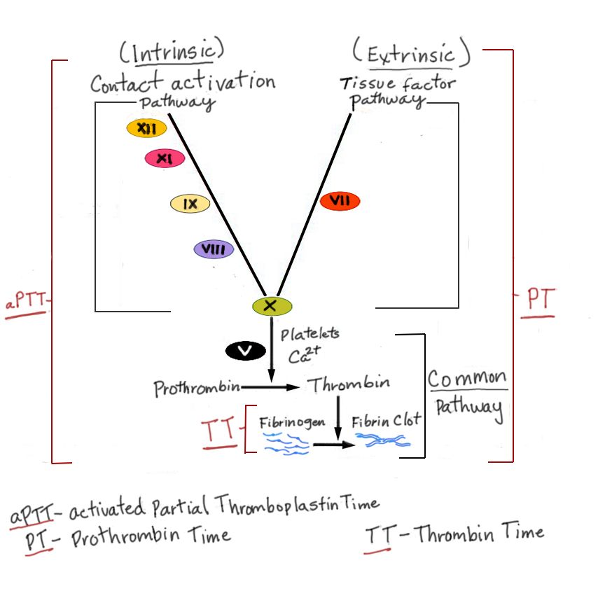
Hemostasis happens in two stages. Primary hemostasis is platelets adhering to injured endothelium and aggregating into a plug — fast, and responsible for controlling small-vessel bleeding in skin and mucous membranes. When it fails, the patient shows petechiae, purpura, easy bruising, gum bleeding, epistaxis and heavy menses. Secondary hemostasis is the coagulation cascade, which converts fibrinogen to a fibrin mesh that stabilises the plug. When it fails, the patient shows deep bleeding — hemarthrosis, muscle hematoma, delayed rebleeding after a procedure. That distinction alone tells you where to look before a single result comes back.
Normal platelet count is 150,000 to 400,000/mm³. Spontaneous bleeding is uncommon above 50,000, becomes a real risk below 20,000, and below 10,000 the danger is intracranial hemorrhage. Bleeding precautions begin around 50,000 and become strict below 20,000: electric razor only, soft toothbrush or oral sponges, no flossing if gums bleed, no rectal temperatures, suppositories or enemas, no intramuscular injections, avoid aspirin and NSAIDs, hold pressure at least five minutes after any venipuncture, humidify air and avoid forceful nose blowing, and use a stool softener so the patient does not strain.
The cascade is monitored by two tests. Prothrombin time (PT, normal 11 to 12.5 seconds) and its standardised expression the INR (normal 0.8 to 1.1) reflect the extrinsic and common pathways — the warfarin and liver-failure test. Activated partial thromboplastin time (aPTT, normal 30 to 40 seconds) reflects the intrinsic and common pathways — the unfractionated heparin and hemophilia test. Fibrinogen (normal 200 to 400 mg/dL) is the substrate; D-dimer (normal under 0.5 mcg/mL FEU) is the breakdown product and rises whenever clot is being lysed anywhere in the body.
The four thrombocytopenias you must tell apart
Immune thrombocytopenia (ITP) is autoantibody-mediated platelet destruction in the spleen. The patient is otherwise well but covered in petechiae with a platelet count that may be under 20,000; PT and aPTT are normal. Treatment is corticosteroids, intravenous immunoglobulin when the count must rise quickly, rituximab, thrombopoietin-receptor agonists such as eltrombopag or romiplostim, and splenectomy for refractory disease. Platelet transfusion is reserved for life-threatening bleeding because transfused platelets are destroyed as fast as native ones.
Heparin-induced thrombocytopenia (HIT) is the trap. Antibodies against the heparin–platelet factor 4 complex activate platelets, so despite a falling count the patient clots rather than bleeds. Suspect it when the platelet count falls more than 50 percent from baseline five to ten days after heparin exposure, or sooner with recent prior exposure. The nursing action is immediate and absolute: stop all heparin — infusions, subcutaneous prophylaxis, flushes, heparin-bonded catheters, dialysis circuits — and notify the provider. Anticoagulation continues with a non-heparin agent such as argatroban, bivalirudin or fondaparinux. Do not transfuse platelets, and do not start warfarin until the platelet count recovers above 150,000, because warfarin alone in acute HIT can cause venous limb gangrene.
Thrombotic thrombocytopenic purpura (TTP) results from deficient ADAMTS13 activity, leaving ultra-large von Willebrand multimers that shear red cells and consume platelets. The classic pentad is fever, thrombocytopenia, microangiopathic hemolytic anemia with schistocytes on smear, renal impairment and fluctuating neurologic changes. Treatment is urgent therapeutic plasma exchange; platelet transfusion is contraindicated because it fuels further microthrombi. Disseminated intravascular coagulation (DIC) is the fourth: widespread activation of coagulation, usually triggered by sepsis, obstetric catastrophe, major trauma, burns or malignancy, that consumes platelets and clotting factors until the patient bleeds from every puncture site while simultaneously infarcting organs.
| Test | DIC | ITP | HIT | Hemophilia A |
|---|---|---|---|---|
| Platelets | Low | Very low | Falling >50% from baseline | Normal |
| PT / INR | Prolonged | Normal | Normal | Normal |
| aPTT | Prolonged | Normal | Normal | Prolonged |
| Fibrinogen | Low | Normal | Normal | Normal |
| D-dimer | Markedly elevated | Normal | May be elevated | Normal |
| Clinical picture | Bleeding and clotting | Mucocutaneous bleeding | Thrombosis | Hemarthrosis, deep bleeds |
| First action | Treat the underlying cause; replace products | Corticosteroids / IVIG | Stop all heparin immediately | Factor VIII replacement |
Inherited bleeding disorders
Hemophilia A is deficiency of factor VIII, hemophilia B of factor IX; both are X-linked recessive, so males are affected and females are usually carriers. The aPTT is prolonged with a normal PT and normal platelet count. The characteristic bleed is hemarthrosis — a warm, swollen, exquisitely painful joint, most often knee, elbow or ankle — and repeated bleeds destroy cartilage and produce chronic arthropathy. Acute management is factor concentrate replacement first, then rest, ice, compression and elevation; the joint is immobilised briefly and then mobilised with physical therapy, because prolonged immobility causes contracture. Desmopressin releases stored factor VIII and von Willebrand factor and is useful in mild hemophilia A only. Aspirin and NSAIDs are contraindicated; acetaminophen is the analgesic of choice for minor pain.
Von Willebrand disease is the most common inherited bleeding disorder and behaves like a platelet problem — mucocutaneous bleeding, epistaxis, menorrhagia, prolonged bleeding after dental work — because von Willebrand factor is what lets platelets adhere. Management is desmopressin for mild disease, von Willebrand factor–containing concentrate for severe disease or surgery, and antifibrinolytics such as tranexamic acid for mucosal bleeding. Teach both groups to carry medical identification, avoid contact sports, use soft toothbrushes and electric razors, and notify every dentist and surgeon before any procedure.
Anticoagulants: the right test, the right antidote
Unfractionated heparin potentiates antithrombin and is monitored by aPTT (therapeutic range about 1.5 to 2.5 times control, roughly 60 to 100 seconds depending on the laboratory) or by anti-factor Xa level. It works within minutes, has a short half-life, and is reversed with protamine sulfate. Low-molecular-weight heparin such as enoxaparin gives a predictable response and needs no routine monitoring except anti-Xa levels in renal impairment, obesity or pregnancy. Give it deep subcutaneously in the abdomen at least two inches from the umbilicus, do not expel the air bubble in the prefilled syringe, do not aspirate, and do not massage the site. Both heparins can cause HIT, so a platelet count baseline and periodic recheck are standard.
Warfarin blocks vitamin K–dependent synthesis of factors II, VII, IX and X and takes three to five days for full effect, which is why it overlaps with a parenteral agent at initiation. It is monitored by INR: therapeutic range is 2 to 3 for atrial fibrillation and venous thromboembolism, and 2.5 to 3.5 for a mechanical mitral valve. The antidote is vitamin K (phytonadione), with prothrombin complex concentrate or fresh frozen plasma for serious bleeding. Teach dietary consistency rather than avoidance — the patient does not have to give up spinach, but the amount should not swing week to week — and review the enormous list of interacting drugs, antibiotics and herbals, including ginkgo, ginger, garlic and St. John’s wort. Direct oral anticoagulants (apixaban, rivaroxaban and edoxaban inhibit factor Xa; dabigatran inhibits thrombin directly) need no routine monitoring, are dose-adjusted for renal function, and are reversed with andexanet alfa for the Xa inhibitors and idarucizumab for dabigatran.
Platelet problems bleed into skin and mucous membranes while factor problems bleed into joints and muscle — and in heparin-induced thrombocytopenia the falling platelet count means the patient is about to clot, so stopping every source of heparin is the first action.
Section 3Transfusion Therapy and Reactions
Components and what each one actually does
Whole blood is rarely used outside massive hemorrhage protocols; modern practice is component therapy. One unit of packed red blood cells, roughly 250 to 350 mL, raises hemoglobin by about 1 g/dL and hematocrit by about 3 percent in an average adult who is not actively bleeding — a fact worth remembering when the post-transfusion CBC comes back and you need to judge whether it makes sense. Platelets are given for counts below 10,000, for counts below 20,000 with fever or sepsis, for counts below 50,000 before an invasive procedure, and for active bleeding; one apheresis unit typically raises the count by 30,000 to 50,000.
Fresh frozen plasma contains all the clotting factors and is used for multiple factor deficiencies, urgent warfarin reversal when prothrombin complex concentrate is unavailable, and dilutional coagulopathy in massive transfusion. Cryoprecipitate is the fibrinogen product — give it when fibrinogen falls below 100 mg/dL, and for factor VIII, factor XIII and von Willebrand factor replacement when specific concentrates are not available. Leukocyte-reduced products lower the rate of febrile nonhemolytic reactions and cytomegalovirus transmission. Irradiated products prevent transfusion-associated graft-versus-host disease in severely immunocompromised recipients, including stem cell transplant patients. Washed red cells remove plasma proteins and are used for recipients with IgA deficiency or recurrent severe allergic reactions.
The procedure, step by step
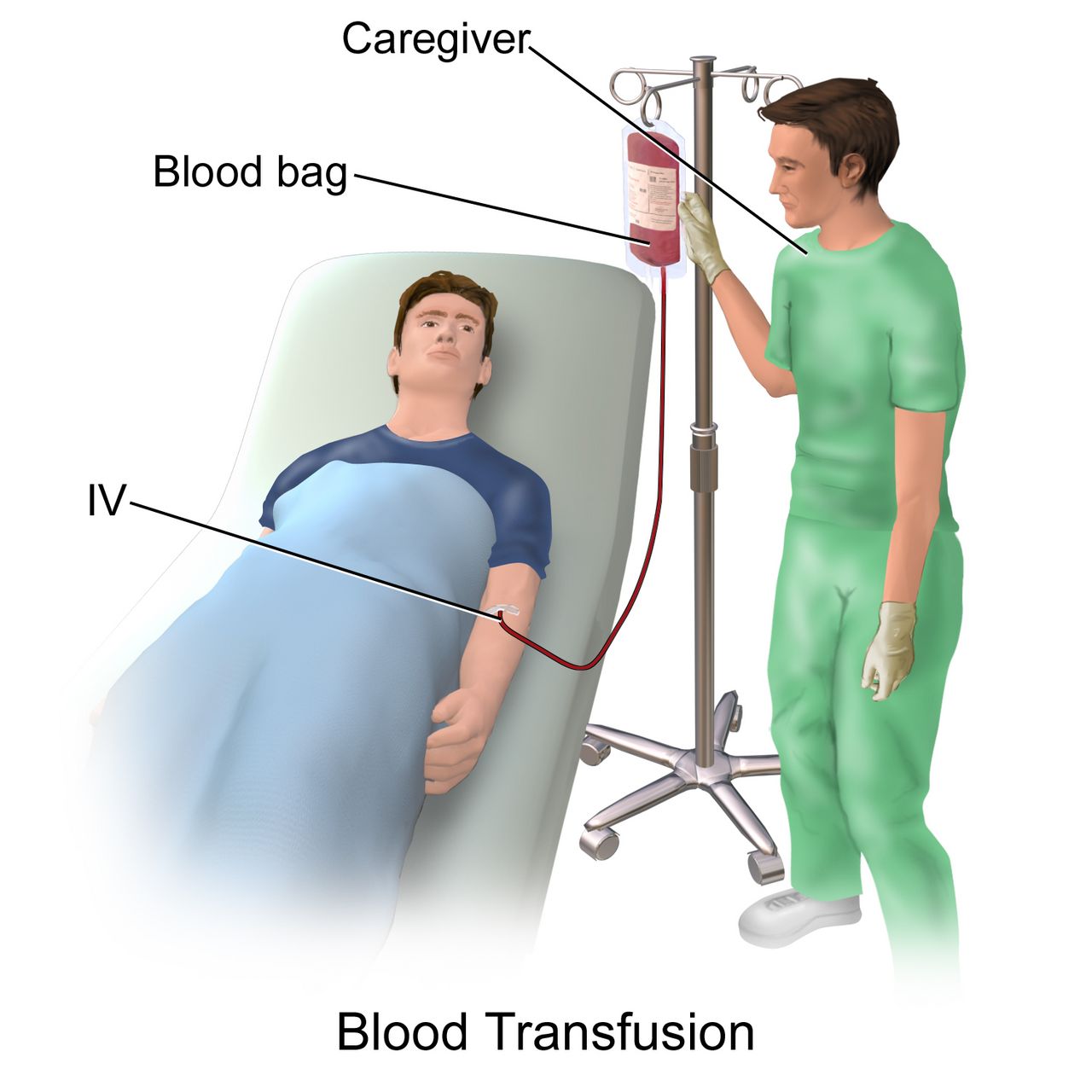
Verify a signed informed consent and a current type and crossmatch. Establish patent intravenous access — an 18- or 20-gauge catheter is preferred for red cells; smaller bores risk hemolysis and slow flow, though platelets and plasma tolerate a 22-gauge. Take a full set of baseline vital signs including temperature; if the patient is already febrile, report it before the product is released, because a fever after the transfusion starts is then impossible to interpret. Prime the blood administration set with 0.9 percent sodium chloride only. Never hang blood with dextrose solutions, which lyse red cells, or lactated Ringer’s, whose calcium can clot the line; no medication is ever added to a blood product or infused through the same lumen.
Two licensed personnel verify at the bedside, against the patient’s identification band and the unit tag, the patient name and second identifier, the ABO and Rh type, the unit number and expiration, and the crossmatch compatibility. Start the infusion within 30 minutes of release from the blood bank. Run it slowly for the first 15 minutes — about 2 mL per minute — and stay with the patient, because the most catastrophic reactions declare themselves in the first 15 minutes and as little as 10 to 15 mL of incompatible blood can be fatal. Recheck vital signs at 15 minutes, then per policy, and again at completion. A unit must finish within four hours because of bacterial growth risk at room temperature; if the patient cannot tolerate that rate, ask the blood bank to split the unit.
Delegation is tightly drawn here. Only the RN may initiate a transfusion, perform the verification, complete the first 15-minute assessment and evaluate a suspected reaction. The UAP may obtain vital signs once the transfusion is under way and stable, and must be told explicitly what to report immediately: any temperature rise of one degree Celsius or more, chills, rash, itching, back pain, shortness of breath or a feeling of impending doom. LPN scope for transfusion varies by state and facility — know your board of nursing rule rather than assuming.
| Reaction | Timing and cause | Manifestations | Management |
|---|---|---|---|
| Acute hemolytic | Within minutes; ABO incompatibility, almost always a clerical or identification error | Fever, chills, low back or flank pain, chest tightness, tachycardia, hypotension, dark or red urine, oozing from IV sites | Stop immediately, disconnect tubing at the hub, infuse 0.9% NaCl with new tubing, notify provider and blood bank, send the bag, tubing and blood and urine specimens; support blood pressure and urine output |
| Febrile nonhemolytic | Within 1 to 6 hours; recipient antibodies to donor leukocytes — the most common reaction | Temperature rise of 1°C or more, chills, headache, flushing; no hypotension | Stop and evaluate; antipyretic; use leukocyte-reduced products in future |
| Mild allergic | Minutes to hours; donor plasma proteins | Urticaria, flushing, pruritus only | Stop, give antihistamine; may resume slowly per policy if symptoms resolve and there is no other sign |
| Anaphylactic | Immediate; often IgA deficiency in the recipient | Wheezing, stridor, angioedema, hypotension, shock | Stop, maintain airway, epinephrine, fluids; future transfusions use washed or IgA-deficient products |
| TACO (circulatory overload) | During or within 6 hours; volume faster than the heart can handle | Dyspnea, orthopnea, crackles, distended neck veins, hypertension, rapid weight gain | Stop or slow, sit the patient upright with legs dependent, oxygen, diuretic per order |
| TRALI | Within 6 hours; donor antibodies injure pulmonary capillaries | Acute hypoxemia, bilateral infiltrates, fever, hypotension, no jugular venous distension | Stop, oxygen and often mechanical ventilation, supportive care; diuretics do not help |
| Bacterial contamination / sepsis | Rapid; highest risk with platelets, stored at room temperature | High fever, rigors, vomiting, marked hypotension | Stop, culture patient and product, broad-spectrum antibiotics, hemodynamic support |
| Delayed hemolytic | 1 to 14 days later; anamnestic antibody response | Unexplained falling hemoglobin, mild jaundice, low-grade fever | Monitor CBC and bilirubin; report to blood bank for antibody identification |
Massive transfusion and special considerations
Massive transfusion — replacement of the patient’s entire blood volume within 24 hours, or 10 or more units of red cells — brings its own physiology. Citrate anticoagulant in the stored product binds calcium, so watch for hypocalcemia with perioral tingling, tetany, positive Chvostek and Trousseau signs and QT prolongation; ionized calcium (normal 4.5 to 5.6 mg/dL) is monitored. Potassium leaks from stored cells, so hyperkalemia is a risk, particularly in renal impairment. Cold product causes hypothermia, which itself worsens coagulopathy — use an approved blood warmer, never a microwave or hot water. And replacing red cells without plasma and platelets produces dilutional coagulopathy, which is why trauma protocols give red cells, plasma and platelets in balanced ratios.
Some patients decline transfusion on religious grounds, most commonly Jehovah’s Witnesses. The competent adult’s refusal must be honoured, documented and communicated to the whole team; the nursing role is to support the decision without coercion and to advocate for alternatives — erythropoiesis-stimulating agents, intravenous iron, cell salvage, tranexamic acid, minimising phlebotomy volumes. Older adults are at particular risk of TACO because of reduced ventricular compliance; transfuse them slowly, over three to four hours per unit when the clinical situation permits, and assess lung sounds before, during and after.
Stay with the patient for the first 15 minutes of every unit, and when any reaction is suspected the first action is always to stop the transfusion and keep the line open with 0.9 percent sodium chloride through new tubing.
Section 4Leukemia, Lymphoma, Myeloma and the Neutropenic Patient
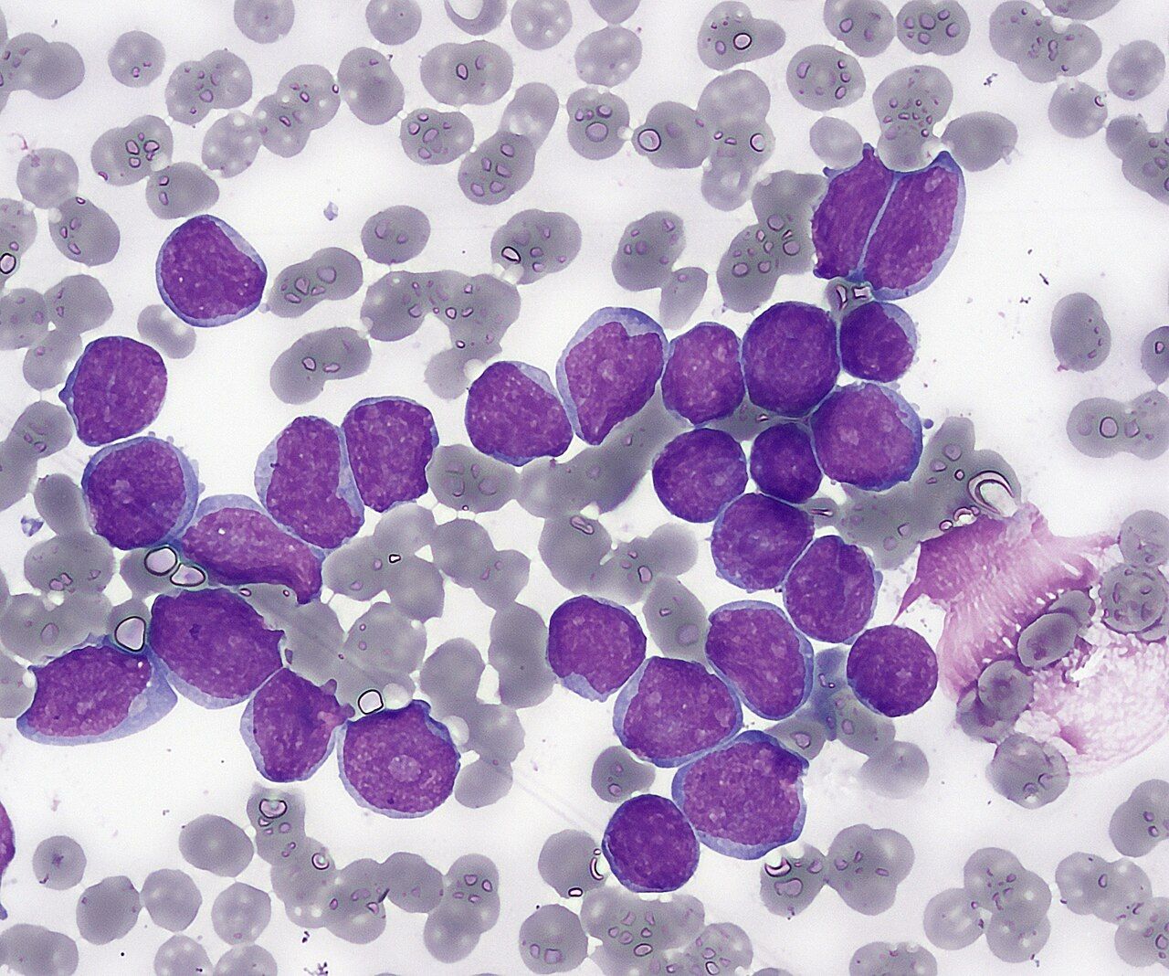
Leukemia: one mechanism, three consequences
Leukemia is malignant proliferation of white cell precursors in the bone marrow. The malignant clone does not work — a blast cannot phagocytose — and it crowds out normal hematopoiesis. That single mechanism explains the entire presentation. Loss of red cell production gives fatigue, pallor and dyspnea. Loss of platelet production gives petechiae, ecchymoses, epistaxis and gum bleeding. Loss of functional neutrophils gives recurrent infection and fever. Add bone pain from marrow expansion, lymphadenopathy, hepatosplenomegaly, weight loss and night sweats, and you have the classic picture. The white cell count may be strikingly high, normal or low; what matters is the differential and the blast percentage, confirmed by bone marrow aspiration and biopsy, usually from the posterior iliac crest.
Four types dominate. Acute lymphocytic leukemia (ALL) is the most common childhood cancer and carries a risk of central nervous system involvement, which is why intrathecal chemotherapy is standard. Acute myelogenous leukemia (AML) is the most common acute leukemia in adults; Auer rods on smear are characteristic, and disseminated intravascular coagulation is a recognised complication of the promyelocytic subtype. Chronic lymphocytic leukemia (CLL) occurs in older adults, is often found incidentally on a routine CBC with lymphocytosis, and may be observed rather than treated for years. Chronic myelogenous leukemia (CML) carries the Philadelphia chromosome and its BCR-ABL fusion protein, and is treated with tyrosine kinase inhibitors such as imatinib — a genuine success story of targeted therapy.
Treatment of acute leukemia proceeds in phases: induction to achieve remission, consolidation to eliminate residual disease, and maintenance. Induction deliberately destroys the marrow, so the nadir — the lowest blood counts — occurs roughly 7 to 14 days after therapy, and that is the window when the patient is most likely to become septic or bleed. Hematopoietic stem cell transplantation follows conditioning chemotherapy with or without total body irradiation; the transplant recipient is profoundly aplastic for weeks and is at risk for graft-versus-host disease, which attacks skin (maculopapular rash), gut (voluminous diarrhea) and liver (rising bilirubin).
Lymphoma and multiple myeloma
Lymphomas arise in lymphoid tissue rather than marrow. Hodgkin lymphoma is identified by Reed-Sternberg cells, has a bimodal age distribution in young adults and adults over 55, spreads predictably from one nodal group to the next, and is among the most curable of cancers. The typical presentation is a painless, firm, enlarged cervical or supraclavicular node, sometimes with the B symptoms — fever, drenching night sweats and unintentional loss of more than 10 percent of body weight — that carry prognostic weight. Non-Hodgkin lymphoma is far more common, occurs mostly in older adults, spreads unpredictably and is often widespread at diagnosis. Late effects of treatment matter for teaching: secondary malignancy, cardiomyopathy from anthracyclines, pulmonary fibrosis from bleomycin, hypothyroidism and infertility.
Multiple myeloma is malignant proliferation of plasma cells that secrete a monoclonal immunoglobulin and activate osteoclasts. The acronym CRAB captures it: hyperCalcemia, Renal insufficiency, Anemia and Bone lesions. Serum protein electrophoresis shows an M spike, urine may contain Bence Jones protein, and skeletal survey shows punched-out lytic lesions with diffuse osteoporosis. Nursing care runs on three rails. Hydration — at least 3 litres daily unless contraindicated — to protect the kidneys from light chains and calcium. Safety — the bones are structurally weak, so use gentle handling, avoid lifting under the arms, assess any new back pain for pathologic fracture, and never apply traction to an unassessed limb. Pain and calcium management — bone-modifying agents reduce skeletal events, and the two available agents belong to different drug classes. Zoledronic acid is a bisphosphonate; denosumab is not — it is a monoclonal antibody against RANK ligand. Denosumab is preferred when renal function is impaired, because it needs no renal dose adjustment, whereas zoledronic acid must be dose-reduced or withheld as creatinine clearance falls and can itself cause acute kidney injury. Monitor calcium on either agent and especially on denosumab, which commonly causes symptomatic hypocalcemia requiring calcium and vitamin D repletion. NSAIDs are avoided because of the renal risk.
Neutropenia, ANC and the fever that must not wait
The absolute neutrophil count, not the total white cell count, defines infection risk. Calculate it as total WBC multiplied by the sum of the percentage of segmented neutrophils and bands, divided by 100. A patient with a WBC of 2,000/mm³, 30 percent segs and 5 percent bands has an ANC of 700 — moderately neutropenic. An ANC below 1,500 is neutropenia, below 1,000 is moderate risk, and below 500 is severe neutropenia with high risk of life-threatening infection.
Here is the part students underestimate: without neutrophils, the patient cannot mount the signs of infection you were taught to look for. There is no purulent drainage, no infiltrate on chest film, no redness around the line site, because all of those are made of neutrophils. Fever may be the only manifestation — and in the older adult or the patient on corticosteroids, even fever may be blunted, so new confusion, unexplained tachycardia, hypotension or simply “not acting right” deserves the same response. A single oral temperature of 38.3°C (101°F), or 38.0°C (100.4°F) sustained for one hour, in a patient with an ANC below 500 is febrile neutropenia: an oncologic emergency.
The sequence is fixed and time-critical. Assess and take vital signs, notify the provider immediately, obtain blood cultures from a peripheral site and from every lumen of any central line plus urine and any other indicated culture, and administer broad-spectrum intravenous antibiotics within 60 minutes of the fever being identified. Do not wait for culture results. Do not give acetaminophen before cultures are drawn, because masking the fever delays recognition. Delegation during this event: the UAP may obtain vital signs and gather supplies; the RN draws cultures from central lines, administers the antibiotics and reassesses. Priority among several patients is straightforward — the neutropenic patient with a new fever outranks the stable patient with chronic pain or a scheduled dressing change, every time.
In the neutropenic patient fever may be the only sign of overwhelming infection, and broad-spectrum antibiotics must be hanging within 60 minutes of that fever — cultures first, but never at the cost of delay.
Section 5Cancer Treatment, Toxicity and Oncologic Emergencies
How the treatments work and what they cost the patient
Cytotoxic chemotherapy kills rapidly dividing cells. It cannot distinguish a tumour cell from any other fast-cycling cell, which is precisely why the toxicities are so predictable: bone marrow (myelosuppression), gastrointestinal mucosa from mouth to anus (mucositis, diarrhea, nausea), hair follicles (alopecia) and gonads (infertility). Certain agents carry signature organ toxicities that drive specific monitoring. Doxorubicin and the other anthracyclines are cardiotoxic in a cumulative, dose-dependent way, so ejection fraction is measured by echocardiogram or MUGA scan before and during therapy. Cisplatin is nephrotoxic and ototoxic — hydrate aggressively, monitor creatinine, and ask about tinnitus and hearing change. Vincristine causes peripheral neuropathy and is fatal if given intrathecally, so it is labelled for intravenous use only. Cyclophosphamide causes hemorrhagic cystitis, prevented with vigorous hydration, frequent voiding and mesna. Bleomycin causes pulmonary fibrosis; a new dry cough or exertional dyspnea is reported, not watched.
Chemotherapy-induced nausea and vomiting has three patterns — acute within 24 hours, delayed from day 2 to day 5, and anticipatory, a conditioned response triggered by the smell of the infusion suite. Management is scheduled and preventive, not rescue: a serotonin antagonist such as ondansetron, an NK-1 antagonist such as aprepitant, dexamethasone, and olanzapine for highly emetogenic regimens, with lorazepam for the anticipatory component. Mucositis is managed with a soft brush or sponge, saline or sodium bicarbonate rinses, no alcohol-containing mouthwash, no hot spicy or acidic food, and topical or systemic analgesia; oral cryotherapy during certain infusions reduces its severity.
Radiation damages DNA locally, so its toxicities are confined to the treatment field plus systemic fatigue. Teach skin care: wash gently with lukewarm water and mild soap, pat dry, use only the moisturiser the radiation oncology team approves, no powders or metal-containing products, never apply heat or ice to the field, wear loose cotton, protect from sun, and do not scrub off the treatment markings. With sealed-source brachytherapy the patient emits radiation: apply time, distance and shielding, assign the patient a private room, wear a dosimeter badge, limit each visitor to about 30 minutes per day at six feet, exclude pregnant staff and visitors and children under 18, and if an implant dislodges never touch it with your hands — use long-handled forceps to place it in the lead container kept in the room and call radiation safety.
Immunotherapy changes the toxicity profile entirely. Checkpoint inhibitors such as nivolumab and pembrolizumab release the brakes on T cells and produce immune-related adverse events — colitis with severe diarrhea, pneumonitis with cough and dyspnea, hepatitis, dermatitis, thyroiditis and adrenal insufficiency — managed with corticosteroids and, if severe, drug discontinuation. CAR-T cell therapy can trigger cytokine release syndrome with high fever, hypotension and hypoxia within days, treated with tocilizumab, and neurotoxicity that begins as a subtle handwriting or naming deficit. Monoclonal antibodies commonly cause infusion reactions during the first exposure; premedication and a slow initial rate are standard, and the nurse stays close.
The oncologic emergencies
These are the events that turn a scheduled admission into a code, and they are heavily tested because the first action separates a safe nurse from an unsafe one.
| Emergency | Mechanism | Recognise it by | First actions |
|---|---|---|---|
| Febrile neutropenia | ANC <500 with no capacity to localise infection | Temp 38.3°C once or 38.0°C for 1 hour; may be only sign | Cultures then broad-spectrum antibiotics within 60 minutes |
| Tumour lysis syndrome | Massive cell breakdown, usually beginning 12 to 72 hours after cytotoxic therapy starts — and occasionally spontaneously before any treatment in bulky, rapidly proliferating tumours — dumps intracellular contents | Potassium >5.0 mEq/L, phosphorus >4.5 mg/dL, uric acid >7.0 mg/dL, calcium low, rising creatinine; peaked T waves, tetany, oliguria | Aggressive IV fluids, cardiac monitoring, treat hyperkalemia. The two urate drugs are not interchangeable: allopurinol blocks xanthine oxidase and so only prevents formation of new uric acid — a prophylactic agent started before and during cytoreduction; rasburicase is urate oxidase and enzymatically degrades urate already circulating, dropping the level within hours, so it is the agent for established hyperuricemia with a rising creatinine. Check G6PD status before rasburicase — it causes severe hemolysis and methemoglobinemia in G6PD deficiency. Prevention beats rescue |
| Spinal cord compression | Tumour or vertebral collapse compresses the cord | Back pain is the first symptom, then weakness, sensory level, then bowel or bladder incontinence | Dexamethasone immediately, urgent MRI, flat bed rest and log roll, radiation or surgical decompression |
| Superior vena cava syndrome | Tumour obstructs venous return from head, neck and arms | Facial and periorbital edema worse in the morning, distended neck and chest veins, dyspnea, hoarseness, headache | Elevate head of bed, oxygen, no upper-extremity IVs or blood pressures, steroids, radiation or stent |
| Hypercalcemia of malignancy | Osteolysis and tumour-secreted PTH-related peptide | Calcium >12 mg/dL (normal 8.6 to 10.2); lethargy, confusion, constipation, polyuria, nausea, shortened QT | Isotonic saline hydration first, calcitonin for rapid effect, bisphosphonate or denosumab, mobilise, fall precautions |
| Cardiac tamponade | Malignant pericardial effusion | Muffled heart sounds, distended neck veins, hypotension, pulsus paradoxus, narrowed pulse pressure | Oxygen, fluids, prepare for pericardiocentesis |
| SIADH | Ectopic ADH, classically small cell lung cancer | Sodium <135 mEq/L with concentrated urine; headache, confusion, seizures | Fluid restriction, seizure precautions, hypertonic saline only for severe symptomatic hyponatremia with slow correction |
Teaching, survivorship and the older adult
Discharge teaching for the patient on active cancer therapy has a short non-negotiable core. Take your temperature and call for anything at or above 100.4°F (38°C) — do not take an antipyretic first and see how it goes. Call for bleeding that will not stop after ten minutes of pressure, black or bloody stools, new bruising or a severe headache. Call for inability to keep fluids down for 24 hours, for diarrhea more than four or five times above your normal, or for a new cough or shortness of breath. Wash your hands, keep away from anyone who is sick, avoid crowds during the nadir, and do not receive live vaccines or let household members receive live oral polio or the live-attenuated intranasal influenza vaccine. Handle body fluids as hazardous for 48 hours after chemotherapy — flush the toilet twice with the lid down, wash soiled linen separately, and have caregivers wear gloves.
Older adults tolerate cancer therapy differently. Renal function falls with age even when creatinine looks normal, so drugs cleared renally accumulate and doses are adjusted by estimated glomerular filtration rate rather than by creatinine alone. Marrow reserve is smaller, so the nadir is deeper and longer. Baseline cognitive impairment plus opioids plus antiemetics plus hypercalcemia produces delirium, which is frequently the first sign of infection, dehydration or metabolic derangement in this group — investigate it, do not sedate it. Polypharmacy and drug interactions, transportation barriers, financial toxicity and social isolation affect adherence as much as any pharmacologic factor, and a case management or social work referral is a clinical intervention, not an afterthought. Finally, palliative care is not hospice and is not a surrender; introduce it early, alongside curative treatment, because symptom control and goals-of-care conversations improve both quality of life and, in several studies, survival.
Chemotherapy toxicity is predictable from the drug, and the oncologic emergencies are survivable only when the nurse acts on the first quiet finding — the new back pain, the single fever, the potassium of 6.1 — rather than waiting for the picture to become obvious.
The chapter in one breath
Blood does three jobs and this chapter followed each one into failure. Carrying oxygen fails in the anemias, which you sort with MCV and the reticulocyte count into loss, destruction and failed production — microcytic points to iron, macrocytic to B12 or folate, and a low reticulocyte count means the marrow itself is the problem. Controlling clot fails in two directions: platelet disorders bleed into skin and mucous membranes, factor disorders bleed into joints and muscle, and heparin-induced thrombocytopenia paradoxically clots, which is why stopping every source of heparin is the first action. Anticoagulants are safe only when matched to the right test and the right antidote — aPTT and protamine for heparin, INR and vitamin K for warfarin. Transfusion places the nurse as the final safety check: two-person bedside verification, normal saline only, stay for the first fifteen minutes, and stop the infusion at the first sign of reaction. Defence fails in neutropenia, where fever may be the only sign of sepsis and antibiotics must hang within sixty minutes. And in oncology the emergencies — tumour lysis, cord compression, superior vena cava syndrome, hypercalcemia and extravasation — are quiet at onset and are survivable only if you act on the first small finding.
Words worth knowing
| Term | What it means |
|---|---|
| Absolute neutrophil count (ANC) | WBC multiplied by the percentage of segmented neutrophils plus bands, divided by 100; below 500/mm³ is severe neutropenia and high infection risk |
| Reticulocyte | Immature red blood cell; normal 0.5 to 2 percent, and a rising count is the earliest evidence that the marrow is responding to therapy |
| MCV | Mean corpuscular volume, average red cell size; normal 80 to 100 fL, low in iron deficiency, high in B12 or folate deficiency |
| Ferritin | Stored iron; below 30 ng/mL confirms iron deficiency but the value is falsely raised by inflammation |
| Pernicious anemia | B12 deficiency from loss of intrinsic factor; produces macrocytosis plus neurologic deficits and requires lifelong intramuscular cobalamin |
| Vaso-occlusive crisis | Ischemic pain episode in sickle cell disease caused by rigid sickled cells obstructing the microcirculation |
| Acute chest syndrome | New pulmonary infiltrate with fever, chest pain and hypoxia in sickle cell disease; the leading cause of death |
| Thrombocytopenia | Platelet count below 150,000/mm³; spontaneous bleeding risk rises sharply below 20,000 |
| Heparin-induced thrombocytopenia | Antibody-mediated platelet activation 5 to 10 days after heparin causing a falling count and paradoxical thrombosis; all heparin must stop immediately |
| Disseminated intravascular coagulation | Simultaneous widespread clotting and bleeding from consumption of platelets and factors; low fibrinogen with markedly elevated D-dimer |
| Hemarthrosis | Bleeding into a joint, the hallmark bleed of hemophilia; treated with factor replacement before ice and elevation |
| INR | Standardised prothrombin time; therapeutic range 2 to 3 for most indications, 2.5 to 3.5 for a mechanical mitral valve |
| TACO | Transfusion-associated circulatory overload; dyspnea with hypertension, distended neck veins and crackles, relieved by slowing the infusion, upright positioning and diuresis |
| TRALI | Transfusion-related acute lung injury; hypoxemia with bilateral infiltrates and hypotension within six hours, treated supportively — diuretics do not help |
| Cryoprecipitate | Blood component rich in fibrinogen, factor VIII, factor XIII and von Willebrand factor; given when fibrinogen falls below 100 mg/dL |
| Nadir | The lowest point of blood counts after chemotherapy, typically 7 to 14 days, when infection and bleeding risk peak |
| Blast | Immature, non-functional white cell precursor; a high blast percentage on marrow biopsy defines acute leukemia |
| CRAB | The multiple myeloma tetrad — hyperCalcemia, Renal insufficiency, Anemia and Bone lesions |
| Tumour lysis syndrome | Metabolic emergency usually beginning 12 to 72 hours after cytotoxic therapy starts — and occasionally spontaneously in bulky, rapidly proliferating tumours — with high potassium, phosphorus and uric acid but low calcium and acute kidney injury |
| Vesicant | A drug that causes tissue necrosis if it leaks outside the vein; extravasation requires stopping, aspirating and drug-specific cold or warm compresses |