Gastrointestinal Disorders
The gastrointestinal tract is thirty feet of muscular tube whose only jobs are to move contents forward, absorb what the body needs and contain a staggering population of bacteria. Nearly every disorder in this chapter is a failure of one of those three jobs: motility fails and the bowel obstructs or ileus sets in; absorption fails and the patient wastes despite eating; containment fails and the gut bleeds, perforates or seeds the peritoneum. That framework matters on a busy floor because gastrointestinal complaints are common, mostly benign and occasionally lethal within the hour — the same abdominal pain that means constipation in one patient means a perforated ulcer in the next. Your work is triage disguised as assessment: recognise the bleed before the blood pressure falls, recognise the rigid abdomen before the sepsis, recognise the dusky stoma before the bowel dies. This chapter teaches the mechanisms, the numbers and the “what do you do first” logic that both the NCLEX and your patients require.
By the end of this chapter you can
- Perform and interpret a focused abdominal assessment in the correct sequence, distinguishing visceral, parietal and referred pain.
- Differentiate upper from lower gastrointestinal bleeding by presentation and laboratory pattern, and state the first four nursing actions for an actively bleeding patient.
- Explain the pathophysiology of gastroesophageal reflux disease, peptic ulcer disease and Helicobacter pylori infection, and describe the drug regimens used to treat them.
- Compare Crohn disease and ulcerative colitis across location, depth, stool pattern, complications and surgical outcome.
- Recognise the manifestations of appendicitis, peritonitis, diverticulitis and Clostridioides difficile infection, and identify the findings that require immediate provider notification.
- Distinguish small-bowel from large-bowel obstruction and from paralytic ileus, including the expected acid-base disturbance of each.
- Describe colorectal cancer screening intervals, staging concepts and the nursing care of a new stoma.
- Manage nasogastric tubes and enteral nutrition safely, including placement verification, aspiration prevention and recognition of refeeding syndrome.
- Apply delegation, prioritisation and SBAR communication to gastrointestinal emergencies, and adjust assessment expectations for the older adult.
Section 1Assessment of the Abdomen and Gastrointestinal Bleeding
Reading the abdomen: sequence, pain patterns and what each finding means
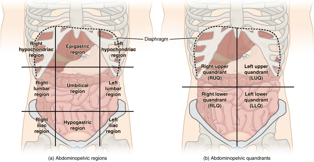
Abdominal assessment is the one examination where the usual order is deliberately broken. You inspect, then auscultate, then percuss, then palpate. Percussion and palpation stimulate the bowel and alter what you would have heard, so listening must come first. Inspect for contour (flat, rounded, distended, scaphoid), symmetry, visible peristalsis, surgical scars, hernias, stoma and skin changes. Auscultate all four quadrants with the diaphragm; normal bowel sounds occur roughly five to thirty times per minute and are irregular. Before you chart “absent,” you must listen for a full five minutes in each quadrant. High-pitched, rushing, hyperactive sounds suggest early mechanical obstruction; a silent abdomen suggests ileus, late obstruction or peritonitis. Palpate lightly first (about one centimetre) in all quadrants, saving the painful quadrant for last, and stop if the abdomen is rigid.
Pain tells you more than any other single finding, and it comes in three flavours. Visceral pain arises from stretch or distention of a hollow organ; it is dull, crampy, poorly localised and often felt near the midline — the periumbilical ache of early appendicitis, the colic of obstruction. Parietal (somatic) pain arises when inflammation reaches the parietal peritoneum; it is sharp, constant, well localised and made worse by movement, coughing or a bumped stretcher — the right-lower-quadrant point tenderness of appendicitis several hours later. Referred pain is felt at a distance because of shared spinal segments: diaphragmatic irritation from a perforation or a post-laparoscopic gas is felt in the shoulder; biliary pain refers to the right scapula. A patient who moves from vague midline pain to focal pain that hurts when the bed is jostled has moved from irritated to inflamed, and that trajectory is the assessment.
Certain physical findings carry disproportionate weight. Rebound tenderness (pain worse on release than on pressure), involuntary guarding and a board-like rigid abdomen together mean peritoneal irritation until proven otherwise. Absent bowel sounds with distention and vomiting suggests obstruction or ileus. Palpable pulsatile mass in a hypotensive older adult suggests abdominal aortic aneurysm — do not palpate deeply again; notify. Also assess what the abdomen produces: last bowel movement, stool colour and consistency, emesis character, oral intake, weight change and abdominal girth measured at the same marked spot each time. Girth increasing by several centimetres in a shift with a tense abdomen is data, not a nuisance.
Upper versus lower gastrointestinal bleeding
Bleeding proximal to the ligament of Treitz is upper gastrointestinal bleeding; the usual culprits are peptic ulcer disease, erosive gastritis, esophageal or gastric varices and Mallory-Weiss tears from forceful retching. Blood exposed to gastric acid is denatured to a coffee-ground appearance, and haemoglobin degraded through the gut turns stool black, tarry and characteristically foul: melena. Hematemesis — bright red or coffee-ground vomitus — is upper by definition. Bleeding distal to the ligament of Treitz is lower; diverticular bleeding, angiodysplasia, haemorrhoids, ischaemic colitis, inflammatory bowel disease and colorectal cancer dominate, and the stool is bright red or maroon blood (hematochezia). One caution flips the rule: a brisk upper bleed can move so fast that it presents as hematochezia, and such a patient is usually the sickest one in the unit.
Laboratory findings follow the physiology. Haemoglobin (normal roughly 14–18 g/dL in men, 12–16 g/dL in women) and haematocrit (42–52 percent and 37–47 percent) may be normal in the first hours of a large bleed because whole blood is lost — the ratio does not change until plasma is replaced or fluid is given. Do not be reassured by an early haemoglobin. In upper bleeding, digested blood is absorbed as protein and the BUN rises out of proportion to creatinine, giving a BUN-to-creatinine ratio above 20:1 and often above 30:1 (normal BUN 10–20 mg/dL, creatinine 0.6–1.2 mg/dL). Draw a type and crossmatch, coagulation studies (INR normally 0.8–1.1), platelets (150,000–400,000/mm3) and a lactate; a lactate above 2 mmol/L suggests hypoperfusion and above 4 mmol/L is an emergency. Upper endoscopy within twenty-four hours is both diagnostic and therapeutic, allowing clipping, thermal coagulation or epinephrine injection.
Management is resuscitation before diagnosis. Two large-bore peripheral IVs (18 gauge or larger — a 22 gauge will not deliver blood at speed), isotonic crystalloid, supplemental oxygen, continuous monitoring and NPO status come first. Packed red cells are transfused using a restrictive threshold: haemoglobin below 7 g/dL for most patients, below 8 g/dL for those with coronary artery disease; one unit raises haemoglobin about 1 g/dL. A high-dose proton pump inhibitor (pantoprazole IV bolus followed by infusion) raises gastric pH so a clot can stabilise. If varices are suspected, octreotide reduces splanchnic flow and prophylactic antibiotics are given. Reversal is considered for anticoagulated patients and is drug-specific: vitamin K plus four-factor prothrombin complex concentrate for warfarin, followed by the INR; protamine sulfate for heparin, followed by the aPTT; idarucizumab for dabigatran; and andexanet alfa for apixaban or rivaroxaban — the direct oral anticoagulants are not reversed by vitamin K, and their effect is not tracked by the INR. Hold home NSAIDs, aspirin and anticoagulants pending provider decision — do not resume them on your own.
Priorities, red flags and who does what
When a patient vomits blood, the order of operations is airway first: position upright or lateral to protect against aspiration, suction available, oxygen on. Then circulation: assess level of consciousness, heart rate, blood pressure, capillary refill and urine output (a goal of at least 0.5 mL/kg/hr, or roughly 30 mL/hr in an average adult). Then access and blood products. Then notify. A common test trap asks whether to insert the nasogastric tube, send labs or start fluids first — the answer is nearly always the intervention that supports oxygenation and perfusion. Never insert or blindly reposition a nasogastric tube in a patient with known varices, recent esophageal surgery, facial trauma or a suspected basilar skull fracture without a specific order.
SBAR — escalating an acute bleed. Situation: “This is Maya Ortiz, RN, on 4 East. Mr. Delgado in 412, sixty-eight, admitted with anaemia, just had 400 mL of coffee-ground emesis.” Background: “He has osteoarthritis, takes daily ibuprofen and apixaban for atrial fibrillation. Admission haemoglobin was 9.1.” Assessment: “Heart rate 118, blood pressure 92/54 down from 138/78, he is pale and diaphoretic, alert but anxious. Two 18-gauge IVs are in, normal saline is running, type and cross is drawn.” Recommendation: “I think this is an active upper GI bleed. I need you to come see him now. Can I have orders for IV pantoprazole, transfusion parameters and a gastroenterology consult for urgent endoscopy?”
| Task on a GI bleed patient | Who may do it | Why |
|---|---|---|
| Vital signs on a stable patient, intake and output, daily weights, oral care, ambulation | UAP | Standardised, non-assessment tasks; results are reported to the RN, who interprets them |
| Obtaining a stool specimen, emptying drainage, positioning, hygiene after melena | UAP | Task-based; the RN inspects and documents stool character |
| Administering scheduled oral or enteral medications, reinforcing teaching already begun, monitoring a stable patient | LPN/LVN | Within scope for predictable patients; the RN retains assessment and evaluation |
| Initial and ongoing assessment, interpreting a rising heart rate, blood transfusion initiation and monitoring, IV push medications, patient teaching, care of the unstable patient | RN only | Requires nursing judgement and cannot be delegated |
Older-adult variation. Older adults bleed silently. Reduced gastric mucosal defence, widespread NSAID and anticoagulant use, and blunted autonomic responses mean the presentation is often fatigue, dyspnoea on exertion, a fall or new delirium rather than pain or frank vomiting. Anaemia is frequently the first laboratory clue. Because thirst perception is blunted and renal concentrating ability is reduced, they decompensate with smaller volume losses. Treat new confusion in an older adult with any gastrointestinal complaint as a haemodynamic sign until you have proved otherwise.
Assess the abdomen in the order inspect–auscultate–percuss–palpate, and in any suspected gastrointestinal bleed treat perfusion first — oxygen, two large-bore IVs, fluids and type and cross — because the initial haemoglobin will not tell you how much blood has been lost.
Section 2Esophageal and Gastric Disorders
Gastroesophageal reflux disease and hiatal hernia
The lower esophageal sphincter is a zone of tonic contraction, not a true valve. When resting tone falls or transient relaxations become frequent, gastric contents move into an esophagus that has no protective mucus layer — and the acid burns. That single mechanism explains the whole syndrome: pyrosis (heartburn) thirty to sixty minutes after meals, worse lying flat or bending forward; regurgitation of sour fluid; water brash; dysphagia if the chronic inflammation has produced a stricture; chronic cough, hoarseness, sore throat and asthma exacerbations when refluxate reaches the larynx or airway. Anything that lowers sphincter tone or raises intra-abdominal pressure aggravates it: caffeine, chocolate, peppermint, alcohol, nicotine, fatty meals, calcium channel blockers, nitrates, anticholinergics, obesity, pregnancy and large late meals.
Diagnosis is often clinical, confirmed by response to therapy. Upper endoscopy is indicated for alarm features — dysphagia, odynophagia, weight loss, anaemia, bleeding, vomiting or age over sixty with new symptoms — and for surveillance of Barrett esophagus, in which chronic acid injury converts squamous epithelium to intestinal-type columnar epithelium. Barrett is a premalignant change carrying meaningful risk of esophageal adenocarcinoma, so those patients need scheduled endoscopic surveillance and lifelong acid suppression. Ambulatory pH monitoring and manometry are reserved for unclear cases or pre-surgical evaluation. Nursing after endoscopy is simple and non-negotiable: keep the patient NPO until the gag reflex returns, monitor for bleeding, crepitus and severe pain that could signal perforation.
Management begins with behaviour. Teach the patient to elevate the head of the bed six to eight inches on blocks (extra pillows bend the trunk and worsen reflux), to finish eating three hours before lying down, to eat smaller meals, to lose weight, to stop smoking and to avoid tight waistbands. Proton pump inhibitors such as pantoprazole and omeprazole irreversibly block the gastric proton pump and are taken thirty to sixty minutes before the first meal of the day; long-term use is associated with Clostridioides difficile infection, community-acquired pneumonia, hypomagnesaemia, vitamin B12 deficiency and fractures, so they are prescribed at the lowest effective dose. Histamine-2 receptor antagonists (famotidine) are weaker and useful for breakthrough symptoms. Antacids neutralise acid already present, work in minutes, and must be separated from other medications by one to two hours; aluminium salts constipate, magnesium salts cause diarrhoea, and magnesium-containing antacids are avoided in renal impairment.
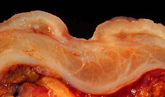
A hiatal hernia is herniation of stomach through the diaphragmatic hiatus. The common sliding (type I) hernia moves the gastroesophageal junction upward and simply worsens reflux. The uncommon paraesophageal (type II) hernia lets the fundus roll up beside the esophagus, where it may become incarcerated or strangulated — a surgical emergency signalled by severe chest or epigastric pain, retching without vomiting and inability to pass a nasogastric tube. Antireflux surgery (Nissen fundoplication, often laparoscopic) wraps the fundus around the distal esophagus. Teach these patients that early dysphagia and gas-bloat are expected, that they should avoid carbonated beverages and straws, chew thoroughly, and understand that they may be unable to belch or vomit — persistent nausea needs a call, not a home remedy.
Peptic ulcer disease and Helicobacter pylori
Ulcers form when aggressive factors overwhelm mucosal defence. Two aggressors account for nearly all of them. Helicobacter pylori is a spiral, urease-producing bacterium that colonises gastric mucus, neutralises its microenvironment with ammonia and provokes chronic inflammation. NSAIDs and aspirin inhibit cyclooxygenase-1, cutting the prostaglandins that maintain mucus, bicarbonate and mucosal blood flow — which is why an enteric coating does not protect the stomach; the injury is systemic, not topical. Corticosteroids, smoking, alcohol and severe physiological stress (the stress-related mucosal injury of ventilated or coagulopathic ICU patients) compound the risk. Zollinger-Ellison syndrome, a gastrin-secreting tumour, produces multiple refractory ulcers and is the rare exception.
Location shapes the story. Duodenal ulcers are more common, occur with normal or high acid output, and produce burning epigastric pain two to three hours after eating and often at one or two in the morning; food or antacids relieve it, and weight is stable or increased. Gastric ulcers occur with normal or low acid output and defective mucosa, produce pain thirty to sixty minutes after eating that food makes worse, and lead to anorexia and weight loss; they are more common after age fifty and, unlike duodenal ulcers, may be malignant, so gastric ulcers are biopsied and followed to healing. Diagnosis uses endoscopy with biopsy plus H. pylori testing by urea breath test, stool antigen or biopsy urease test. Both breath and stool testing require holding proton pump inhibitors for two weeks and antibiotics and bismuth for four weeks beforehand, or the test will be falsely negative.
Eradication uses combination therapy for fourteen days, most often bismuth quadruple therapy: a proton pump inhibitor plus bismuth subsalicylate plus metronidazole plus tetracycline. Teach three things. First, finish the entire course — partial treatment breeds resistance and the ulcer returns. Second, bismuth turns the stool and tongue black; that is harmless and is not melena, but the patient must know the difference. Third, metronidazole with alcohol produces a disulfiram-like reaction of flushing, vomiting and tachycardia, so no alcohol during therapy and for three days after. Sucralfate forms a physical barrier over the ulcer base and must be given on an empty stomach, one hour before meals and at bedtime, separated from other drugs and antacids. Misoprostol restores prostaglandin for high-risk NSAID users and is absolutely contraindicated in pregnancy because it induces uterine contractions.
Gastric surgery, gastric cancer and dumping syndrome
Gastric adenocarcinoma is insidious: vague epigastric discomfort, early satiety, anorexia, unexplained weight loss and iron-deficiency anaemia that appear only after the tumour is advanced. Risk rises with H. pylori infection, atrophic gastritis, pernicious anaemia, heavy salt-preserved and smoked food intake, smoking and a family history. Endoscopy with biopsy is definitive; CT and endoscopic ultrasound stage it. Treatment is surgical resection (partial or total gastrectomy) with chemotherapy, and the nursing focus is nutritional. After any gastrectomy the patient loses intrinsic factor and therefore cannot absorb vitamin B12 from the diet — lifelong parenteral or high-dose oral B12 replacement is required, and failure to teach that produces a preventable megaloblastic anaemia with irreversible neuropathy years later. Iron, calcium and folate deficiencies also follow.
Whenever the pylorus is bypassed or removed — gastrectomy, gastrojejunostomy, and also Roux-en-Y gastric bypass — hyperosmolar chyme dumps into the jejunum. Early dumping syndrome occurs fifteen to thirty minutes after eating: the osmotic load pulls fluid into the lumen, producing cramping, borborygmi, nausea, urgent diarrhoea, and simultaneously reduces circulating volume, producing tachycardia, palpitations, diaphoresis, dizziness and pallor. Late dumping occurs one and a half to three hours after eating: the rapid glucose bolus triggers excessive insulin release and the patient becomes hypoglycaemic, with sweating, shakiness, confusion and weakness. Teach small, frequent meals; high protein and moderate fat; low simple carbohydrate; fluids between meals rather than with them; and lying down or reclining for twenty to thirty minutes after eating to slow gastric emptying — the opposite of reflux teaching, which is exactly why students confuse the two.
After bariatric surgery specifically, the emergency to recognise is an anastomotic leak, typically in the first days: sustained heart rate above 120, restlessness or a sense of impending doom, fever, left shoulder pain, and increasing abdominal or back pain. Tachycardia in a fresh bariatric patient is a leak until proven otherwise — notify the surgeon rather than medicating for anxiety. Routine post-gastric-surgery care includes keeping the nasogastric tube patent but never repositioning or irrigating it without a specific order (the tip sits at a fresh suture line), monitoring drainage that should change from bloody to yellow-green within the first day, early ambulation, and slow advancement from sips of clear liquid to small soft meals.
Reflux and ulcer disease are both failures of mucosal defence — treat the cause (acid suppression, H. pylori eradication, stopping NSAIDs), teach the position that helps (upright after meals for reflux, recumbent after meals for dumping) and watch for the sudden rigid abdomen that means perforation.
Section 3Inflammatory and Infectious Disorders of the Bowel
Appendicitis and peritonitis
Appendicitis begins with obstruction of the appendiceal lumen by a fecalith, lymphoid hyperplasia or, in older adults, a tumour. Mucus continues to be secreted behind the obstruction, pressure rises, venous drainage fails, bacteria invade the wall and the appendix becomes ischaemic and eventually perforates. That sequence explains the classic migration of pain: distention of a hollow viscus produces vague periumbilical visceral pain with anorexia and nausea, and once inflammation reaches the parietal peritoneum the pain moves to a sharp, focal point at McBurney point, one-third of the way from the right anterior superior iliac spine to the umbilicus. Look for rebound tenderness, guarding, pain on coughing or heel-strike, Rovsing sign (right-lower-quadrant pain when the left lower quadrant is pressed), a low-grade fever of 99–101 °F and a white blood cell count of 10,000–18,000/mm3 (normal 5,000–10,000/mm3); a count above 20,000/mm3 suggests perforation.
Nursing care before surgery is a list of prohibitions, and each one has a reason. Nothing by mouth. No heat to the abdomen — vasodilation accelerates rupture. No enemas or laxatives — increased peristalsis and pressure accelerate rupture. Position of comfort is usually semi-Fowler with knees flexed. Give IV fluids and antibiotics as ordered, and monitor closely. The single most important observation: if severe pain suddenly stops, the appendix has likely ruptured and released the pressure; a brief calm is followed by diffuse pain, rigidity, rising fever and tachycardia. That sudden relief is a red flag, not good news, and it warrants immediate notification. Definitive management is appendectomy, usually laparoscopic; perforated cases may need drain placement and several days of antibiotics.
Peritonitis is the common endpoint of perforation from any cause — ruptured appendix or diverticulum, perforated ulcer, ischaemic bowel, anastomotic leak, or peritoneal dialysis catheter contamination. Bacteria and chemical irritants inflame a membrane with enormous surface area, so fluid shifts into the peritoneal cavity (third spacing), circulating volume falls, and sepsis follows. Findings: severe constant abdominal pain, board-like rigidity, rebound tenderness, absent bowel sounds, shallow rapid breathing because deep breaths hurt, the patient lying still with knees flexed, fever, tachycardia and hypotension. Treatment is broad-spectrum antibiotics, aggressive fluid resuscitation, nasogastric decompression, NPO status, analgesia and surgical source control. Nursing priorities follow ABC and perfusion: oxygen, fluids, hourly urine output, serial lactate and blood cultures before antibiotics whenever that does not delay them.
Diverticular disease
Diverticulosis is the presence of small herniations of mucosa through weak points in the colonic muscular wall, overwhelmingly in the sigmoid colon where luminal pressure is highest. It is a disease of low dietary fibre and ageing; more than half of adults over sixty have it, and most never know. It becomes clinically important two ways. It is the most common cause of brisk painless lower gastrointestinal bleeding in older adults, because a diverticulum sits directly over a penetrating arteriole. And it can inflame.
Diverticulitis occurs when a diverticulum becomes obstructed and micro-perforates. The patient reports steady left-lower-quadrant pain (the mirror image of appendicitis), with fever, leukocytosis, nausea and a change in bowel habit. The diagnostic study is CT of the abdomen and pelvis with contrast. Note carefully what is contraindicated during the acute phase: colonoscopy and barium enema, because insufflation or contrast pressure can convert a contained micro-perforation into a free one. Colonoscopy is done six to eight weeks after recovery to exclude cancer, which can mimic diverticulitis exactly.
Mild uncomplicated cases are managed at home with clear liquids and often oral antibiotics; more ill patients are admitted for bowel rest, IV fluids and IV antibiotics covering gut flora (commonly ciprofloxacin plus metronidazole, or a beta-lactam/beta-lactamase inhibitor). Teach the diet sequence and the reason: low-fibre or clear liquids during the flare to rest the colon, then advance to a high-fibre diet (25–35 grams daily) with generous fluids once inflammation resolves, to prevent recurrence. The old instruction to avoid nuts, seeds, popcorn and corn has been abandoned — evidence does not support it. Complications requiring surgery include abscess, free perforation with peritonitis, obstruction from stricture, and fistula — a colovesical fistula announces itself with pneumaturia and fecaluria, which the patient may describe only as “bubbles when I urinate” unless you ask directly.
Inflammatory bowel disease, celiac disease, irritable bowel syndrome and Clostridioides difficile
Inflammatory bowel disease is immune-mediated chronic inflammation in a genetically susceptible host. Distinguishing its two forms is worth more test points than almost anything else in this chapter, and the distinction is anatomical: how much of the bowel and how deep.
| Feature | Crohn disease | Ulcerative colitis |
|---|---|---|
| Location | Anywhere from mouth to anus; terminal ileum most common; skip lesions with normal bowel between | Rectum extending proximally in a continuous pattern; colon only |
| Depth | Transmural — all layers; cobblestone mucosa | Mucosa and submucosa only |
| Stool | Three to five soft or loose stools daily, steatorrhoea, usually without gross blood | Ten to twenty liquid stools daily with blood and mucus, urgency, tenesmus |
| Pain | Cramping right lower quadrant, often with a palpable mass | Left lower quadrant cramping relieved by defecation |
| Characteristic complications | Fistula, abscess, stricture and obstruction; perianal disease; B12 and fat-soluble vitamin malabsorption after ileal disease or resection | Toxic megacolon, massive haemorrhage, perforation; higher colorectal cancer risk |
| Surgery | Not curative; disease recurs at the anastomosis; resections are kept short | Total proctocolectomy is curative |
Both share extraintestinal manifestations — arthritis, erythema nodosum, uveitis, aphthous ulcers, primary sclerosing cholangitis — and both cause weight loss, fatigue, iron-deficiency anaemia, hypoalbuminaemia (normal albumin 3.5–5.0 g/dL) and electrolyte loss. Useful labs include CBC, C-reactive protein, erythrocyte sedimentation rate, albumin, iron studies, vitamin B12 and fecal calprotectin, which is elevated in inflammatory bowel disease and normal in irritable bowel syndrome. Colonoscopy with biopsy establishes the diagnosis; CT or MR enterography maps small-bowel Crohn disease.
Drug therapy is stepwise by mechanism. Aminosalicylates (mesalamine, sulfasalazine) deliver topical anti-inflammatory effect to the mucosa and are the backbone for mild ulcerative colitis; sulfasalazine is a sulfonamide, so ask about sulfa allergy, expect orange-yellow discoloration of urine and skin, and supplement folic acid because absorption is impaired. Corticosteroids induce remission in flares and are tapered, never used for maintenance because of osteoporosis, hyperglycaemia, infection and adrenal suppression. Immunomodulators (azathioprine, 6-mercaptopurine, methotrexate) are steroid-sparing and require serial CBC and liver function monitoring; methotrexate demands folate supplementation and reliable contraception. Biologics — anti-tumour necrosis factor agents such as infliximab and adalimumab, plus vedolizumab and ustekinumab — are potent immunosuppressants; before the first dose the patient must be screened for latent tuberculosis and hepatitis B, and during therapy live vaccines are avoided and any fever is treated as potentially serious. Antidiarrhoeals are used cautiously and are contraindicated in a severe flare because slowing the colon can precipitate toxic megacolon.
Celiac disease is an immune reaction to gluten that flattens small-intestinal villi, producing diarrhoea, steatorrhoea, bloating, weight loss, iron and folate deficiency anaemia, osteoporosis and the intensely itchy vesicular rash dermatitis herpetiformis. Diagnosis uses tissue transglutaminase IgA antibodies drawn while the patient is still eating gluten, confirmed by duodenal biopsy. The only treatment is lifelong strict elimination of wheat, barley and rye; teach label reading, cross-contamination in shared toasters and fryers, and that rice, corn, quinoa and certified uncontaminated oats are safe. Irritable bowel syndrome, by contrast, is a disorder of gut-brain interaction with normal structure and normal labs: recurrent abdominal pain related to defecation with altered stool form or frequency, no bleeding, no weight loss, no nocturnal symptoms. Management is dietary (soluble fibre, a trial of a low-FODMAP plan), stress reduction, antispasmodics such as dicyclomine, and agents targeted to the predominant pattern. Any alarm feature — bleeding, anaemia, weight loss, onset after age fifty, nocturnal diarrhoea — means it is not irritable bowel syndrome and requires workup.
Clostridioides difficile infection follows disruption of normal flora by antibiotics — classically clindamycin, fluoroquinolones and cephalosporins — allowing a spore-forming anaerobe to release toxins A and B that inflame the colon. Risk rises with age over sixty-five, hospitalisation, tube feeding and proton pump inhibitor use. The patient has frequent watery, foul-smelling stools, cramping, fever and leukocytosis that can be striking (a white count above 15,000/mm3 is a severity marker), with rising creatinine and falling albumin in severe disease. Diagnosis is stool nucleic acid amplification and toxin testing on liquid stool only; do not send formed stool and do not send a test of cure. Treatment is oral vancomycin or fidaxomicin; recurrent disease may be treated with faecal microbiota transplantation. The nursing actions that matter most are infection control: contact precautions in a private room, dedicated equipment, cleaning with a sporicidal bleach agent, and handwashing with soap and water — alcohol-based gel does not kill spores. Discontinue antidiarrhoeals, replace fluid and potassium (normal 3.5–5.0 mEq/L), and review whether the inciting antibiotic and the proton pump inhibitor can be stopped.
Localise and go deeper: right-lower-quadrant pain that migrated means appendicitis, left-lower-quadrant pain with fever means diverticulitis, bloody mucoid stools from a continuously inflamed rectum mean ulcerative colitis, and any of them can end in a rigid abdomen — while distention with a falling stool count in colitis means toxic megacolon and an immediate call.
Section 4Obstruction, Ischaemia and Colorectal Cancer
Mechanical obstruction and paralytic ileus
Obstruction is either mechanical (something blocks the lumen) or functional (the bowel stops moving). In mechanical obstruction, gas and fluid accumulate above the blockage; the bowel distends, secretes more fluid, absorbs less, and litres of extracellular fluid are sequestered in the lumen. That third spacing produces hypovolaemia, electrolyte loss and, if pressure exceeds capillary perfusion pressure, strangulation — ischaemic bowel that necroses, perforates and seeds peritonitis. Adhesions from prior surgery cause most small-bowel obstructions; hernias and Crohn strictures follow. In the large bowel, colorectal cancer, volvulus and diverticular stricture dominate.
| Feature | Small-bowel obstruction | Large-bowel obstruction | Paralytic ileus |
|---|---|---|---|
| Onset | Rapid | Gradual, over days | Follows surgery, opioids, hypokalaemia, peritonitis, immobility |
| Pain | Colicky, periumbilical, waves every few minutes | Lower abdominal cramping, less intense | Diffuse discomfort, distention more than pain |
| Vomiting | Early, profuse; bilious then feculent if distal | Late; may be feculent | Possible, usually with a high nasogastric output |
| Distention | Minimal early if proximal, marked if distal | Marked; obstipation is prominent | Marked, tympanic, diffuse |
| Bowel sounds | High-pitched and hyperactive early, absent late | Hypoactive to absent | Absent or hypoactive from the start |
| Acid-base pattern | Metabolic alkalosis from vomiting gastric hydrochloric acid | Metabolic acidosis is more likely with distal loss and ischaemia | Depends on cause and losses |
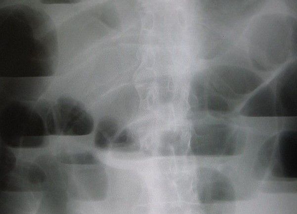
Assessment is serial and quantitative: vital signs, abdominal girth at a marked line, bowel sounds, character and volume of emesis and nasogastric output, strict intake and output, daily weight, and electrolytes — especially potassium (3.5–5.0 mEq/L), sodium (135–145 mEq/L) and chloride, all of which fall with prolonged suction and vomiting. Abdominal CT identifies the level, cause and any ischaemia; plain films show dilated loops with air-fluid levels. Non-strangulating adhesive obstruction is often managed conservatively: NPO, nasogastric tube to low intermittent suction, isotonic fluid replacement with potassium once urine output is adequate, analgesia, and serial examination. Volvulus may be reduced endoscopically; cancer, strangulation, closed-loop obstruction and failure of conservative therapy go to the operating room.
Escalate immediately for fever, tachycardia, rising or persistent pain that becomes constant rather than colicky, localised tenderness with rebound, rigidity, a rising white count or lactate, hypotension, or bloody nasogastric or rectal output. Those signal strangulation or perforation. Paralytic ileus, by contrast, is usually a post-operative or metabolic problem: manage the cause. Replace potassium, minimise opioids in favour of multimodal analgesia, get the patient walking, and consider chewing gum, which has good evidence for shortening post-operative ileus. Mesenteric ischaemia deserves its own mention because it is missed: an older adult with atrial fibrillation or atherosclerosis who has severe pain out of proportion to a soft, benign-feeling abdomen, often with a rising lactate, may have an occluded mesenteric artery. That combination is an emergency.
Colorectal cancer, screening and stoma creation
Most colorectal cancers arise from adenomatous polyps over roughly ten years, which is why screening works — removing a polyp prevents a cancer. Risk factors include age, a personal or family history of colorectal cancer or adenomas, inflammatory bowel disease of long duration, hereditary syndromes (familial adenomatous polyposis, Lynch syndrome), obesity, physical inactivity, smoking, heavy alcohol and a diet high in processed and red meat and low in fibre. For average-risk adults, screening now begins at age 45 and continues to 75: colonoscopy every ten years, or a faecal immunochemical test annually, or stool DNA testing every three years, or CT colonography every five years. A positive non-invasive test always requires a diagnostic colonoscopy.
Manifestations depend on side. Right-sided tumours present with occult bleeding and iron-deficiency anaemia, fatigue and vague pain, because stool there is liquid and the lumen is wide. Left-sided and rectal tumours present with a change in bowel habit, narrowed “pencil” stools, tenesmus, hematochezia and obstruction. Any unexplained iron-deficiency anaemia in an adult over fifty is colorectal cancer until proven otherwise. Colonoscopy with biopsy confirms; CT of the chest, abdomen and pelvis stages the disease by the TNM system, in which the depth the tumour has invaded through the bowel wall (T), the number of positive regional lymph nodes (N) and the presence of distant metastasis (M, most often liver or lung) together determine stage and prognosis; carcinoembryonic antigen (CEA, usually less than about 3 ng/mL in a non-smoker) is not a screening test but is used to monitor for recurrence after resection. Treatment combines surgical resection with chemotherapy and, for rectal cancer, radiation. Nursing preparation for colonoscopy is its own teaching moment: clear liquids with nothing red or purple, the full bowel preparation solution taken as scheduled even though it is unpleasant, and arrangements for a driver because of sedation.
When bowel is resected, a stoma may be created — temporary to protect a distal anastomosis, or permanent when the rectum is removed. Output depends on location. An ileostomy begins draining within 24 hours, produces 500–1,000 mL daily of liquid to pasty effluent rich in sodium and potassium and containing digestive enzymes that excoriate skin instantly; those patients dehydrate easily and must not take enteric-coated or extended-release tablets, which pass through unabsorbed. A descending or sigmoid colostomy produces formed stool on a semi-predictable schedule. Assess the stoma every shift: it should be pink to beefy red, moist and slightly raised, with mild oedema for the first week. A pale stoma suggests anaemia; a dusky, purple, dark blue or black stoma means ischaemia and must be reported immediately. Report also a stoma that retracts below skin level, prolapses, bleeds beyond a small ooze, or produces no output with distention and cramping — that is obstruction.
Older-adult variation. Older adults with obstruction or peritonitis often lack fever and leukocytosis, and delirium may be the presenting sign. They are more likely to have a large-bowel obstruction from cancer, more likely to be on anticholinergics, calcium channel blockers and opioids that cause ileus, and less able to tolerate the fluid shifts of third spacing. Do not accept “he is just confused today” in a patient who has not passed stool or flatus for three days and has a tympanic, distended abdomen.
In obstruction the nursing job is decompress, replace and watch — nasogastric suction, isotonic fluid with potassium, strict output and girth — while pain that turns from colicky to constant, fever, rising lactate or a dusky stoma means the bowel is dying and the provider is called now.
Section 5Nutrition, Elimination and the Post-operative Gastrointestinal Patient
Nasogastric tubes and enteral nutrition
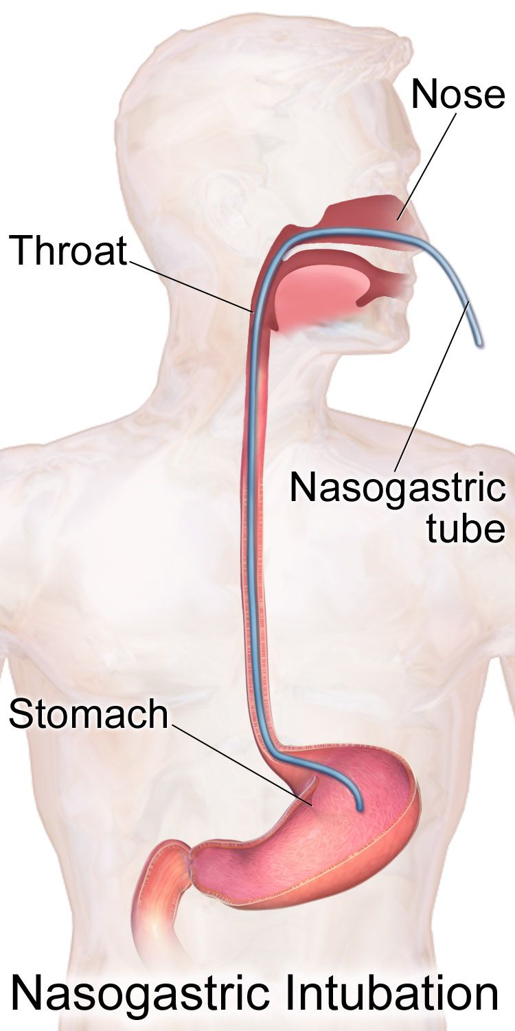
A nasogastric tube does one of two things: it takes contents out (decompression for obstruction, ileus or post-operative distention, usually a double-lumen Salem sump on low intermittent suction with the blue air vent kept above the level of the stomach and never clamped) or it puts nutrition in (a small-bore feeding tube). To insert, place the patient in high Fowler position, measure from the tip of the nose to the earlobe to the xiphoid process, mark the tube, lubricate with water-soluble gel, advance along the floor of the nostril, and have the patient flex the chin toward the chest and sip water as you pass the tube — chin flexion closes the airway and opens the esophagus. Stop and withdraw for coughing, choking, gasping or a change in phonation.
Verification is where patients are harmed. Radiographic confirmation is required before the first use of any newly placed blind feeding tube — auscultating an air bolus is not acceptable practice and cannot distinguish stomach from lung. After confirmation, mark the exit point at the nostril, document the external length, and before every feeding and medication and at least every four hours during continuous feeding, verify by checking that the marked length is unchanged and by aspirating gastric contents, which should be grassy-green, tan or bloody with a pH of 5.5 or less; small-bowel aspirate is typically golden and above 6, and respiratory secretions are also above 6. Any doubt means stop and re-image. Never reposition or irrigate a tube placed at surgery near a fresh anastomosis without an explicit order.
Enteral feeding is preferred over parenteral whenever the gut works, because it maintains mucosal integrity and reduces infection. The dominant risk is aspiration: keep the head of bed elevated 30–45 degrees during feeding and for at least 30–60 minutes after intermittent feeds, hold feeding for procedures requiring the patient to lie flat, and provide oral care every four hours. Flush with 30 mL of water before and after each medication, between medications and every four hours during continuous feeding to maintain patency. Give liquid formulations when available; never crush enteric-coated or extended-release tablets, and never mix medications together in the syringe. Monitor gastric residual volumes per facility policy — a single elevated value is not a reason to stop feeding, but a residual above 500 mL together with distention, vomiting or discomfort means hold the feeding and notify. Diarrhoea during tube feeding is usually caused by sorbitol-containing liquid medications, antibiotics, hyperosmolar formula or C. difficile, not by the formula itself; investigate before you stop the nutrition.
Constipation, diarrhoea and the drugs that treat them
Constipation is not a diagnosis; it is a symptom of low fibre, low fluid, immobility, ignored urges, opioids, anticholinergics, iron, calcium channel blockers, hypothyroidism, hypokalaemia or obstruction. Assess the patient’s baseline pattern rather than a textbook number, and screen for the emergency masquerading as constipation: a faecal impaction presents with paradoxical liquid stool leaking around a hard mass, abdominal distention, anorexia and, in an older adult, confusion or urinary retention. Non-drug therapy comes first — 25–35 grams of fibre daily, 1.5–2 litres of fluid unless restricted, activity, and honouring the urge, ideally after breakfast when the gastrocolic reflex is strongest.
Know laxatives by mechanism. Bulk-forming agents (psyllium, methylcellulose) draw water into stool and must be taken with a full glass of fluid or they can obstruct. Osmotic agents (polyethylene glycol, lactulose, magnesium citrate) hold water in the lumen; magnesium products are avoided in renal impairment. Stimulants (senna, bisacodyl) increase peristalsis; do not crush enteric-coated bisacodyl and do not take it within one hour of milk or antacids, which dissolve the coating and cause gastric irritation. Stool softeners (docusate) are surfactants and are weak on their own. For opioid-induced constipation, a stimulant plus softener is started prophylactically with the opioid, and peripherally acting mu-antagonists such as methylnaltrexone are available when that fails.
Diarrhoea threatens volume and potassium before it threatens anything else. Assess frequency, volume, blood, recent antibiotics, travel, tube feeding and new medications; check orthostatic vital signs, mucous membranes, urine output, potassium, sodium and creatinine. Loperamide and diphenoxylate slow transit but are contraindicated in suspected C. difficile, in bloody diarrhoea with fever and in severe colitis, because retaining toxin risks toxic megacolon. Antiemetics have their own cautions: ondansetron prolongs the QT interval, promethazine is a vesicant that causes severe tissue injury and is given deeply and dilutely if at all, and metoclopramide carries a black-box warning for tardive dyskinesia and should not be used beyond twelve weeks. Protect perineal skin with a barrier cream and cleanse gently; incontinence-associated dermatitis is a preventable injury.
The post-operative gastrointestinal patient and discharge teaching
Modern enhanced-recovery practice has changed post-operative abdominal care: routine nasogastric tubes are avoided, clear liquids often begin the day of surgery, multimodal analgesia limits opioids, and patients are out of bed the first evening. Your surveillance targets three complications. Ileus — distention, no flatus, absent sounds — is managed with ambulation, potassium repletion, opioid reduction and gum chewing. Anastomotic leak — classically around post-operative day five to seven — presents with fever, tachycardia, increasing rather than decreasing pain, purulent or feculent drain output and peritoneal signs; it is a surgical emergency. Surgical-site and intra-abdominal infection presents with fever, wound erythema and drainage, or a persistent leukocytosis. Wound evisceration — viscera protruding through a separated incision — is handled by placing the patient supine with knees flexed, covering the viscera with sterile saline-moistened gauze, keeping them NPO and calling the surgeon immediately; never attempt to replace the bowel.
Discharge teaching should be specific enough to act on. Every gastrointestinal patient goes home knowing the red flags: fever above 100.4 °F, vomiting that prevents fluids, severe or worsening abdominal pain, a rigid abdomen, no bowel movement or flatus with distention, black tarry or bloody stools, dizziness on standing, and incision redness, warmth or drainage. Ostomy patients additionally need to know to report output above roughly 1,000–1,500 mL a day from an ileostomy or no output for several hours with cramping, to chew high-residue foods (nuts, popcorn, corn, mushrooms, celery, raw cabbage) thoroughly to prevent blockage, to replace fluid and salt in hot weather or illness, and where their supplies and their ostomy nurse are. Reflux and ulcer patients need the medication timing, the NSAID conversation and the follow-up endoscopy date. Inflammatory bowel disease patients on biologics need to know to report fever and to keep vaccinations current with live vaccines avoided.
Delegation reminder. The unlicensed assistive personnel may empty and measure an established ostomy pouch, record output, assist with ambulation and hygiene, and take vital signs on a stable patient. The LPN/LVN may perform routine appliance changes on a healed stoma, administer most enteral and oral medications, reinforce teaching you have started and monitor a predictable patient. The registered nurse assesses the stoma and the abdomen, verifies feeding-tube placement decisions, initiates blood and parenteral nutrition, evaluates response, teaches the new ostomy patient and manages anyone whose condition is changing.
Feed the gut when it works, but prove tube placement radiographically before the first use, keep the head of bed at 30–45 degrees, watch phosphorus in the malnourished patient who is being refed, and treat new fever with increasing pain after abdominal surgery as an anastomotic leak until the surgeon says otherwise.
The chapter in one breath
Gastrointestinal disease is a set of failures in motility, absorption and containment, and reading it that way turns memorisation into reasoning. Assess the abdomen by inspecting, auscultating, percussing and palpating in that order, and follow pain as it converts from vague visceral ache to sharp parietal tenderness — that conversion is inflammation reaching the peritoneum. In bleeding, treat perfusion before diagnosis: oxygen, two large-bore lines, isotonic fluid, type and cross, and remember that the first haemoglobin lies while an elevated BUN-to-creatinine ratio points upstream. Reflux and peptic ulcer disease are defence failures treated by suppressing acid, eradicating Helicobacter pylori and stopping NSAIDs, with perforation and its rigid abdomen as the emergency. Appendicitis, diverticulitis, inflammatory bowel disease and C. difficile each localise differently but converge on peritonitis and toxic megacolon if missed. Obstruction is decompressed, replaced and watched, with pain that turns constant, fever or a rising lactate signalling strangulation. Colorectal cancer is prevented by screening from age 45 and survived through resection, and a new stoma must stay pink and moist. Feed the gut when it works, prove tube placement with a film, elevate the head of the bed, and in every scenario ask the same question first: airway, breathing, circulation, then what is most likely to kill this patient in the next hour.
Words worth knowing
| Term | What it means |
|---|---|
| Melena | Black, tarry, strongly foul stool from blood digested during transit; indicates upper gastrointestinal bleeding. |
| Hematochezia | Bright red or maroon blood passed rectally; usually lower gastrointestinal bleeding, but can indicate a very brisk upper bleed. |
| Pyrosis | Heartburn — substernal burning caused by gastric contents refluxing into the esophagus. |
| Barrett esophagus | Replacement of esophageal squamous epithelium by columnar epithelium after chronic acid injury; premalignant for adenocarcinoma. |
| McBurney point | Point one-third of the way from the right anterior superior iliac spine to the umbilicus; maximal tenderness in appendicitis. |
| Rebound tenderness | Pain that is worse when pressure is released than when applied; a sign of peritoneal irritation. |
| Peritonitis | Inflammation of the peritoneum from perforation or infection, producing rigidity, absent bowel sounds, third spacing and sepsis. |
| Skip lesions | Patches of diseased bowel separated by normal bowel; characteristic of Crohn disease. |
| Tenesmus | Painful, ineffective straining with a persistent urge to defecate; typical of rectal inflammation in ulcerative colitis. |
| Toxic megacolon | Acute colonic dilation with systemic toxicity in severe colitis; distention, absent sounds, fever, tachycardia and falling stool frequency. |
| Steatorrhoea | Bulky, greasy, foul-smelling, floating stool caused by fat malabsorption. |
| Diverticulitis | Inflammation or micro-perforation of a colonic diverticulum, producing left-lower-quadrant pain, fever and leukocytosis. |
| Obstipation | Complete inability to pass stool or flatus; a hallmark of large-bowel obstruction. |
| Strangulation | Obstruction severe enough to cut off blood supply to the bowel wall, leading to necrosis and perforation. |
| Paralytic ileus | Functional cessation of peristalsis without a mechanical blockage; commonly post-operative, opioid-related or from hypokalaemia. |
| Dumping syndrome | Rapid emptying of hyperosmolar chyme into the jejunum after gastric surgery, causing early vasomotor symptoms and late reactive hypoglycaemia. |
| Stoma | Surgically created opening of bowel onto the abdominal wall; should appear pink to red, moist and slightly raised. |
| Effluent | The stool output from an ostomy; liquid and enzyme-rich from an ileostomy, formed from a sigmoid colostomy. |
| Refeeding syndrome | Dangerous intracellular shift of phosphate, potassium and magnesium when a malnourished patient is fed again. |
| Carcinoembryonic antigen (CEA) | Tumour marker used to monitor for colorectal cancer recurrence after treatment; not a screening test. |