Postpartum Complications
The hours and weeks after birth are the most dangerous stretch of the childbearing year, and they are the stretch we watch least closely. A birthing parent who walked into the unit healthy can bleed a litre in ten minutes with a blood pressure that has not yet moved, develop severe-range hypertension five days after discharge, throw a pulmonary embolus at three weeks, or be quietly overwhelmed by intrusive thoughts at six. Most of these deaths are judged preventable, and what usually fails is not knowledge of the disease but the speed and order of the response. This chapter is therefore built around sequences: what you assess, what you do first, what you do next, and what you say when you escalate. Massage the fundus before you reach for a drug. Stop the magnesium before you draw up the antidote. Ask about the thoughts before the family leaves the room.
By the end of this chapter you can
- Define early and late postpartum haemorrhage using cumulative quantitative blood loss, and explain why pregnancy’s hypervolaemia delays hypotension.
- Perform the first-response sequence for uterine atony in order — massage the fundus first — and justify each subsequent step.
- Compare the uterotonics (oxytocin, methylergonovine, carboprost, misoprostol) and tranexamic acid by dose, route, timing and absolute contraindication.
- Distinguish the four Ts at the bedside, including genital tract haematoma, retained placental fragments, disseminated intravascular coagulation and uterine inversion.
- Recognise endometritis, mastitis and maternal sepsis, and calculate weight-based antibiotic and fluid doses, showing the arithmetic and the independent double check.
- Manage venous thromboembolism, postpartum preeclampsia and magnesium sulfate toxicity, naming the antidote calcium gluconate and the order in which toxicity appears.
- Differentiate postpartum blues, perinatal depression and anxiety and postpartum psychosis, and act on a positive self-harm item.
- Escalate any of the above using SBAR, and apply delegation limits so that assessment, teaching, evaluation and the unstable patient stay with the registered nurse.
- Teach the POST-BIRTH warning signs to a family in plain, non-blaming language before discharge.
Section 1Early Postpartum Haemorrhage and the First Ten Minutes
Why the numbers deceive you
Postpartum haemorrhage is defined as cumulative blood loss of 1000 mL or more, or blood loss of any volume accompanied by signs or symptoms of hypovolaemia, within 24 hours of birth — regardless of whether the birth was vaginal or caesarean. That second clause matters more than the number. A small person who has lost 700 mL and is suddenly restless, yawning and tachycardic is haemorrhaging; a large person who has lost 1100 mL and is chatting comfortably may not yet be. Loss of more than 500 mL after a vaginal birth, even if it does not meet the definition, should still prompt you to look for a cause. Haemorrhage occurring after the first 24 hours and up to 12 weeks postpartum is called late or secondary haemorrhage and has a different cause list.
Visual estimation of blood loss is unreliable, and the error runs in the dangerous direction: clinicians underestimate large losses and overestimate small ones. Quantitative blood loss (QBL) replaces the guess with a scale. Weigh the blood-soaked pads, drapes, chux and lap sponges, subtract the known dry weight, and convert — one gram equals approximately one millilitre. Use a calibrated under-buttocks drape at vaginal birth and read the graduated pouch, subtracting amniotic fluid volume collected before the placenta delivers. In theatre, measure canister volume and subtract irrigation. QBL is cumulative and is recorded at defined intervals, not once at the end.
The deeper trap is physiological. Pregnancy expands plasma volume by roughly 40 to 50 per cent, which is nature’s own transfusion in anticipation of birth. A previously healthy birthing person can therefore lose 1000 to 1500 mL before the systolic blood pressure falls at all. Blood pressure is a late sign. Tachycardia, narrowing pulse pressure, rising respiratory rate, cool clammy skin, delayed capillary refill and above all a change in mental state — anxiety, air hunger, “something is wrong” — arrive first. Trust the trend and the person, not the single number on the monitor.
Two tools sharpen the eye. The shock index is heart rate divided by systolic blood pressure; a normal postpartum value is 0.5 to 0.7, a value of 0.9 or above should trigger a second look and 1.4 or above signals severe compromise. The maternal early warning criteria flag any of the following for immediate bedside evaluation by a clinician who can escalate: systolic pressure below 90 or above 160 mmHg, diastolic above 100, heart rate below 50 or above 120, respiratory rate below 10 or above 30, oxygen saturation below 95 per cent on room air, urine output under 35 mL per hour for two hours or more, or altered mental status, agitation, unrelieved headache or breathlessness.
The four Ts, and why tone comes first
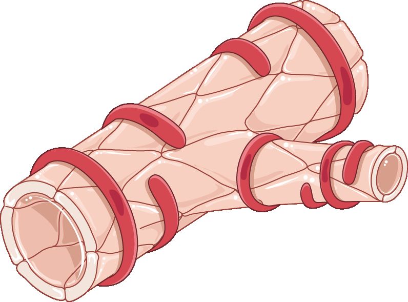
Haemostasis at the placental site is mechanical, not chemical. The myometrium is arranged in interlacing bundles that surround the spiral arterioles; when those bundles contract, they strangle the vessels shut. Clinicians call them the living ligatures. Clotting factors help, but a uterus that will not contract cannot be clotted into safety. This is why the whole first-line management of haemorrhage is aimed at making the uterus squeeze.
Causes sort into four Ts. Tone — uterine atony — accounts for roughly 70 to 80 per cent of cases. Trauma (lacerations, haematoma, rupture, inversion) accounts for most of the remainder, around 20 per cent. Tissue means retained placenta or membranes and includes the placenta accreta spectrum. Thrombin means a coagulation problem, whether inherited, acquired in pregnancy, or consumed by the haemorrhage itself. In practice you assess tone first because it is both the most likely cause and the one you can treat with your hands in the first five seconds.
Risk factors for atony cluster around an overstretched, exhausted or chemically relaxed uterus: multiple gestation, polyhydramnios, a large baby, high parity, a long labour or a very rapid one, prolonged oxytocin exposure during labour, chorioamnionitis, magnesium sulfate infusion, general anaesthesia with halogenated agents, and retained placental tissue. Risk assessment is repeated at three defined points — at prenatal booking, again on admission in labour, and again immediately after birth — because the category changes as labour unfolds. But remember the humbling statistic: a substantial proportion of haemorrhages occur in people categorised as low risk. Every birth gets a fundal check.
One reversible cause hides in plain sight. A full bladder sits directly beneath the lower uterine segment, pushes the uterus up and to one side, and mechanically prevents it from contracting down. A fundus that is boggy, high and deviated laterally — classically to the right — is a bladder problem until you have emptied the bladder. Have the patient void; if they cannot, catheterise. Document the volume.
What you do first
The single most common examination error in postpartum haemorrhage is reaching for a syringe before reaching for the fundus. Massage is faster than any drug, needs no order, and works on the cause of four out of five haemorrhages. Place one hand flat above the symphysis pubis to support and anchor the lower uterine segment — this is not optional, because massaging an unsupported uterus can invert it — and with the other hand cup the fundus and massage firmly in a circular motion until it firms to the consistency of a tennis ball. Expel clots only once the uterus is firm; expressing clots from a boggy uterus simply pushes the fundus further up and can invert it.
Then the sequence unfolds in parallel, not one at a time, because the team you called in step two does several of these at once.
| Order | Action | Why this, why now |
|---|---|---|
| 1 | Massage the fundus with lower-segment support | Mechanical contraction is the definitive haemostat; works in seconds without an order |
| 2 | Call for help — activate the haemorrhage protocol | One nurse cannot massage, give drugs, quantify loss and document; this is a team event |
| 3 | Increase or start the oxytocin infusion | First-line uterotonic, already hanging in most units |
| 4 | Empty the bladder (void or catheterise) | A distended bladder mechanically prevents involution |
| 5 | Two large-bore IV lines (18 gauge or larger); warmed isotonic crystalloid | Volume and a route for blood products; small cannulae will not transfuse fast |
| 6 | Oxygen 10 to 15 L per minute by non-rebreather mask (below 10 L the reservoir bag collapses); keep the patient flat and warm | Maximises oxygen carriage on the red cells that remain; hypothermia worsens coagulopathy |
| 7 | Bloods: full blood count, type and crossmatch, coagulation screen, fibrinogen, lactate | Guides transfusion; fibrinogen predicts severity |
| 8 | Second-line uterotonic if bleeding continues; tranexamic acid | Escalate by drug class rather than repeating a failed one |
| 9 | Quantify blood loss continuously; vital signs every 5 minutes; indwelling catheter for hourly output | Urine output under 30 mL per hour signals inadequate perfusion |
| 10 | Tamponade device, then surgical management | Escalate before the patient becomes coagulopathic, not after |
Notice what is not first. Not the blood pressure cuff, not the call to pharmacy, not fetching the emergency box. Hands on the fundus, voice calling for help. Everything else follows.
Uterotonics, tranexamic acid and what comes after the drugs
Each uterotonic has a contraindication that can kill the patient you are trying to save, and each of those contraindications describes a patient you will meet often. Learn the pairs: methylergonovine with hypertension, carboprost with asthma.
| Drug | Dose and route | Watch for | Do not give if |
|---|---|---|---|
| Oxytocin | 10 to 40 units in 500 to 1000 mL isotonic crystalloid IV infusion, titrated; or 10 units IM | Hypotension and tachycardia if pushed fast; water intoxication with large volumes | Never give undiluted as a rapid IV bolus |
| Methylergonovine | 0.2 mg IM, may repeat every 2 to 4 hours; oral 0.2 mg every 6 to 8 hours for maintenance | Sudden severe hypertension, headache, chest pain | Hypertension, preeclampsia, cardiac disease; avoid the IV route |
| Carboprost tromethamine | 250 micrograms IM or intramyometrial, repeat every 15 to 90 minutes, maximum 8 doses (2 mg total) | Diarrhoea, vomiting, fever, flushing — pre-medicate with an antiemetic and antidiarrhoeal | Asthma (bronchospasm); caution in hepatic, renal or cardiac disease |
| Misoprostol | 600 to 1000 micrograms sublingual, oral or rectal, single dose | Shivering and pyrexia — expected, not infection | Few absolute contraindications; useful where refrigeration is unavailable |
| Tranexamic acid | 1 g IV over 10 minutes, ideally within 3 hours of birth; may repeat 1 g if bleeding persists after 30 minutes or restarts within 24 hours | Hypotension if infused faster than 1 mL per minute; seizures at high dose | Known thromboembolic disease; benefit falls sharply after 3 hours |
Tranexamic acid is an antifibrinolytic, not a uterotonic. It does not make the uterus contract; it stops the clot that forms from being dissolved. Its mortality benefit is time-dependent, which is why it belongs in the early bundle rather than the salvage phase. Give it in addition to uterotonics, never instead of them.
When drugs fail, the next tier is mechanical. Intrauterine balloon tamponade — a fluid-filled balloon inflated inside the uterine cavity, commonly to 300 to 500 mL — presses the endometrial surface against the myometrium. A newer vacuum-induced device does the opposite, applying low-level suction so the uterus collapses inward on itself. Both buy time. While a device is in place, keep an indwelling urinary catheter, watch for concealed bleeding above the balloon (the fundal height rising with a firm uterus and a falling haematocrit), and record drainage-port output as part of QBL. Surgical options follow: uterine compression sutures such as the B-Lynch, uterine or internal iliac artery ligation, uterine artery embolisation by interventional radiology, and finally hysterectomy.
Massive haemorrhage becomes a coagulopathy if you replace only red cells and crystalloid. Massive transfusion protocols deliver packed red cells, fresh frozen plasma and platelets in a balanced ratio, often 1:1:1. Fibrinogen deserves special attention in obstetrics: pregnancy raises it to roughly 350 to 650 mg/dL, so a “normal-looking” level of 250 is actually low for a pregnant patient, and a level below 200 mg/dL predicts severe haemorrhage and calls for cryoprecipitate or fibrinogen concentrate. Keep the patient warm and correct acidosis and hypocalcaemia — the lethal triad of hypothermia, acidosis and coagulopathy is what turns a survivable bleed into an unsurvivable one.
In postpartum haemorrhage the first action is a firm fundal massage with the lower uterine segment supported, because uterine tone — not a drug and not the blood pressure reading — is what stops the bleeding.
Section 2Trauma, Tissue, Thrombin and Uterine Inversion
Trauma: lacerations and the haematoma you cannot see
The clinical discriminator is simple enough to teach in one line: a boggy fundus is atony; a firm fundus with continued bleeding is trauma. If you have massaged the uterus to the firmness of a tennis ball, emptied the bladder, given oxytocin, and blood is still running — often a steady bright red trickle rather than dark clots — the source is a tear. Someone must inspect the cervix, vagina and perineum under good light with adequate analgesia. That is a provider task, but you set it up, position the patient, and keep quantifying.
Genital tract haematoma is the version of trauma that bleeds where you cannot see it. Blood collects in the vulvar, vaginal or retroperitoneal space while the perineal pad stays deceptively dry. The presentation is pain — severe, unrelenting perineal, vaginal or rectal pressure that is out of proportion to the repair and is not relieved by ice or the usual analgesia. On inspection there may be a tense, bluish-purple, unilateral bulging mass; on the vaginal side there may be nothing visible at all. Meanwhile the pulse climbs, the haematocrit falls and the fundus stays firm. A patient who says “I feel like I need to have a bowel movement” an hour after a repair is describing a haematoma until proven otherwise.
Small, non-expanding haematomas of roughly 3 to 5 cm are managed conservatively with ice for the first 12 hours, analgesia, careful marking of the borders and serial reassessment. Larger or expanding collections require incision, evacuation of clot, ligation of the bleeding vessel and often a drain, with a urinary catheter because oedema obstructs the urethra. Retroperitoneal haematoma is rare, is usually associated with caesarean or uterine artery injury, and presents as shock with no external bleeding whatsoever; it is diagnosed by imaging and managed surgically or by embolisation.
Tissue: retained fragments, accreta and late haemorrhage
The placenta is normally delivered within 5 to 30 minutes of the baby. Beyond 30 minutes it is called retained, and the cavity cannot contract around the adherent tissue. Every placenta is inspected for completeness — missing cotyledons or a torn vessel running to the edge of the membranes suggests a succenturiate lobe still inside. Management is manual removal under analgesia or ultrasound-guided curettage, with antibiotic cover, and the patient needs vigilant fundal and bleeding checks afterwards.
The placenta accreta spectrum — accreta (adherent to myometrium), increta (invading myometrium) and percreta (through the serosa, sometimes into bladder) — is the modern face of catastrophic obstetric haemorrhage, and it tracks the caesarean rate. Suspect it when a prior caesarean is combined with a placenta praevia. Where it is diagnosed antenatally the birth is planned in a centre with blood banking, interventional radiology and surgical backup, and the plan is usually caesarean hysterectomy with the placenta left in situ. An accreta discovered at the time of birth, in a unit that has not prepared for it, is a genuine emergency: do not tug on the cord, activate massive transfusion, and get help.
Retained fragments are also the leading cause of late (secondary) postpartum haemorrhage, occurring between 24 hours and 12 weeks. The typical story is a patient who returns on day 7 to 14 with heavier bleeding than she had at day 3, sometimes with clots and cramping. Related is subinvolution: the fundus is higher and softer than the day count predicts, and lochia rubra persists past the usual 3 to 4 days or returns after becoming serosa. Normal involution descends the fundus about 1 cm (one fingerbreadth) per day, from the level of the umbilicus in the first 12 hours to non-palpable abdominally by day 10 to 14. Evaluation is by ultrasound; treatment is uterotonics such as oral methylergonovine 0.2 mg every 6 to 8 hours, antibiotics if infection is present, and curettage if tissue is retained.
| Finding | Suggests | Your next move |
|---|---|---|
| Boggy fundus, dark blood and clots, fundus above umbilicus | Atony | Massage with lower-segment support, then uterotonic |
| Firm fundus, steady bright red trickle | Laceration | Notify for inspection and repair; keep quantifying loss |
| Firm fundus, minimal external loss, severe unilateral perineal or rectal pain, rising pulse | Haematoma | Inspect, mark borders, notify urgently, prepare for evacuation |
| Boggy fundus deviated to one side, high | Distended bladder | Void or catheterise, then reassess tone |
| Fundus not palpable, mass at introitus, profound shock, severe pain | Uterine inversion | Stop uterotonics, call for help, do not remove the placenta |
| Persistent oozing from IV sites and gums, blood not clotting | Coagulopathy or DIC | Send fibrinogen and coagulation screen, activate massive transfusion |
Thrombin: coagulopathy, DIC and amniotic fluid embolism
Some patients arrive with a bleeding tendency. Von Willebrand disease is the commonest inherited bleeding disorder in people who menstruate; factor levels rise physiologically during pregnancy and then fall back within one to two weeks of birth, which is exactly why these patients characteristically bleed late rather than immediately. Immune thrombocytopenia, liver disease, and therapeutic anticoagulation are the other common set-ups.
More often, coagulopathy is acquired. Disseminated intravascular coagulation in obstetrics follows placental abruption, severe preeclampsia and HELLP syndrome, intrauterine fetal demise, sepsis, amniotic fluid embolism, and any haemorrhage large enough to consume its own clotting factors. Clinically you see bleeding from every breached surface at once: oozing from cannula sites and the gums, petechiae, bruising at the blood-pressure cuff, haematuria, and blood in the drain that will not clot. Laboratory findings are a falling platelet count, prolonged prothrombin and activated partial thromboplastin times, rising D-dimer and a falling fibrinogen. Treatment is to stop the bleeding source and replace what has been consumed — plasma, platelets, cryoprecipitate — while supporting perfusion.
Amniotic fluid embolism is rare, unpredictable and unpreventable. It presents as sudden cardiorespiratory collapse during labour, at birth, or within about 30 minutes after, classically as a triad of abrupt hypoxia, hypotension and coagulopathy, often with seizure. There is no test that makes the diagnosis in real time and no specific treatment. Management is high-quality resuscitation: airway and 100 per cent oxygen, chest compressions with manual left uterine displacement if still undelivered, early massive transfusion for the coagulopathy, and immediate obstetric and critical care presence. Your role is to recognise it in seconds and call the code.
Uterine inversion: the emergency where you stop the uterotonic
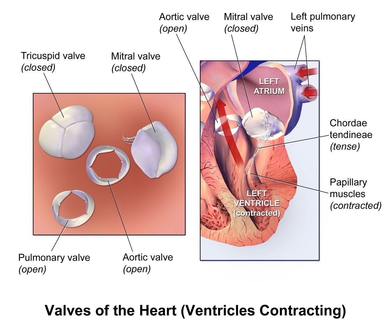
Uterine inversion — the fundus turning inside out and descending through the cervix — is rare, occurring in perhaps 1 in 2000 to 1 in 20 000 births, and it is the one obstetric haemorrhage where the reflex to give a uterotonic is exactly wrong. It is usually caused by excessive cord traction on an uncontracted uterus or by fundal pressure, which is one reason cord traction is only ever applied with counter-pressure on the lower segment and only with a contracted uterus.
The presentation is unmistakable once you have seen it: sudden severe lower abdominal pain, a dark red-purple mass appearing at or protruding from the introitus, a fundus that cannot be palpated abdominally or feels like a dimple or crater, and shock that is out of proportion to the visible blood loss because vagal stimulation from traction on the peritoneum adds neurogenic shock to the haemorrhagic shock.
The sequence is: call for help and anaesthesia immediately; stop all uterotonics, because a contracting cervical ring makes replacement impossible; do not attempt to remove the placenta if it is still attached, because separating it increases blood loss before the uterus is back; establish large-bore access and start volume resuscitation; anticipate a uterine relaxant such as terbutaline, nitroglycerin, magnesium sulfate or a halogenated inhalational agent; and support the provider’s manual replacement, in which the fundus is pushed back through the cervix. Only after the uterus is replaced do you restart uterotonics, vigorously, to keep it there — and then remove the placenta. If manual replacement fails, laparotomy follows.
When the fundus is firm and the patient is still bleeding, still hurting or still tachycardic, the problem is trauma, retained tissue, coagulopathy or inversion — and in inversion you stop the uterotonic, leave the placenta alone and get the uterus back first.
Section 3Puerperal Infection, Mastitis and Maternal Sepsis
Defining puerperal infection and recognising endometritis
Puerperal infection is classically defined as an oral temperature of 38.0°C (100.4°F) or higher on any two of the first ten days postpartum, excluding the first 24 hours, with temperatures taken at least four times a day. The exclusion of the first day matters: a transient low-grade temperature in the first 24 hours is common and usually reflects dehydration and the work of labour, and it responds to fluids. A temperature on day two or later does not get explained away.
Endometritis is the commonest puerperal infection and is polymicrobial, arising from ascending genital tract flora. Risk rises steeply with caesarean birth, particularly an unscheduled caesarean after labour and ruptured membranes, and with prolonged rupture of membranes, multiple vaginal examinations, internal monitoring, manual removal of the placenta, chorioamnionitis in labour, prolonged labour, retained tissue, anaemia and diabetes. Prophylactic antibiotics given within 60 minutes before caesarean incision substantially reduce it.
Assessment findings appear on day two to day five: fever, chills, tachycardia that tracks the temperature, lower abdominal or uterine tenderness that is exquisite on fundal palpation, lochia that is foul-smelling or profuse (though endometritis caused by group A streptococcus may have scant, odourless lochia), malaise and anorexia. Treatment is intravenous broad-spectrum antibiotics — the classic regimen is clindamycin 900 mg IV every 8 hours plus gentamicin, dosed by weight — continued until the patient has been afebrile and asymptomatic for 24 to 48 hours, at which point oral antibiotics are usually unnecessary. Nursing care adds fluids, antipyretics, Fowler position to promote lochial drainage, meticulous perineal hygiene, and continued support for feeding, because most of these antibiotics are compatible with lactation.
If the fever persists after 48 to 72 hours of appropriate antibiotics, the differential widens: abscess, retained products, wound infection, septic pelvic thrombophlebitis (a diagnosis of exclusion in a patient who looks surprisingly well between fever spikes), or a resistant organism. Persistent fever on adequate therapy is an escalation trigger, not a reason to wait another day.
Wound, perineal and urinary infection
Caesarean wound infection usually presents on day four to seven with erythema extending beyond the incision line, induration, warmth, tenderness, purulent or serosanguineous drainage and fever. Management is drainage and opening of the wound where needed, antibiotics, and packing with dressing changes; teach the family to report increasing pain, spreading redness, new drainage or wound edges separating. Perineal and episiotomy infections present with disproportionate pain, oedema, erythema at the repair, and sometimes wound breakdown; sitz baths, analgesia and antibiotics are the mainstay.
The rare but lethal variant is necrotising fasciitis. The clue is pain far out of proportion to the visible wound, rapid progression over hours, dusky or bronze skin discoloration, bullae, crepitus, anaesthesia over the affected skin, and systemic toxicity. This is a surgical emergency requiring immediate wide debridement; antibiotics alone will not save the patient. If a postpartum wound looks mild but the patient is in agony and looks unwell, escalate immediately rather than reassess in an hour.
Urinary tract infection is common because of birth trauma to the bladder, epidural-related retention, catheterisation and postpartum diuresis. Cystitis presents with frequency, urgency, dysuria and suprapubic tenderness; pyelonephritis adds high fever, rigors, flank or costovertebral angle tenderness, nausea and vomiting, and requires intravenous antibiotics and hydration. Prevention is unglamorous and effective: encourage voiding within 4 to 6 hours of birth, scan or palpate for retention, remove catheters as early as possible, and encourage fluids.
Mastitis and the feeding relationship
Lactational mastitis typically appears in the second to fourth week postpartum and is usually unilateral. The lactating parent describes a hard, hot, red, wedge-shaped area of the breast, aching muscles, chills and a fever often above 38.5°C — many say it feels like influenza arriving in an hour. Risk factors are milk stasis from missed or shortened feeds, a poor latch, oversupply, pressure from a tight bra or a bag strap, and nipple trauma providing a portal of entry, most often for Staphylococcus aureus.
The single most important teaching point is that feeding or expressing must continue. Milk from an infected breast is safe for a healthy term infant, and stopping abruptly worsens the stasis that caused the problem and raises the risk of abscess. Current guidance is to feed on demand rather than to try to drain the breast: extra pumping and deliberate over-emptying drive hyperlactation and worsen the inflammation. Apply cold — ice packs between and after feeds — and do not apply heat before feeds or use deep massage, both of which increase oedema and inflammation; gentle lymphatic stroking towards the axilla is the only manual treatment. Alongside that, the plan is rest, fluids, analgesia and an anti-inflammatory such as ibuprofen, and, if there is no improvement within 24 to 48 hours or the parent is systemically unwell, a 10 to 14 day antibiotic course — dicloxacillin 500 mg orally four times daily or cephalexin 500 mg four times daily, with trimethoprim-sulfamethoxazole or clindamycin where meticillin-resistant S. aureus is suspected. Symptoms should improve within 48 to 72 hours; if they do not, or a fluctuant mass is palpable, ultrasound looks for an abscess requiring needle or surgical drainage.
Language matters here as much as pharmacology. A parent with mastitis is frequently exhausted, in pain and being told by well-meaning relatives to stop feeding. Nothing in that story is a failure. Describe what is happening mechanically — milk is not moving, and the breast is inflamed — and offer the fix. Never label an infant “a difficult feeder” or a parent “non-compliant”; describe the latch, the transfer and the plan.
Maternal sepsis: the hour that counts, and doing the arithmetic
Sepsis is life-threatening organ dysfunction caused by a dysregulated response to infection, and in obstetrics it is doubly dangerous because the young, physiologically compensated patient looks well until she does not. Standard adult screening tools underperform in pregnancy and the puerperium because tachycardia, tachypnoea and a modestly raised white cell count are all normal. Use obstetric-specific triggers: temperature above 38.0°C or below 36.0°C, heart rate above 120, respiratory rate above 30 (or above 25 with any other trigger), systolic pressure below 90, oxygen saturation below 95 per cent, and any altered mental state. Lactate of 2 mmol/L or more raises concern; 4 mmol/L or more is severe.
The bundle is time-defined. Draw blood cultures before antibiotics but do not let culture collection delay them; administer broad-spectrum antibiotics within one hour of recognition; measure lactate and repeat it if elevated; give a crystalloid fluid bolus of 30 mL per kilogram for hypotension or lactate of 4 mmol/L or more; and start vasopressors if the mean arterial pressure remains below 65 mmHg after fluid. Also find and remove the source — retained tissue, an abscess, an infected wound.
Two of those steps are weight-based, and weight-based dosing is where arithmetic errors become patient harm. Show your work every time.
A postpartum temperature of 38.0°C or higher after the first 24 hours is infection until proven otherwise, and once sepsis is suspected the clock is one hour to antibiotics with every weight-based dose calculated, written and independently checked.
Section 4Thromboembolism, Postpartum Hypertension and the Failing Heart
Venous thromboembolism: highest risk, lowest vigilance
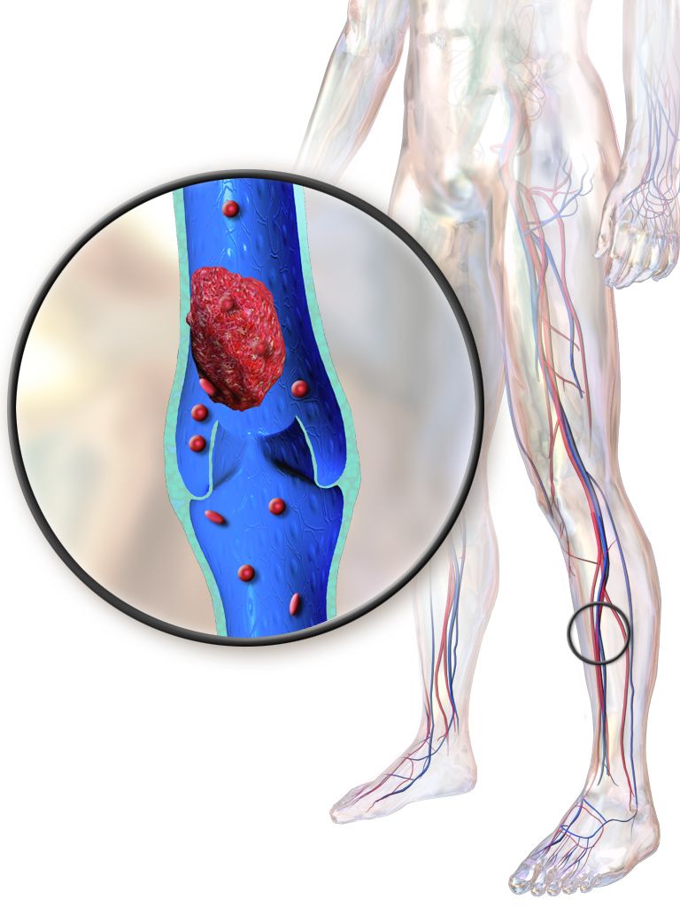
Pregnancy satisfies all three limbs of Virchow’s triad simultaneously: venous stasis from the gravid uterus and reduced venous tone, endothelial injury at birth or caesarean, and a physiologically hypercoagulable state that evolved to protect against haemorrhage. That protective adaptation does not switch off at birth. Thrombotic risk is roughly four to five times baseline in pregnancy but is at its highest in the first three weeks after birth — by some estimates twenty-fold or more — and remains raised for six to twelve weeks. This is precisely the window when the patient is at home, sleep-deprived, sitting still to feed, and least likely to be assessed.
Deep vein thrombosis presents with unilateral leg pain, swelling, warmth, erythema and sometimes a palpable cord; a calf circumference difference of more than 2 cm between legs is meaningful. Left-sided DVT predominates in pregnancy. Do not use the Homans sign — dorsiflexing the foot to elicit calf pain — it is neither sensitive nor specific and may dislodge clot. Diagnosis is by compression ultrasonography. Superficial thrombophlebitis is tender and erythematous along a visible vein and is managed with warmth, elevation, compression and anti-inflammatories.
Pulmonary embolism is the killer. Sudden dyspnoea, pleuritic chest pain, tachypnoea, tachycardia, apprehension or a sense of impending doom, cough, haemoptysis and hypoxaemia. In a postpartum patient, sudden breathlessness is never “just anxiety” until an embolus has been excluded. The first actions are: stay with the patient, sit her upright, apply high-flow oxygen, call the rapid response team, obtain vital signs including oxygen saturation, and establish or confirm intravenous access — then anticoagulation, imaging and, for massive embolism with haemodynamic collapse, thrombolysis or embolectomy. Notice again that the first action is oxygenation and calling for help, not fetching the heparin.
Anticoagulation and the arithmetic that goes with it
Low-molecular-weight heparin is the agent of choice in pregnancy and lactation. It does not cross the placenta, it is not meaningfully excreted in milk, it has predictable pharmacokinetics and it does not require routine monitoring. Therapeutic enoxaparin is 1 mg per kilogram subcutaneously every 12 hours; prophylactic dosing is typically 40 mg subcutaneously once daily, or 30 mg every 12 hours in some protocols. For an 82 kg patient the therapeutic dose is 82 × 1 = 82 mg subcutaneously every 12 hours — a calculation that should be written out and checked, not estimated from a vial size.
Unfractionated heparin is used when rapid reversibility matters, when renal function is poor, or around the time of neuraxial anaesthesia, because its short half-life and complete reversal with protamine sulfate (approximately 1 mg per 100 units of heparin) make timing easier; protamine only partially reverses low-molecular-weight heparin. Warfarin is teratogenic and is avoided in pregnancy but is acceptable during lactation, and it is bridged with heparin until the international normalised ratio is therapeutic. Direct oral anticoagulants are generally avoided while breastfeeding or chestfeeding because of limited safety data.
Nursing care wraps around the drug. Teach subcutaneous injection into the abdominal fat pad at least 5 cm from the umbilicus, alternating sides, without expelling the air bubble and without rubbing the site. Teach the bleeding precautions — soft toothbrush, electric razor, no non-steroidal anti-inflammatory drugs unless prescribed, report bruising, nosebleeds, blood in urine or stool, and heavier lochia. Reinforce mobility and hydration, apply graduated compression or sequential devices, and time anticoagulant doses carefully in anyone with an epidural catheter, because spinal haematoma is the feared complication of getting that interval wrong.
Postpartum preeclampsia, eclampsia and magnesium sulfate toxicity
Hypertensive disease does not end with the placenta. Postpartum preeclampsia can present up to six weeks after birth, most commonly between 48 hours and seven days, and in a meaningful number of patients it is new — there was no hypertension in pregnancy at all. Severe features are systolic pressure of 160 mmHg or more or diastolic of 110 mmHg or more on two occasions, a headache unrelieved by analgesia, visual disturbance, right upper quadrant or epigastric pain, platelets below 100 000 per microlitre, creatinine above 1.1 mg/dL or doubled from baseline, transaminases at twice the upper limit of normal, or pulmonary oedema. HELLP syndrome is the laboratory-defined severe variant, and its three criteria are in its name: haemolysis (schistocytes on the smear, lactate dehydrogenase of 600 units/L or more, rising indirect bilirubin), elevated liver enzymes at twice the upper limit of normal or higher, and low platelets below 100 000 per microlitre. HELLP can occur with only mildly raised, or occasionally normal, blood pressure, which is why right upper quadrant or epigastric pain in a postpartum patient earns a full panel rather than an antacid. A patient telephoning on day five with a headache that will not go away and “spots” in her vision needs a blood pressure measured today, not an appointment next week.
Acute severe-range hypertension must be treated within 30 to 60 minutes of confirmation. First-line agents are intravenous labetalol 20 mg over 2 minutes, escalating to 40 mg then 80 mg at 10-minute intervals to a cumulative maximum of 300 mg; intravenous hydralazine 5 to 10 mg over 2 minutes, repeated every 20 minutes; or immediate-release oral nifedipine 10 mg. Avoid labetalol in asthma and in bradycardia. Magnesium sulfate is given for seizure prophylaxis in severe features and for treatment of eclampsia; it is not an antihypertensive and does not lower blood pressure.
Magnesium sulfate is loaded at 4 to 6 g intravenously over 15 to 30 minutes and maintained at 1 to 2 g per hour, always on a pump, always as a secondary line. Therapeutic serum levels are approximately 4 to 7 mEq/L, which is 5 to 8 mg/dL (multiply mEq/L by 1.2 to get mg/dL). Because magnesium is cleared renally, oliguria means accumulation, and urine output is a toxicity parameter, not just a fluid parameter. Assess before and hourly during the infusion: deep tendon reflexes, respiratory rate, urine output, level of consciousness, blood pressure, and continuous oximetry.
Toxicity appears in a fixed order, and the order is the whole lesson. Deep tendon reflexes disappear first, at roughly 8 to 12 mg/dL, before the respiratory rate falls at around 12 to 15 mg/dL, before ECG changes, before cardiac arrest above roughly 25 to 30 mg/dL. The absent patellar reflex is therefore your early warning, which is why you check reflexes hourly on a patient who otherwise looks perfectly comfortable. If reflexes are absent, the respiratory rate is below 12 per minute, urine output is under 30 mL per hour, or the patient is newly drowsy or slurring: stop the infusion first, then call for help, then give the antidote — calcium gluconate 1 g intravenously, which is 10 mL of a 10 per cent solution, administered slowly over about 3 minutes. Keep calcium gluconate and a bag-valve mask at the bedside of every patient on magnesium.
| Serum magnesium (mg/dL) | What you see | Action |
|---|---|---|
| 5 to 8 | Therapeutic; flushing, warmth, feeling hot, mild drowsiness and nausea may be normal at this level. New slurred speech is not — it is early central nervous system depression | Continue, reassure, keep monitoring hourly; if speech becomes slurred, stop the infusion and draw a level |
| 8 to 12 | Deep tendon reflexes diminished then absent; new drowsiness, slurred speech | Stop infusion, notify provider, draw a level |
| 12 to 15 | Respiratory rate below 12, respiratory depression | Stop infusion, oxygen and support ventilation, calcium gluconate 1 g IV over 3 minutes |
| Above 15 to 25 | ECG changes, hypotension, altered consciousness | Emergency response, calcium gluconate, intensive care |
| Above 25 to 30 | Cardiac arrest | Resuscitation with calcium gluconate given during the code |
Peripartum cardiomyopathy and the heart that fails quietly
Peripartum cardiomyopathy is systolic heart failure with an ejection fraction below 45 per cent, arising in the last month of pregnancy or within five months of birth, without another identifiable cause. It is easy to miss because every symptom is also a normal postpartum complaint: fatigue, breathlessness, ankle swelling, a cough. What distinguishes it is the pattern — breathlessness lying flat (orthopnoea), waking at night gasping (paroxysmal nocturnal dyspnoea), a persistent dry cough, palpitations, and oedema that is worsening rather than resolving in the second week when postpartum diuresis should have cleared it. Risk is higher with hypertensive disease of pregnancy, multiple gestation, advanced maternal age and Black race, the last reflecting structural inequities in access and care rather than biology.
Assessment includes crackles, a third heart sound, jugular venous distension, tachycardia, hypoxaemia and weight gain; diagnosis is by echocardiography with elevated natriuretic peptides. Management is standard heart-failure therapy adapted for lactation: diuretics for congestion, beta-blockade, afterload reduction with hydralazine and nitrates if still pregnant, and angiotensin-converting enzyme inhibitors postpartum using lactation-compatible agents such as enalapril or captopril. Anticoagulation is considered when the ejection fraction is severely reduced, because of thrombus risk. Counselling about future pregnancy is essential, since recurrence risk is significant and highest when function has not normalised.
All three of the conditions in this section share one nursing failure mode: the symptom is attributed to normal recovery. Breathlessness, a headache that will not lift, a swollen calf, oedema that is getting worse — each has a benign explanation that is usually right and occasionally lethal. Assess, do not assume, and escalate with structure.
Magnesium toxicity announces itself by the loss of deep tendon reflexes before respiratory depression, so hourly reflex checks are the monitor — and if toxicity appears you stop the infusion first and then give calcium gluconate 1 g intravenously.
Section 5Perinatal Mood, Psychosis and the Fourth Trimester
Blues, depression, anxiety — telling them apart
Postpartum blues affects roughly half to four-fifths of birthing parents. It begins around day three to five as oestrogen and progesterone fall precipitously, peaks around day five, and resolves without treatment by day ten to fourteen. The picture is tearfulness that arrives without an obvious trigger, mood lability, irritability, anxiety and poor sleep — but crucially the parent still functions, still cares for the infant, and still enjoys moments of the day. Treatment is sleep, food, practical help, honest reassurance that this is common and self-limiting, and a clear instruction about when to call: if it lasts beyond two weeks, if it deepens, or if the parent cannot care for herself or the baby.
Perinatal depression affects about one in seven and may begin in pregnancy or at any point in the first year, most commonly around four to six weeks postpartum. It is distinguished from the blues by duration beyond two weeks, by severity, and by functional impairment. Listen for persistent low mood or numbness, loss of interest, guilt and worthlessness (“the baby would be better off with someone else”), inability to sleep even when the baby sleeps, appetite change, poor concentration, feeling detached from the infant, and thoughts of death or self-harm. Depression in the non-birthing parent is also real, affecting roughly one in ten partners, and is worth asking about.
Anxiety disorders are at least as common as depression and are under-recognised because vigilance is culturally expected of new parents. Perinatal generalised anxiety, panic disorder, and obsessive-compulsive presentations with intrusive thoughts are all seen. Intrusive thoughts — sudden, vivid, unwanted images of harm coming to the baby — are horrifying to the parent, who typically hides them for fear of losing the child. The clinical distinction is critical and reassuring: in obsessive-compulsive presentations the thoughts are ego-dystonic, meaning the parent is repelled by them, has no wish to act, and avoids the trigger. In psychosis the thoughts are ego-syntonic, driven by delusion, and carry real risk. Post-traumatic stress after a frightening birth or an intensive-care admission is also common and responds to trauma-focused therapy.
Screening as a clinical act
Screening is recommended at least once in the perinatal period and in practice at several points: at the first prenatal visit, later in pregnancy, before discharge, and at the comprehensive postpartum visit; paediatric practices additionally screen the parent at the 1, 2, 4 and 6-month well-child visits, because that is where the family actually appears. The Edinburgh Postnatal Depression Scale is the most widely used instrument — ten items, each scored 0 to 3, maximum 30, with a score of 10 to 13 or above prompting further evaluation. It deliberately excludes somatic items such as fatigue and sleep disturbance, which are universal after birth and would otherwise inflate every score. The Patient Health Questionnaire-9 is a common alternative.
The item that must never be totalled away is item 10 of the Edinburgh scale, which asks about thoughts of self-harm. Any response other than “never” requires immediate evaluation, regardless of the total score. A parent can score 6 overall and still be in danger. Ask directly, in plain words, without euphemism: “Have you had thoughts of hurting yourself, or thoughts that your family would be better off without you? Have you had thoughts of harming the baby?” Asking does not plant the idea. Not asking leaves it unspoken.
Screening is an assessment, and assessment is registered-nurse work. Administering a form can be supported by others, but interpreting the score, acting on item 10, teaching the family and evaluating the response cannot be delegated. Neither can the initial assessment of any postpartum patient, the assessment of an unstable patient, the titration of oxytocin or magnesium, blood product administration, or discharge teaching. Assistive personnel can take vital signs on a stable patient, assist with ambulation and hygiene, apply ice packs, weigh pads for you to quantify and interpret, and support feeding position — and they must be told exactly what to report back and how quickly.
Treatment is effective and should be offered without delay. Psychotherapy — cognitive behavioural and interpersonal therapy in particular — is first-line for mild to moderate illness. Selective serotonin reuptake inhibitors are used for moderate to severe illness, with sertraline commonly chosen during lactation because milk transfer is low; if a parent did well on a particular agent before, restarting that agent is usually preferable to switching. Two neuroactive steroid treatments specific to postpartum depression now exist: zuranolone, taken orally 50 mg once daily for 14 days, and brexanolone, given as a 60-hour intravenous infusion under a monitoring programme because of the risk of excessive sedation and sudden loss of consciousness. Both carry sedation warnings that affect infant care and driving, and both require a conversation about who is holding the baby.
Postpartum psychosis is an emergency
Postpartum psychosis occurs in approximately 1 to 2 births per 1000 and typically begins early — often within the first two weeks, frequently between days three and ten. The strongest risk factor is bipolar disorder, personal or family, and a previous episode of postpartum psychosis carries a high recurrence risk. The presentation is dramatic and fluctuating: insomnia with no sense of needing sleep, agitation, rapid mood swings, confusion and disorganised thinking that waxes and wanes hour to hour, hallucinations, and delusions that very often centre on the infant — that the baby is possessed, defective, not hers, or in danger from which only she can save him. The risk of infanticide is estimated at around 4 per cent and of suicide around 5 per cent, which is why this is treated as an emergency without exception.
What you do first: do not leave the parent alone, and do not leave the parent alone with the infant. Stay, assign continuous observation, remove obvious means of harm from the room, and call for immediate psychiatric evaluation. Notify the provider, involve the family in the safety plan, arrange for another responsible adult to care for the infant, and expect inpatient admission with mood stabilisation, antipsychotics and sometimes electroconvulsive therapy, which is highly effective here. Speak calmly, do not argue with the delusion and do not agree with it; orient to what is real and safe, and keep the environment quiet and low-stimulus.
Frame this correctly for the family. Postpartum psychosis is a medical illness of the brain in the days after birth. It is not a character flaw, not a parenting failure, and not evidence that this person cannot ever parent. Most recover fully with treatment. The family will remember the words you use in the first hour for the rest of their lives.
The fourth trimester: discharge, follow-up and the whole family
Maternal mortality in high-income systems is now weighted towards the postpartum period, and mental health conditions including suicide and overdose, together with cardiovascular conditions, are leading causes of death in the year after birth. Most of these deaths occur after discharge. That makes discharge teaching a clinical intervention, not paperwork. Contemporary guidance replaces the single six-week visit with ongoing contact: a check within the first three weeks, additional contacts as needed, and a comprehensive visit no later than twelve weeks that covers mood, bleeding, blood pressure, feeding, pain, sexuality, contraception, chronic disease follow-up and future pregnancy planning.
Teach the warning signs by symptom, in plain language, and give them in writing to more than one adult in the household, because a parent who is bleeding, febrile or depressed is not the best keeper of her own instructions. The POST-BIRTH warning signs give the list an order that families remember: Pain in the chest, Obstructed breathing or shortness of breath, Seizures, Thoughts of hurting yourself or the baby, Bleeding that soaks more than one pad an hour or clots the size of an egg or larger, Incision that is not healing, Red or swollen leg that is painful or warm, Temperature of 38.0°C (100.4°F) or higher, and a Headache that will not go away or comes with vision changes.
| Warning sign to teach | What it may mean | What the family does |
|---|---|---|
| Chest pain, trouble breathing, or a seizure | Pulmonary embolism, cardiomyopathy, eclampsia | Call emergency services immediately |
| Thoughts of harming yourself or the baby | Perinatal depression or psychosis | Call emergency services or the crisis line immediately; do not stay alone |
| Soaking more than one pad an hour, or clots the size of an egg or larger | Late postpartum haemorrhage | Call emergency services immediately |
| Temperature of 38.0°C (100.4°F) or higher | Endometritis, wound infection, mastitis, sepsis | Call the provider the same day |
| An incision or tear that is red, draining, opening or increasingly painful | Wound infection | Call the provider the same day |
| A red, swollen, warm or painful leg | Deep vein thrombosis | Call the provider the same day; do not massage the leg |
| A headache that will not go away, or a bad headache with vision changes | Postpartum preeclampsia | Get the blood pressure checked the same day |
Finally, hold the language. Describe the labour pattern rather than saying anyone failed to progress. Describe the latch and the milk transfer rather than calling an infant a difficult feeder. Describe barriers — transport, childcare, cost, fear, exhaustion — rather than calling a parent non-compliant. Use “birthing parent” and “chestfeeding” where they fit and “mother” and “breastfeeding” where they fit, and follow the family’s lead. For families experiencing loss, offer choices rather than assumptions about seeing and holding, use the baby’s name, provide memory-making, and remember that grief and postpartum physiology occur in the same body at the same time — the fundal checks, the bleeding, the milk coming in and the mood screening all still apply.
Perinatal mental health is assessed with a scored tool and acted on immediately — any positive self-harm item is an emergency regardless of the total, and postpartum psychosis means the parent is never left alone with the infant.
The chapter in one breath
Postpartum complications kill quickly and quietly, and almost every one of them turns on a sequence rather than a fact. Haemorrhage is defined by 1000 mL of quantified loss or by any loss with signs of hypovolaemia, and because pregnancy expands plasma volume by nearly half, the pulse, the respiratory rate and the patient’s own words change long before the blood pressure does. Tone causes most of it, so the first action is a firm fundal massage with the lower segment supported, then help, oxytocin, an empty bladder, two large-bore lines, and tranexamic acid within three hours — escalating through methylergonovine (never in hypertension) and carboprost (never in asthma) to tamponade and surgery. A firm fundus that keeps bleeding means a laceration, a concealed haematoma announced by disproportionate pain, retained tissue, coagulopathy, or an inversion in which you stop the uterotonics and leave the placenta alone. Fever after the first day is infection until disproved, and sepsis gives you one hour to antibiotics with every weight-based dose written out and checked. Thromboembolism peaks in the first three weeks; postpartum preeclampsia can arrive after discharge; magnesium toxicity takes the reflexes before it takes the breathing, and the antidote is calcium gluconate after you stop the infusion. And in the fourth trimester, a scored mood screen and a positive self-harm item outrank every other task in the room.
Words worth knowing
| Term | What it means |
|---|---|
| Postpartum haemorrhage | Cumulative blood loss of 1000 mL or more, or any loss with signs or symptoms of hypovolaemia, within 24 hours of birth by any route. |
| Quantitative blood loss (QBL) | Measured rather than estimated loss, using calibrated drapes and weighed items where 1 gram equals about 1 millilitre. |
| Shock index | Heart rate divided by systolic blood pressure; normally 0.5 to 0.7 postpartum, concerning at 0.9 or above. |
| Uterine atony | Failure of the myometrium to contract after birth, leaving the spiral arterioles open; cause of 70 to 80 per cent of haemorrhage. |
| Four Ts | Tone, trauma, tissue and thrombin — the working differential for postpartum haemorrhage. |
| Uterotonic | A drug that stimulates uterine contraction: oxytocin, methylergonovine, carboprost, misoprostol. |
| Tranexamic acid | An antifibrinolytic given as 1 g intravenously over 10 minutes, ideally within 3 hours of birth, in addition to uterotonics. |
| Genital tract haematoma | Concealed bleeding into vulvar, vaginal or retroperitoneal tissue, presenting as severe unrelieved pain with a firm fundus. |
| Subinvolution | Delayed return of the uterus to pre-pregnant size, with a fundus higher and softer than expected and prolonged lochia rubra. |
| Uterine inversion | The fundus turning inside out through the cervix; management stops uterotonics, leaves the placenta attached and replaces the uterus first. |
| Placenta accreta spectrum | Abnormally adherent or invasive placentation (accreta, increta, percreta), strongly associated with prior caesarean and praevia. |
| Disseminated intravascular coagulation | Consumption of clotting factors producing simultaneous clotting and bleeding, with falling platelets and fibrinogen and rising D-dimer. |
| Puerperal infection | Temperature of 38.0°C or higher on any two of the first ten postpartum days, excluding the first 24 hours. |
| Endometritis | Infection of the uterine lining, commonest after caesarean, presenting with fever, uterine tenderness and abnormal lochia. |
| Mastitis | Unilateral wedge-shaped breast inflammation with systemic symptoms; treated with continued on-demand feeding, cold application and anti-inflammatories, plus antibiotics if not improving. |
| Magnesium sulfate toxicity | Loss of deep tendon reflexes, then respiratory depression, then cardiac arrest; reversed with calcium gluconate 1 g intravenously. |
| Peripartum cardiomyopathy | Systolic heart failure with ejection fraction below 45 per cent from the last month of pregnancy to five months postpartum. |
| Edinburgh Postnatal Depression Scale | Ten-item screening tool scored 0 to 30; item 10 asks about self-harm and any positive answer requires immediate evaluation. |
| Postpartum psychosis | Rare early-onset emergency with delusions often centred on the infant; the parent is never left alone with the baby. |