The Newborn: Transition, Assessment and Care
In the first ten minutes of life a fetus becomes a newborn, and almost every organ system has to change its job at once. Lungs that were fluid-filled must aerate and absorb; a circulation built around a placenta must reroute itself through the pulmonary bed; a body that never had to make its own heat or its own glucose must suddenly do both. Most newborns manage this without help. The ones who do not deteriorate quietly — a temperature drifting down, a glucose falling, a bilirubin climbing — and the deterioration is almost always visible to a nurse who knows the numbers and looks on schedule. This chapter walks the transition physiologically, then teaches the first assessment (APGAR component by component, gestational-age scoring, head-to-toe), then the three metabolic vulnerabilities that fill newborn nurseries: cold stress, hypoglycaemia and jaundice. It ends with daily care, weight-based dosing and the discharge teaching that keeps babies alive at home.
By the end of this chapter you can
- Explain the mechanics and chemistry of the first breath, including alveolar fluid clearance and the role of surfactant, and trace the closure of the ductus arteriosus, foramen ovale and ductus venosus with their time frames.
- Score an APGAR component by component at 1 and 5 minutes, state what each total range triggers, and explain why the APGAR score never delays resuscitation.
- Perform a systematic newborn assessment: measurements, gestational-age (Ballard) scoring, size classification (SGA, AGA, LGA), primitive reflexes, and the distinction between caput succedaneum, cephalhaematoma and subgaleal haemorrhage.
- Describe non-shivering thermogenesis in brown adipose tissue, list the four routes of heat loss, and outline the metabolic cascade of cold stress.
- Apply operational hypoglycaemia thresholds by hour of life, identify at-risk newborns, and calculate a dextrose 10% bolus and a 40% dextrose gel dose for a given weight.
- Differentiate physiologic from pathologic jaundice using the 24-hour rule, rate of rise and direct bilirubin fraction, and manage phototherapy safely.
- Calculate weight-based paediatric doses and maintenance fluids using the 4-2-1 rule, and check every paediatric dose against the maximum adult dose.
- State the first nursing action in postpartum haemorrhage, magnesium sulfate toxicity, umbilical cord prolapse, shoulder dystocia and uterine rupture, and escalate newborn red flags using SBAR.
Section 1Transition to Extrauterine Life
The first breath: fluid out, air in
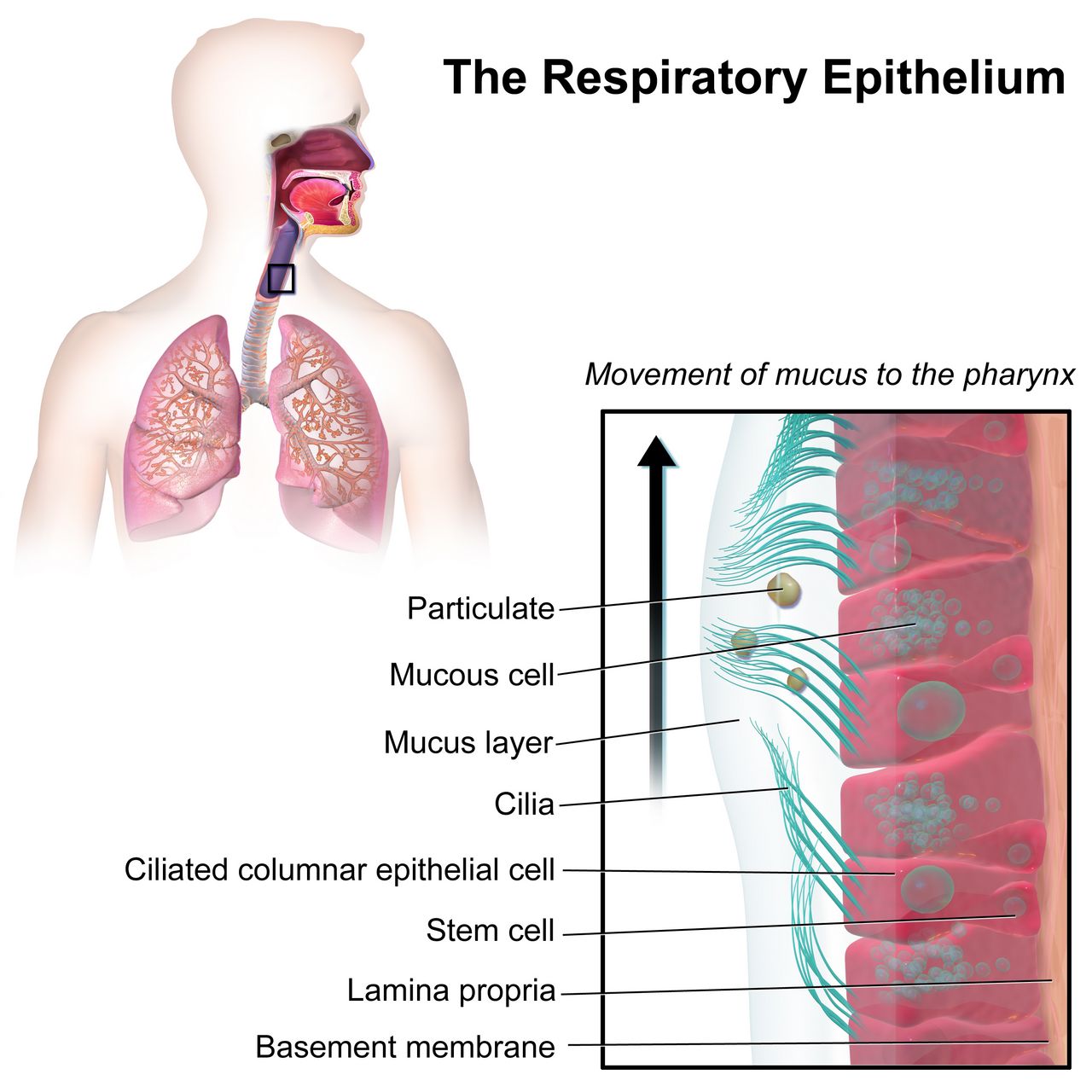
Before birth the lung is not collapsed — it is full. Roughly 20 to 30 mL/kg of lung liquid, secreted by the pulmonary epithelium itself, keeps the airways distended and lets the lung grow. That fluid has to disappear in minutes. The old teaching that vaginal squeeze empties the chest explains only a small fraction of it. The dominant mechanism is chemical: in the days and hours before birth, the surge of fetal cortisol, thyroid hormone and catecholamines flips the alveolar epithelium from chloride secretion to sodium absorption through epithelial sodium channels (ENaC). Water follows sodium out of the airspace into the interstitium and then into the pulmonary capillaries and lymphatics. This is why a baby born by scheduled caesarean before labour begins — without the catecholamine surge that labour supplies — is at higher risk for transient tachypnoea of the newborn. Labour is not an inconvenience the fetus survives; it is preparation the fetus needs.
The first inspiratory effort is triggered by a convergence of stimuli. Cord clamping removes placental gas exchange, so carbon dioxide rises and oxygen falls; the resulting mild hypercapnia and acidosis stimulate the medullary respiratory centre. Cold air on wet skin, gravity, light, sound and tactile stimulation add to the drive. That first breath demands enormous negative pressure — opening pressures of 40 to 80 cm H₂O are typical, far above the 5 to 10 cm H₂O a settled newborn uses — because the infant must overcome the surface tension of fluid-lined alveoli and the resistance of remaining lung liquid. Surfactant, produced by type II pneumocytes from about 24 weeks and reaching mature quantities near 34 to 36 weeks, lowers that surface tension and, crucially, keeps small alveoli from emptying into large ones at end-expiration. Without adequate surfactant the lung re-collapses with every breath, and the infant works progressively harder for progressively less gas exchange — the mechanism of respiratory distress syndrome.
Normal newborn respirations are 30 to 60 breaths per minute, irregular, diaphragmatic and predominantly nasal. Newborns are obligate nose breathers for the first several months, which is why nasal obstruction is never trivial. Brief pauses of up to 15 seconds without colour or heart-rate change are periodic breathing and are normal. A pause of 20 seconds or more, or any pause accompanied by cyanosis, pallor or bradycardia, is apnoea and is not.
The circulatory switch: three shunts close
Fetal circulation is designed to bypass two organs the fetus does not need: the lungs and, largely, the liver. Oxygenated blood arrives from the placenta in the umbilical vein, and roughly half of it is shunted past the hepatic sinusoids through the ductus venosus into the inferior vena cava. In the right atrium, the foramen ovale lets the better-oxygenated stream cross directly to the left atrium and on to the coronary and cerebral circulations. Whatever blood does enter the right ventricle meets a pulmonary vascular bed held in intense hypoxic vasoconstriction, so about 90% of it is diverted through the ductus arteriosus into the descending aorta.
Birth reverses the pressure gradients that hold that pattern in place. Lung expansion, rising alveolar oxygen tension and rising pH cause pulmonary vascular resistance to fall steeply, so pulmonary blood flow increases eight- to tenfold within minutes. Simultaneously, clamping the cord removes the placenta — a very large low-resistance circuit — so systemic vascular resistance rises. Left atrial pressure now exceeds right atrial pressure and the flap of the foramen ovale is pressed shut; functional closure occurs within minutes to hours, though anatomic fusion takes months and a probe-patent foramen persists in roughly a quarter of adults. The ductus arteriosus constricts in response to two changes: rising arterial oxygen tension, which is a direct constrictor of ductal smooth muscle, and the abrupt fall in prostaglandin E₂, which the placenta had been supplying. Functional closure usually occurs by 12 to 24 hours and is essentially complete by 72 hours; anatomic obliteration to the ligamentum arteriosum takes 2 to 3 weeks. The ductus venosus closes functionally within minutes of cord clamping and becomes the ligamentum venosum; the umbilical vein becomes the ligamentum teres and the umbilical arteries become the medial umbilical ligaments.
Two clinical consequences follow directly. First, hypoxia and acidosis reopen the ductus arteriosus and re-constrict the pulmonary bed — the newborn can fall back into a fetal circulatory pattern. This is the physiology behind persistent pulmonary hypertension of the newborn, and it is why cold stress, hypoglycaemia and delayed resuscitation are dangerous well beyond their immediate effects. Second, in duct-dependent congenital heart lesions the closing ductus is what kills the baby, typically at 24 hours to 2 weeks of age when a previously well newborn suddenly becomes grey, mottled and shocked. Those infants need the ductus held open with prostaglandin E₁ (alprostadil) at 0.05 to 0.1 mcg/kg/min; apnoea is the expected adverse effect, so intubation equipment stays at the bedside.
What the birth itself hands you: obstetric emergencies and the newborn
The newborn nurse inherits whatever happened in the last ten minutes of labour. In each of the emergencies below the correct first action is often not the intuitive one, and the sequence itself is what saves the life. Learn these as sequences, not as lists.
| Emergency | Recognition | What you do FIRST | What follows — and what the newborn needs |
|---|---|---|---|
| Postpartum haemorrhage (cumulative loss ≥1000 mL, or any loss with signs of hypovolaemia, within 24 h) | Boggy uterus displaced above and to the right of the umbilicus; saturating a pad in under 15 minutes; rising pulse before falling pressure | Massage the fundus. Uterine atony causes 70 to 80% of postpartum haemorrhage, and a firm fundus stops the bleeding faster than any drug you can draw up | Empty the bladder, call for help, large-bore IV, oxytocin 10 to 40 units in 500 to 1000 mL infusion; methylergonovine 0.2 mg IM (avoid if hypertensive); carboprost 250 mcg IM (avoid in asthma); misoprostol 600 to 1000 mcg; tranexamic acid 1 g IV within 3 hours. The newborn may need a second nurse assigned so care is not interrupted |
| Magnesium sulfate toxicity in the birthing parent | Therapeutic range for seizure prophylaxis is 4 to 8 mg/dL. Above it, deep tendon reflexes disappear before respiratory depression: loss of patellar reflex at roughly 9 to 12 mg/dL, respiratory depression 12 to 18, cardiac arrest above 25 to 30. A lost reflex is your warning that the next thing to go is the breathing | Stop the magnesium infusion. Then give the antidote — calcium gluconate 1 g (10 mL of a 10% solution) IV over about 3 minutes | Monitor respiratory rate (hold for fewer than 12/min), urine output (hold below 30 mL/h), reflexes and level of consciousness. Anticipate a floppy, sleepy, hypotensive newborn with respiratory depression — have full resuscitation ready at the birth |
| Umbilical cord prolapse | Sudden prolonged deceleration or bradycardia, often just after membranes rupture; a pulsating cord palpated or seen at the introitus | Relieve the pressure. With a gloved hand, lift the presenting part off the cord and keep it there; place in knee-chest or Trendelenburg with hips elevated. Never push the cord back in | Call for immediate help, oxygen 10 L/min by non-rebreather, stop oxytocin, prepare for emergency caesarean; cover an exposed cord with saline-moistened sterile gauze. Your hand stays in place through transport and prep. Expect a newborn who may be acidotic and need resuscitation |
| Shoulder dystocia | The turtle sign — the head delivers and retracts against the perineum; restitution fails; gentle traction achieves nothing | Call for help and note the time, then McRoberts (hyperflex the parent’s hips onto the abdomen) plus suprapubic pressure. Never fundal pressure — it drives the shoulder harder into the symphysis and risks uterine rupture | Then Gaskin all-fours, Rubin, Woods screw, delivery of the posterior arm. Assess the newborn for brachial plexus injury (Erb palsy, C5 to C6 — arm adducted, internally rotated, “waiter’s tip” posture, absent Moro on that side), clavicle fracture (crepitus, asymmetric Moro) and hypoxic injury |
| Uterine rupture | Most common sign is an abnormal fetal heart pattern — prolonged deceleration or bradycardia. Sudden tearing abdominal pain, contractions stop, presenting part rises and station is lost, fetal parts palpable abdominally, shock | Stop oxytocin and mobilise for immediate surgical delivery. There is no medical management — this is an operating-room diagnosis | Two large-bore IVs, oxygen, type and crossmatch, notify the neonatal team. Expect a severely asphyxiated newborn: full resuscitation team, cord gases, and screening for therapeutic hypothermia if criteria are met |
Resuscitation in the golden minute
At every birth, ask three questions: is this a term gestation, does the infant have good tone, and is the infant breathing or crying? Three yeses buy skin-to-skin care with the birthing parent, with drying, warming and ongoing observation. Any no sends the infant to the radiant warmer for the initial steps — warm, dry, position the airway, stimulate — and if the infant remains apnoeic or gasping, or the heart rate is below 100 beats per minute, positive-pressure ventilation must be established within the first 60 seconds of life. Ventilation is the single most effective intervention in neonatal resuscitation, and inflating the lungs is what raises the heart rate.
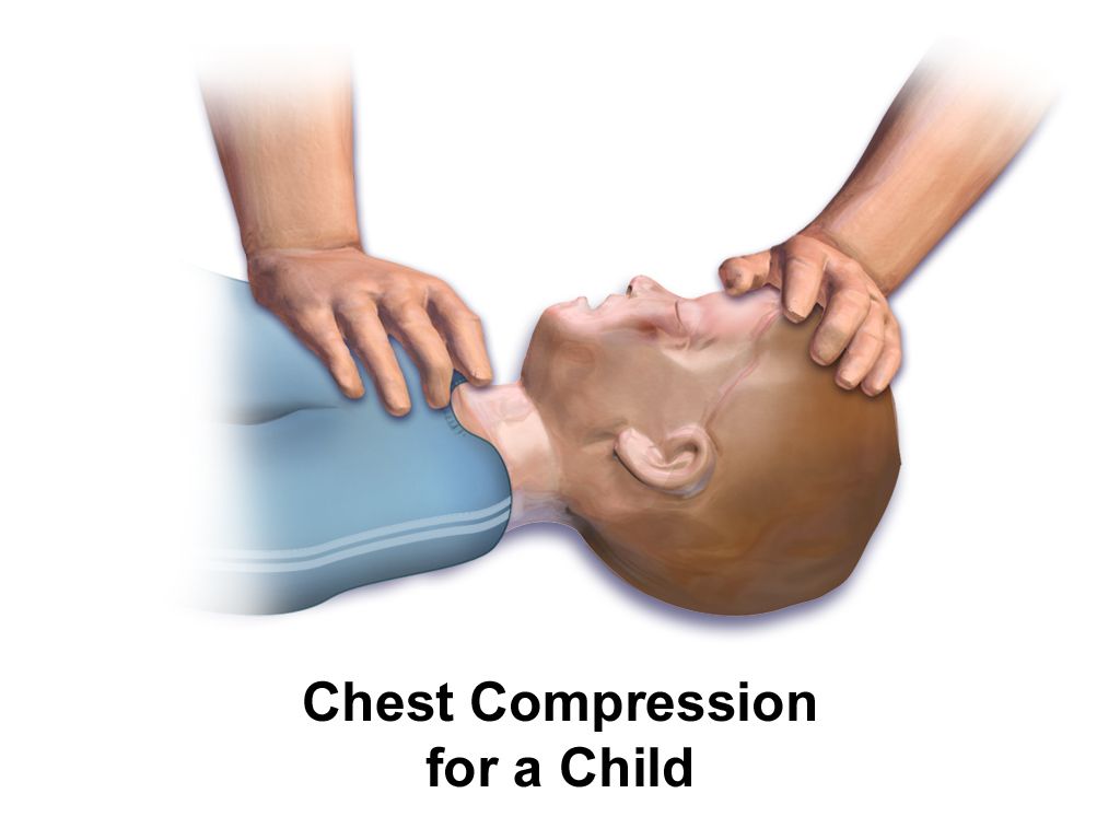
If the heart rate does not rise, the fault is almost always in the ventilation, not the baby. Run the MRSOPA corrective sequence: Mask adjustment, Reposition the airway, Suction mouth then nose, Open the mouth, Pressure increase, Airway alternative (endotracheal tube or laryngeal mask). If the heart rate remains below 60 after 30 seconds of demonstrably effective ventilation through an advanced airway with 100% oxygen, begin chest compressions at a ratio of 3 compressions to 1 breath — 90 compressions plus 30 breaths, that is 120 events per minute. If the heart rate stays below 60 after 60 seconds of coordinated compressions, give epinephrine 0.01 to 0.03 mg/kg IV or intraosseous (0.1 to 0.3 mL/kg of the 0.1 mg/mL concentration, formerly written 1:10,000), preferably by umbilical venous catheter. For hypovolaemia give normal saline or emergency-release O-negative blood at 10 mL/kg over 5 to 10 minutes. Start resuscitation of term infants in 21% oxygen (21 to 30% for infants under 35 weeks) and titrate to targeted preductal saturations: about 60 to 65% at 1 minute, 70 to 75% at 3 minutes, 80 to 85% at 5 minutes and 85 to 95% at 10 minutes. A pink newborn at one minute of life is the exception, not the goal.
Aeration of the lung is the engine of the entire transition — it drops pulmonary vascular resistance, closes the fetal shunts and raises the heart rate — which is why effective ventilation within the first 60 seconds outranks every other resuscitation manoeuvre.
Section 2APGAR, Gestational Age and the Newborn Examination
APGAR, component by component
Virginia Apgar built the score in 1952 to standardise what people meant when they said a baby “looked bad.” It has five components, each scored 0, 1 or 2, for a maximum of 10. Assign it at 1 minute and 5 minutes of life. If the 5-minute score is below 7, repeat it every 5 minutes up to 20 minutes of age.
| Component | 0 | 1 | 2 | What it is telling you |
|---|---|---|---|---|
| A — Appearance (colour) | Blue or pale all over | Acrocyanosis — pink body, blue hands and feet | Completely pink | The least specific component. Acrocyanosis is normal for 24 to 48 hours and reflects peripheral vasomotor instability. Central cyanosis of lips, tongue and trunk is never normal |
| P — Pulse (heart rate) | Absent | Below 100/min | 100/min or above | The most important single component and the one that drives resuscitation decisions. Auscultate at the apex for a full 60 seconds or use ECG leads |
| G — Grimace (reflex irritability) | No response to stimulation | Grimace or feeble cry | Cry, cough, sneeze, active withdrawal | Tests brainstem and cranial-nerve integrity. Elicited by suctioning the nares or flicking the sole |
| A — Activity (muscle tone) | Limp, flaccid | Some flexion of the extremities | Active motion, well flexed | Tone falls early with hypoxia and with maternal magnesium or anaesthesia. A term newborn at rest should be in flexion |
| R — Respiration (effort) | Absent | Slow, irregular, weak cry | Good, vigorous cry | Effort, not rate. Gasping scores the same as apnoea for management purposes — both mean start ventilating |
Interpretation is banded. A total of 7 to 10 is reassuring and calls for routine care — drying, warmth, skin-to-skin and observation. A total of 4 to 6 indicates moderate difficulty: stimulate, clear the airway if needed, provide oxygen or positive-pressure ventilation and reassess. A total of 0 to 3 indicates severe depression and calls for full resuscitation, including ventilation and, by heart-rate criteria, compressions and medication. A 5-minute score of 0 to 3 is associated with increased risk of neurologic injury at a population level, but the score is a poor predictor for any individual baby and must never be used to counsel a family about long-term outcome.
Measurements, gestational age and size classification
Normal term newborn measurements: weight 2500 to 4000 g (average about 3400 g), length 45 to 55 cm (average 50 cm), head circumference 32 to 38 cm (average 34 to 35 cm) and chest circumference 30 to 36 cm, typically 2 to 3 cm less than the head. Measure head circumference at the widest point, just above the eyebrows and over the occipital prominence, and remeasure after moulding resolves. A physiologic weight loss of up to 7 to 10% of birth weight over the first 3 to 5 days is expected, with birth weight regained by 10 to 14 days. A loss exceeding 7% is a signal to evaluate feeding — a latch assessment, a weighed feed, a look at output — not a signal to label the infant or the parent.
Gestational age is scored with the New Ballard Score, which combines six neuromuscular criteria (posture, square window at the wrist, arm recoil, popliteal angle, scarf sign, heel-to-ear) with six physical criteria (skin, lanugo, plantar surface, breast bud, eye and ear, genitals). It is accurate to roughly plus or minus 2 weeks, is most reliable when performed between 12 and 24 hours of age, and spans 20 to 44 weeks; each 5-point increment corresponds to about 2 weeks of gestation. Classify gestation as preterm below 37 0/7 weeks (with late preterm 34 0/7 to 36 6/7), early term 37 0/7 to 38 6/7, full term 39 0/7 to 40 6/7, late term 41 0/7 to 41 6/7 and post-term at 42 0/7 weeks or beyond. Then plot weight for gestational age: small for gestational age (SGA) below the 10th percentile, appropriate (AGA) 10th to 90th, large (LGA) above the 90th. Both SGA and LGA infants are hypoglycaemia risks, for opposite reasons — the SGA infant has minimal glycogen stores, the LGA infant of a parent with diabetes has hyperinsulinaemia that outlasts the maternal glucose supply.
Newborn vital signs
A newborn awake heart rate runs 100 to 180 beats per minute (typically 110 to 160), respirations 30 to 60 breaths per minute, and systolic blood pressure 60 to 90 mmHg; a systolic below 60 mmHg in a term newborn is hypotensive. Normal newborn axillary temperature is 36.5 to 37.5 °C (97.7 to 99.5 °F). Count a newborn heart rate and respiratory rate for a full 60 seconds; both are irregular and a 15-second count multiplied by four is unreliable.
Head to toe: what is normal, what is not
Begin with the head, because three swellings look similar and mean very different things. Caput succedaneum is oedema of the scalp above the periosteum: it is present at birth, feels soft and pits, crosses suture lines, and resolves in 3 to 5 days. Cephalhaematoma is bleeding beneath the periosteum: it appears hours after birth, feels firm and fluctuant, does not cross suture lines because the periosteum is bound at the sutures, resolves over 2 to 8 weeks, and adds a bilirubin load as the blood breaks down. Subgaleal haemorrhage is an emergency: bleeding into the loose potential space beneath the scalp aponeurosis, which is large enough to hold most of a newborn’s blood volume. It is associated with vacuum-assisted birth, presents as a boggy, shifting, fluctuant swelling that crosses sutures and spreads toward the neck and behind the ears, and it causes hypovolaemic shock. Serial head circumference measurements every 15 to 30 minutes, with heart rate and perfusion checks, are the nursing surveillance; escalate immediately for an increase of more than 1 cm.
The anterior fontanel is diamond-shaped, 4 to 5 cm across, and closes at 12 to 18 months; the posterior fontanel is triangular, 0.5 to 1 cm, and closes at 2 to 3 months. Both should be soft and flat, and may bulge transiently with crying. A persistently bulging, tense fontanel suggests raised intracranial pressure; a sunken one suggests dehydration. Eyes may show subconjunctival haemorrhages and transient strabismus; the red reflex must be present and symmetric in both eyes — absence suggests congenital cataract or retinoblastoma and is urgent. Count the umbilical cord vessels: two arteries and one vein. A two-vessel cord warrants evaluation for renal and other anomalies. Auscultate the chest with the infant quiet; murmurs in the first 24 hours are frequently the closing ductus, but a murmur with cyanosis, poor perfusion, tachypnoea or weak femoral pulses is not. Palpate femoral pulses in every newborn — diminished or delayed femoral pulses suggest coarctation of the aorta.
Skin findings that are normal and need explanation, not treatment: acrocyanosis, erythema toxicum neonatorum (blotchy erythema with tiny yellow-white papules, day 1 to 3), milia, vernix caseosa, lanugo, cutis marmorata (mottling with cool), nevus simplex (“stork bite”) and congenital dermal melanocytosis — slate-grey macules over the sacrum and buttocks, most common in infants with darker skin. Document melanocytosis carefully with location and size at the first assessment; it is a normal variant and misreading it as bruising has real consequences for families. Never normal: central cyanosis, pallor, jaundice in the first 24 hours, and petechiae outside the presenting part.
That documentation cuts both ways, and the safeguarding rule is the mirror image of it. A true bruise on an infant who is not yet mobile is a red flag: “those who don’t cruise rarely bruise.” The test you apply to any injury is whether it is plausible for the child’s developmental stage and consistent with the history given — a skull fracture attributed to a roll off a sofa by a two-week-old who cannot roll, an injury whose explanation changes between tellings, or a significant delay in seeking care all fail that test. Other patterns that warrant concern include bruising of the torso, ears or neck in a child under four, and any burn with sharply demarcated margins. Report it. In every jurisdiction the nurse is a mandatory reporter, the duty is triggered by reasonable suspicion and not by certainty, you do not need to prove anything or identify who caused it, and you do not need the permission of a manager or a physician to make the report. Investigating is someone else’s job; reporting is yours. Document objectively — measure and describe what you see, quote the caregiver’s explanation verbatim, and avoid conclusions or accusations in the record.
Primitive reflexes give you a rapid neurologic screen. Moro (startle, symmetric abduction then embrace) fades by 4 to 6 months; asymmetry suggests clavicle fracture or brachial plexus injury. Rooting and sucking fade by 3 to 4 months awake. Palmar grasp fades by 3 to 4 months, plantar grasp by 8 to 12 months. The tonic neck (fencing) reflex fades by 3 to 4 months, stepping by 4 to 8 weeks, and the truncal incurvation (Galant) reflex by about 3 months. An upgoing plantar response (Babinski) is normal until 12 to 24 months. Reflexes that are absent, asymmetric or persist beyond their window are referrals.
The APGAR describes the transition and the physical examination explains it — and the three scalp swellings are the classic worked example: caput crosses sutures and resolves in days, cephalhaematoma respects sutures and adds bilirubin, and subgaleal haemorrhage crosses sutures, expands, and bleeds a newborn into shock.
Section 3Thermoregulation, Cold Stress and Glucose
Why newborns lose heat and how they make it
A newborn is a small, wet body with a large surface area relative to mass, thin subcutaneous fat, limited ability to vasoconstrict, and no capacity to put on a sweater. Heat leaves by four routes, and each has a specific countermeasure. Evaporation — the dominant loss in the first minutes — is heat carried away as amniotic fluid vaporises; the countermeasure is immediate, thorough drying and removal of the wet linen. Conduction is loss to a cold surface in direct contact; pre-warm the scale, the mattress, the stethoscope and your hands. Convection is loss to moving air; close the door, move the cot away from vents, and use the sides of the radiant warmer. Radiation is loss to nearby cold surfaces without contact; move the cot away from exterior windows and walls.
Newborns cannot shiver effectively. They make heat by non-shivering thermogenesis in brown adipose tissue, which is deposited from about 26 to 30 weeks of gestation and sits in the interscapular region, around the neck, in the axillae, in the mediastinum and around the kidneys and adrenals. Cold sensed at the skin triggers a noradrenergic signal; norepinephrine drives lipolysis in brown fat, and the mitochondrial uncoupling protein 1 (thermogenin) dissipates the proton gradient as heat instead of capturing it as ATP. It is metabolically expensive and it is a finite resource — which is exactly why preterm and growth-restricted infants, who laid down little brown fat, decompensate first.
The cold-stress cascade
Cold stress is not simply being chilly. Follow the chain. Cold triggers norepinephrine release, which does two things at once: it burns brown fat, and it constricts pulmonary vessels. Burning brown fat raises oxygen consumption and glucose consumption, which produces hypoglycaemia. Pulmonary vasoconstriction plus rising oxygen demand produces hypoxia, which forces anaerobic metabolism, which generates lactic acid and metabolic acidosis. Acidosis and hypoxia together do two more things: they impair surfactant production and function, worsening respiratory distress, and they reopen the ductus arteriosus and re-constrict the pulmonary bed, pushing the infant back toward fetal circulation. And the free fatty acids released by brown-fat lipolysis compete with bilirubin for albumin binding sites, raising the free bilirubin available to cross into the brain — acidosis compounds this by further weakening bilirubin-albumin binding. A single unaddressed temperature of 36.0 °C can therefore end as respiratory distress, hypoglycaemia, acidosis and worsening jaundice.
Classify hypothermia by axillary or rectal temperature: cold stress or mild hypothermia 36.0 to 36.4 °C, moderate 32.0 to 35.9 °C, severe below 32.0 °C. The nursing goal is a neutral thermal environment — the narrow ambient range in which the infant maintains a normal core temperature with minimal oxygen consumption and minimal metabolic work. Rewarm a cold newborn gradually, aiming to raise the temperature by no more than about 0.5 °C per hour; rapid rewarming causes apnoea and hypotension. When using a radiant warmer, place the servo-control skin probe on the abdomen over the liver, shiny side up, and never over brown fat, a bony prominence or the area under the infant — a probe reading a warm surface will command the warmer off and let the infant cool. Delay the first bath until the temperature has been stable at 36.5 °C or above for at least 2 to 4 hours (many units now wait 12 to 24 hours), keep the room warm during the bath, and dry and dress immediately afterward.
Neonatal hypoglycaemia: who, when and how much
The fetus receives a continuous glucose infusion across the placenta. Cord clamping ends it, glucose falls to a physiologic nadir at about 1 to 2 hours of age, and the newborn then mobilises glycogen, launches gluconeogenesis and begins to use ketones and lactate as alternative brain fuels. Infants who cannot complete that switch become hypoglycaemic. The at-risk groups you screen are: infants of parents with diabetes (any type), LGA infants, SGA or growth-restricted infants, preterm and late preterm infants (34 0/7 to 36 6/7 weeks), infants with perinatal stress or asphyxia, cold-stressed infants, those with polycythaemia or suspected sepsis, infants exposed to maternal beta-blockers or terbutaline, and infants with syndromes such as Beckwith-Wiedemann.
Signs are non-specific and often absent: jitteriness or tremors, a high-pitched or weak cry, poor feeding, lethargy, hypotonia, temperature instability, tachypnoea, apnoea, cyanosis, diaphoresis and, late, seizures. Jitteriness stops when you hold the limb; a seizure does not. Because many hypoglycaemic newborns are asymptomatic, the rule is to screen the at-risk groups on schedule rather than wait for signs. A common protocol screens at 30 minutes to 1 hour after the first feed, then before feeds every 2 to 3 hours for the first 12 to 24 hours (longer for infants of parents with diabetes). Always confirm a low bedside glucometer value with a laboratory plasma glucose — but treat first and send the confirmatory sample second; do not wait on the laboratory to feed a symptomatic baby.
| Hour of life | Action threshold (plasma glucose) | Target | Intervention |
|---|---|---|---|
| First 4 hours | Below 25 mg/dL after a feed, or any symptomatic infant | Maintain 45 mg/dL or above | Intravenous dextrose. Between 25 and 40 mg/dL, feed and recheck in 1 hour |
| 4 to 24 hours | Below 35 mg/dL after a feed, or any symptomatic infant | Maintain 45 mg/dL or above | Intravenous dextrose. Between 35 and 45 mg/dL, refeed and recheck in 1 hour |
| Beyond 48 hours | Persistently below 50 to 60 mg/dL | 60 mg/dL or above | Investigate for hyperinsulinism or a metabolic or endocrine cause |
| Any age, symptomatic | Below 40 mg/dL | Treat immediately | Intravenous dextrose — do not rely on oral feeding alone in a symptomatic infant |
Thresholds vary between guidelines and units; follow the protocol where you work, but know that the operational floor most units use in the first 24 to 48 hours is a plasma glucose of 45 mg/dL (2.5 mmol/L). Management is tiered. An asymptomatic infant with a mildly low value is fed — breast or chestfeed, or supplement with expressed milk or formula — and rechecked in 30 to 60 minutes. 40% oral dextrose gel, massaged into the buccal mucosa immediately before a feed, is an evidence-based first-line adjunct that reduces the need for intravenous therapy and separation from the parent; the dose is 0.5 mL/kg (equal to 200 mg/kg of dextrose). A symptomatic infant, or one below 25 mg/dL, needs intravenous dextrose 10% in water (D10W) 2 mL/kg (also 200 mg/kg) given as a slow push over about 5 minutes, followed by a continuous infusion at a glucose infusion rate of 5 to 8 mg/kg/min so the glucose does not rebound downward.
Dextrose gel 40%: 0.5 mL/kg × 3.2 kg = 1.6 mL buccally, then feed.
D10W bolus: 2 mL/kg × 3.2 kg = 6.4 mL IV over 5 minutes.
Maintenance infusion, checking the glucose infusion rate: GIR in mg/kg/min = (dextrose percent × rate in mL/h) ÷ (6 × weight in kg). D10W at 12 mL/h gives (10 × 12) ÷ (6 × 3.2) = 120 ÷ 19.2 = 6.25 mg/kg/min — inside the 5 to 8 mg/kg/min target. Recheck the glucose 30 minutes after any intervention. Never give undiluted 25% or 50% dextrose to a newborn: the osmotic load causes rebound hypoglycaemia and, through a peripheral vein, tissue necrosis.
Cold stress and hypoglycaemia are the same emergency arriving from two directions — the cold newborn burns brown fat and glucose to make heat, and the hypoglycaemic newborn cannot make heat — so drying, warming and early feeding are simultaneously the thermal and the metabolic intervention.
Section 4Bilirubin: Physiologic versus Pathologic Jaundice
Where the bilirubin comes from
Every newborn has a bilirubin problem for four structural reasons. Newborns are polycythaemic, with haematocrits of 45 to 65%, so there is more haemoglobin to break down. Fetal red cells live only 70 to 90 days rather than 120, so they break down faster. The hepatic enzyme that conjugates bilirubin, uridine diphosphate glucuronosyltransferase (UGT1A1), is immature for the first days of life, so conjugation lags. And the newborn gut is initially sterile and slow, while it contains beta-glucuronidase that deconjugates bilirubin and returns it to the circulation — the enterohepatic circulation. Meconium is loaded with bilirubin; every stool passed is bilirubin leaving the body. This is why feeding frequency, not water, is the intervention for early jaundice.
Unconjugated (indirect) bilirubin is lipid-soluble and travels bound to albumin. Free unconjugated bilirubin — the fraction not bound — can cross the blood-brain barrier and deposit in the basal ganglia and brainstem nuclei. Acidosis, hypoxia, hypothermia, hypoalbuminaemia, sepsis and certain drugs that compete for albumin binding all increase the free fraction, which is why a sick newborn is treated at a lower bilirubin threshold than a well one. Jaundice becomes visible when the total serum bilirubin reaches roughly 5 to 7 mg/dL, and it progresses cephalocaudally — face first, then trunk, then extremities, palms and soles last. Visual estimation is unreliable, especially in infants with darker skin: blanch the skin over the sternum or check the sclerae and gums, and then get a number. Do not manage jaundice by eye.
The 24-hour rule and the other pathologic markers
Jaundice appearing in the first 24 hours of life is pathologic until proven otherwise. There is no exception to this rule and it is the single most important sentence in newborn jaundice care. Early jaundice means haemolysis is running: Rh isoimmunisation, ABO incompatibility (a parent with type O blood and an infant with type A or B, which does not require prior sensitisation and so can affect a first pregnancy), G6PD deficiency, hereditary spherocytosis, or sepsis. Get a total and direct bilirubin, blood type and Rh, direct antiglobulin (Coombs) test, haematocrit and reticulocyte count, and escalate.
| Feature | Physiologic jaundice | Pathologic jaundice |
|---|---|---|
| Onset | After 24 hours of age | Within the first 24 hours — always investigate |
| Peak | Day 3 to 5 in term infants; day 5 to 7 in preterm | Any time; often early and rising fast |
| Rate of rise | Less than 5 mg/dL per day (less than 0.2 mg/dL per hour) | More than 5 mg/dL per day, or more than 0.2 mg/dL per hour |
| Peak level | Usually below 12 to 13 mg/dL (may reach about 15 in exclusively breastfed infants) | Above the hour-specific treatment threshold on the nomogram for that gestational age and risk category |
| Type of bilirubin | Unconjugated (indirect) | A direct (conjugated) bilirubin above 1 mg/dL, or above 20% of the total, is never physiologic — think biliary atresia, neonatal cholestasis or sepsis |
| Duration | Resolves by 1 week (term), 2 weeks (preterm) | Persisting beyond 2 weeks in a term infant requires evaluation, including a direct bilirubin and thyroid screen |
| Other findings | Well infant, feeding, normal output, normal haematocrit | Pallor, hepatosplenomegaly, poor feeding, lethargy, dark urine, pale or clay-coloured stools, positive Coombs test |
Two feeding-related patterns are worth separating because they are managed oppositely. Suboptimal-intake jaundice (historically “breastfeeding jaundice”) appears on days 2 to 5 and is a volume problem: intake is not yet established, stooling is infrequent, and bilirubin is reabsorbed through the enterohepatic circulation. The management is more effective feeding — 8 to 12 feeds in 24 hours, latch assessment, lactation support, expressed milk or supplementation if indicated. Water and dextrose water are contraindicated: they displace milk, do not increase stooling, and risk hyponatraemia. Breast-milk jaundice is different: it begins around day 5 to 7, peaks in the second week, can persist for up to 12 weeks, and occurs in a thriving, well-gaining infant. Factors in the milk increase enterohepatic reabsorption and slow conjugation. Feeding continues; interruption is rarely needed and is not the routine answer.
Escalation, phototherapy and the injury you are preventing
All newborns should have a transcutaneous or serum bilirubin measured before discharge, plotted against the hour-specific nomogram together with gestational age and neurotoxicity risk factors, and paired with a follow-up appointment timed to the risk zone. A transcutaneous reading is a screen, not a diagnosis: confirm with serum when the value approaches the treatment threshold, and note that transcutaneous readings are unreliable during and shortly after phototherapy.
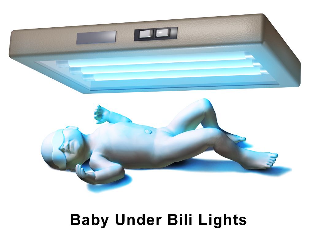
Phototherapy works by photoisomerisation. Blue light in the 460 to 490 nm range converts unconjugated bilirubin in the skin into water-soluble isomers, chiefly lumirubin, which are excreted in bile and urine without needing conjugation. Nursing care is specific: expose the maximum skin surface with only a nappy; apply opaque eye shields and check the eyes at least every shift for discharge or corneal irritation; remove the shields during feeding and parent holding so the infant sees faces; keep the prescribed distance between light and infant; monitor temperature frequently, since both hyperthermia and hypothermia occur; and increase feeding frequency because insensible water losses rise. Expect loose, green stools — that is bilirubin leaving and is a sign of effectiveness, but watch skin integrity and hydration. Do not apply lotions, oils or powders, which can cause burns. Bronzing of the skin (bronze baby syndrome) occurs when phototherapy is used with cholestasis and is a reason to reassess. For isoimmune haemolytic disease with bilirubin approaching exchange level, intravenous immunoglobulin at 0.5 to 1 g/kg may be given; exchange transfusion is reserved for extreme hyperbilirubinaemia or any sign of acute bilirubin encephalopathy.
The injury being prevented is acute bilirubin encephalopathy, which progresses through three phases: early — lethargy, hypotonia, poor suck; intermediate — irritability, a shrill high-pitched cry, fever, and retrocollis and opisthotonos (arching of the neck and trunk); advanced — apnoea, seizures, coma, death. Survivors develop kernicterus: athetoid cerebral palsy, sensorineural hearing loss or auditory neuropathy, upward-gaze palsy and dental enamel dysplasia. It is permanent and it is preventable, which is why the timed bilirubin and the follow-up appointment carry the weight they do.
Jaundice in the first 24 hours of life, a rise faster than 0.2 mg/dL per hour, or a direct bilirubin above 1 mg/dL are each pathologic by definition and demand investigation — everything else is managed by hour-specific bilirubin levels, effective feeding and timed follow-up.
Section 5Daily Care, Weight-Based Dosing and Discharge
Routine prophylaxis and screening — every dose per kilogram
Three medications and three screens are standard for essentially every newborn. Vitamin K (phytonadione) 1 mg intramuscularly for infants weighing 1500 g or more, or 0.5 mg for those under 1500 g, is given within 6 hours of birth into the vastus lateralis with a 25-gauge, 5/8-inch needle at a 90-degree angle. Newborns are born with a sterile gut that cannot yet synthesise vitamin K, and placental transfer is poor; without prophylaxis, vitamin K deficiency bleeding occurs, and the late form presents at 2 to 12 weeks with intracranial haemorrhage. If a family declines, document the discussion and educate about bleeding signs — do not treat the decision as an argument to win. Erythromycin 0.5% ophthalmic ointment, a 1 cm ribbon in each lower conjunctival sac within the first hour (up to 2 hours), prevents gonococcal ophthalmia neonatorum; it does not reliably prevent chlamydial conjunctivitis, which presents at 5 to 14 days and needs systemic treatment. Hepatitis B vaccine 0.5 mL IM is given within 24 hours; if the birthing parent is HBsAg-positive, the infant receives both the vaccine and hepatitis B immune globulin 0.5 mL within 12 hours of birth, at separate sites.
Screens: the newborn metabolic screen is a heel-stick collected after at least 24 hours of feeding (with a repeat per state protocol), warming the heel first and sampling the lateral aspect to avoid the calcaneus and nerves. Hearing screening by automated auditory brainstem response or otoacoustic emissions is done before discharge. Critical congenital heart disease (CCHD) screening is a pulse-oximetry screen performed at 24 hours of age or later, with probes on the right hand (preductal) and either foot (postductal). A pass requires saturation of 95% or above in either limb and a difference of 3% or less between them. Any reading below 90% is an immediate fail requiring urgent evaluation; readings of 90 to 94%, or a difference greater than 3%, are repeated hourly for up to three screens before failing.
Weight-based dosing and maintenance fluids
Children are not small adults, and paediatric dosing is not adult dosing scaled by eye. Every paediatric dose is expressed per kilogram of actual measured weight in kilograms — never a stated weight, never pounds. Weigh the child on admission, record in kilograms only, and remove the pounds display from the scale if your unit permits. Then run three checks on every calculation: is the dose per kilogram within the reference range, is the total dose below the maximum adult dose, and does the volume make sense for the route and the child’s size. High-alert paediatric medications require an independent double-check by a second nurse.
A 22 kg child: 15 mg/kg × 22 kg = 330 mg per dose. Daily maximum 75 × 22 = 1650 mg — four 330 mg doses total 1320 mg, which is within range. Safe.
A 70 kg adolescent: 15 mg/kg × 70 kg = 1050 mg — mathematically correct, but it exceeds the maximum single adult dose of 1000 mg. Give 1000 mg. Once a child’s weight-based calculation exceeds the adult dose, the adult dose becomes the ceiling. This is the check that catches the adolescent overdose.
Maintenance intravenous fluids for infants beyond the newborn period and for children use the 4-2-1 rule: 4 mL/kg/h for the first 10 kg of body weight, plus 2 mL/kg/h for the second 10 kg, plus 1 mL/kg/h for every kilogram above 20. Worked example, a 26 kg child: (10 kg × 4) + (10 kg × 2) + (6 kg × 1) = 40 + 20 + 6 = 66 mL/h. The daily equivalent (the 100-50-20 rule) cross-checks it: (10 × 100) + (10 × 50) + (6 × 20) = 1000 + 500 + 120 = 1620 mL/day, which is 67.5 mL/h — the same answer. Use an isotonic maintenance fluid with dextrose in most children; hypotonic maintenance fluid is a recognised cause of hospital-acquired hyponatraemia. Note the exception that matters in this chapter: the 4-2-1 rule does not apply to the first days of life. Term newborns start at roughly 60 to 80 mL/kg/day on day 1 and advance by 10 to 20 mL/kg/day toward about 150 mL/kg/day by the end of the first week, because renal concentrating ability and insensible losses in the newborn follow a different curve entirely.
Feeding, elimination and the first weeks
Feed on cue, not on a clock. A breast- or chestfed newborn feeds 8 to 12 times in 24 hours; early hunger cues are rooting, hand-to-mouth movements, lip smacking and stirring, and crying is a late cue that makes latching harder. Teach latch by what you can see and hear: a wide gape, more areola visible above the upper lip than below the lower, flanged lips, the chin touching the breast, and rhythmic swallowing that is audible. Assess comfort, transfer and output rather than counting minutes. Formula-fed newborns take about 45 to 90 mL (1.5 to 3 ounces) every 3 to 4 hours in the first week, roughly 150 mL/kg/day; never prop a bottle, never heat formula in a microwave, and discard formula left after a feed. Exclusively or partially breastfed infants need vitamin D 400 IU daily starting in the first days of life.
Output is the most honest measure of intake in the first week. The first void should occur within 24 hours and the first meconium within 24 to 48 hours; failure of either is an escalation. Expect one wet nappy on day 1, increasing to six or more per day by day 4 to 5, with pale straw-coloured urine. Stools progress from meconium (sticky, black-green, first 24 to 48 hours) to transitional (loose, greenish-brown, days 3 to 4) to the yellow, seedy stools of a milk-fed infant by days 4 to 5, at least three to four times a day in a breastfed newborn. Brick-red urate crystals in the nappy in the first days are usually benign but, after day 3, prompt a feeding evaluation.
Cord care is dry cord care: keep it clean and dry, fold the nappy below it, and expect separation at 7 to 14 days. Teach parents to call for purulent drainage, foul odour, periumbilical redness spreading onto the abdomen, or active bleeding. After circumcision, apply petroleum jelly to the glans with each nappy change (except with a Plastibell, where it is not used), expect a yellowish exudate at 24 to 48 hours as normal granulation — not infection — and report bleeding larger than a coin-sized spot or failure to void within 6 to 8 hours. Take temperatures axillary, and teach the family the same.
Safe sleep, safe travel, and when to call
Safe sleep is taught at every contact and modelled in the hospital, because parents copy what they saw the nurse do. Every sleep, the infant goes down supine, on a firm, flat, non-inclined surface with a fitted sheet and nothing else — no bumpers, pillows, blankets, positioners, wedges or soft toys. Room-share without bed-sharing for at least the first 6 months. Avoid overheating and overbundling; use a wearable blanket rather than loose bedding. Offer a pacifier at sleep once feeding is established. Keep the environment smoke-free. Supervised awake tummy time from the first days prevents positional plagiocephaly and builds neck strength. Car-seat teaching is equally concrete: rear-facing in the back seat, harness straps at or below the shoulders in a rear-facing seat, chest clip at armpit level, snug enough that you cannot pinch a horizontal fold of webbing at the shoulder, and no bulky coats or aftermarket inserts between the child and the harness. Keep the child rear-facing for as long as possible — until they reach the highest weight or height allowed by the seat’s manufacturer — rather than turning them forward-facing at a set age.
| Newborn red flag | Why it matters | SBAR escalation — what you say |
|---|---|---|
| Rectal temperature 38.0 °C (100.4 °F) or above, or below 36.0 °C, in an infant under 3 months | Either direction signals sepsis in a newborn; hypothermia is at least as ominous as fever | S: “Baby Ortiz, 26 hours old, rectal temp 38.2.” B: “Term, unremarkable birth, group B strep status unknown.” A: “This meets criteria for sepsis evaluation.” R: “I need you at the bedside now for blood culture, CBC, lumbar puncture and empiric antibiotics.” |
| Central cyanosis, or a failed CCHD screen | Duct-dependent congenital heart disease presents as the ductus closes at 24 hours to 2 weeks | R: “Preductal 88%, postductal 82%. I need urgent evaluation and an echocardiogram, and I want prostaglandin availability confirmed.” |
| Jaundice in the first 24 hours | Haemolysis until proven otherwise | R: “Requesting total and direct bilirubin, type and Rh, direct Coombs, haematocrit and reticulocyte count now.” |
| Respiratory rate above 60 sustained, grunting, nasal flaring, retractions | Respiratory distress; grunting is auto-generated positive end-expiratory pressure and is a late, serious sign | R: “Sustained rate 78 with grunting and subcostal retractions. I need respiratory evaluation and a saturation target order now.” |
| Glucose below 40 mg/dL with symptoms, or below 25 mg/dL at any time | Neuroglycopenia; brain injury is silent until it is not | R: “Glucose 22 at 3 hours, infant jittery. I am feeding and requesting an order for D10W 2 mL/kg IV now.” |
| No void by 24 hours or no stool by 48 hours; weight loss above 7% | Intake problem, obstruction, or renal anomaly | R: “No void at 26 hours. Requesting evaluation and a feeding plan; I have lactation consulting now.” |
| Boggy, expanding scalp swelling that crosses sutures, especially after vacuum-assisted birth | Subgaleal haemorrhage — can exsanguinate a newborn | R: “Head circumference up 1.5 cm in 30 minutes, heart rate 190, pale. I need you at the bedside immediately and blood available.” |
Routine newborn care is a set of exact numbers — vitamin K 1 mg IM within 6 hours, hepatitis B immune globulin within 12 hours for an HBsAg-positive parent, CCHD screening at 24 hours with a 95% and 3% rule, and every paediatric dose calculated per kilogram and then capped at the adult maximum.
The chapter in one breath
Birth is a coordinated physiological handover. The first breath aerates a fluid-filled lung that has already begun absorbing its own liquid through sodium channels primed by labour, and that aeration drops pulmonary vascular resistance so the foramen ovale presses shut within hours and the ductus arteriosus constricts by 12 to 24 hours as oxygen rises and placental prostaglandin disappears. Everything that follows is surveillance of a transition that can reverse: hypoxia, acidosis and cold reopen the shunts. The APGAR at 1 and 5 minutes describes the response but never delays resuscitation, where effective ventilation in the first 60 seconds outranks every other action, compressions run 3:1 for a heart rate below 60, and epinephrine is 0.01 to 0.03 mg/kg. The newborn then faces three metabolic tests. Cold stress burns brown fat through non-shivering thermogenesis and cascades into hypoglycaemia, acidosis and reopened shunts. Hypoglycaemia is screened by hour of life, treated with feeding, 40% dextrose gel at 0.5 mL/kg or D10W at 2 mL/kg. Jaundice is physiologic after 24 hours and pathologic before it, or when the rise exceeds 0.2 mg/dL per hour or the direct fraction exceeds 1 mg/dL. In every obstetric emergency the sequence is the treatment: massage the fundus first, stop the magnesium and give calcium gluconate, lift the presenting part off a prolapsed cord, McRoberts and suprapubic pressure but never fundal pressure.
Words worth knowing
| Term | What it means |
|---|---|
| Acrocyanosis | Blue hands and feet with a pink trunk; a normal vasomotor finding for the first 24 to 48 hours, unlike central cyanosis, which is never normal. |
| APGAR score | A 0 to 10 score of Appearance, Pulse, Grimace, Activity and Respiration assigned at 1 and 5 minutes; describes the transition and never delays resuscitation. |
| Ballard score | Assessment of gestational age using six neuromuscular and six physical maturity criteria; accurate to about plus or minus 2 weeks, best performed at 12 to 24 hours. |
| Brown adipose tissue | Specialised fat laid down from 26 to 30 weeks that generates heat through uncoupling protein 1 rather than storing energy; the substrate of non-shivering thermogenesis. |
| Caput succedaneum | Soft, pitting scalp oedema present at birth that crosses suture lines and resolves in 3 to 5 days. |
| Cephalhaematoma | Subperiosteal bleeding appearing hours after birth that does not cross suture lines, resolves over weeks, and increases the bilirubin load. |
| Cold stress | The metabolic cascade triggered by heat loss: brown-fat burning, rising oxygen and glucose consumption, hypoglycaemia, acidosis and re-opening of fetal shunts. |
| Ductus arteriosus | Fetal vessel from the pulmonary artery to the aorta; closes functionally in 12 to 24 hours as oxygen rises and prostaglandin E₂ falls, becoming the ligamentum arteriosum. |
| Enterohepatic circulation | Reabsorption of deconjugated bilirubin from the newborn gut back into the bloodstream; reduced by frequent feeding and stooling, not by water. |
| Erythema toxicum neonatorum | Blotchy erythema with small yellow-white papules appearing on days 1 to 3; benign, self-limiting and requiring only explanation. |
| Foramen ovale | Fetal opening between the atria; closes functionally within minutes to hours when left atrial pressure exceeds right atrial pressure after birth. |
| Kernicterus | Permanent bilirubin-induced neurological injury — athetoid cerebral palsy, sensorineural hearing loss, upward-gaze palsy and enamel dysplasia. |
| McRoberts manoeuvre | Hyperflexion of the birthing parent’s hips onto the abdomen to flatten the sacral promontory in shoulder dystocia; paired with suprapubic — never fundal — pressure. |
| Neutral thermal environment | The ambient temperature range in which a newborn maintains a normal core temperature with minimal oxygen consumption and metabolic work. |
| Non-shivering thermogenesis | Heat production by noradrenaline-driven lipolysis in brown fat; the newborn’s main heat source because effective shivering is not available. |
| Periodic breathing | Pauses of up to 15 seconds without colour or heart-rate change; normal, and distinct from apnoea, which lasts 20 seconds or more or is accompanied by bradycardia or cyanosis. |
| Physiologic jaundice | Unconjugated hyperbilirubinaemia appearing after 24 hours, peaking on days 3 to 5, rising less than 5 mg/dL per day and resolving within a week at term. |
| Subgaleal haemorrhage | Bleeding into the space beneath the scalp aponeurosis that crosses sutures and expands; an emergency because it can hold most of a newborn’s blood volume. |
| Surfactant | Phospholipid produced by type II pneumocytes from about 24 weeks, mature near 34 to 36 weeks, that lowers alveolar surface tension and prevents end-expiratory collapse. |
| Vitamin K deficiency bleeding | Haemorrhage from absent vitamin K synthesis in the sterile newborn gut; prevented by phytonadione 1 mg intramuscularly within 6 hours of birth. |