Integumentary Disorders and Burns
Skin is the only organ you assess with your eyes and hands every single shift, and it is the organ students most often under-report. It is a barrier, a thermostat, an immune sentinel and a fluid container — and when it fails, the failure is systemic. A cellulitic leg becomes bacteraemia. A sacral pressure injury becomes osteomyelitis. A forty-percent flame burn becomes distributive-plus-hypovolaemic shock inside an hour, and then, three days later, becomes sepsis. This chapter teaches the integument the way it is practised on a busy floor: recognise the lesion, name the depth, escalate the airway, calculate the fluid, protect the tissue that is still salvageable, and feed the patient enough protein to close the wound. Burns get the most space here because burns are where nursing judgement is measured in millilitres per hour and where the wrong priority in the first sixty minutes cannot be undone later.
By the end of this chapter you can
- Describe the layers and functions of the integument, name primary and secondary lesions, and document a wound using length × width × depth, undermining and tunnelling in clock positions.
- Differentiate cellulitis, erysipelas and necrotising fasciitis, and state the finding that makes necrotising fasciitis a surgical emergency.
- Recognise Stevens–Johnson syndrome and toxic epidermal necrolysis, identify the high-risk drug classes, and name the single most important intervention.
- Stage a pressure injury using current NPIAP criteria, apply the Braden Scale, and build a prevention plan that includes what may and may not be delegated.
- Distinguish venous, arterial and neuropathic ulcers by location and appearance, and explain why the ankle–brachial index must precede compression.
- Classify burn depth, estimate extent with the Rule of Nines and the rule of palm, and calculate resuscitation volume with the Parkland/consensus formula, titrating to urine output.
- Identify the airway red flags of inhalation injury, carbon monoxide and cyanide toxicity, and escalate using SBAR before the airway closes.
- Explain why the first 24 hours are about fluid and the period after 48–72 hours is about infection and hypermetabolism, and describe topical antimicrobials, grafting and anti-contracture positioning.
- Modify assessment and management for the older adult, whose skin is thinner, whose burns are deeper and whose infection may present only as confusion.
Section 1The Integument, Skin Assessment and Wound Healing
Structure that explains the clinical picture
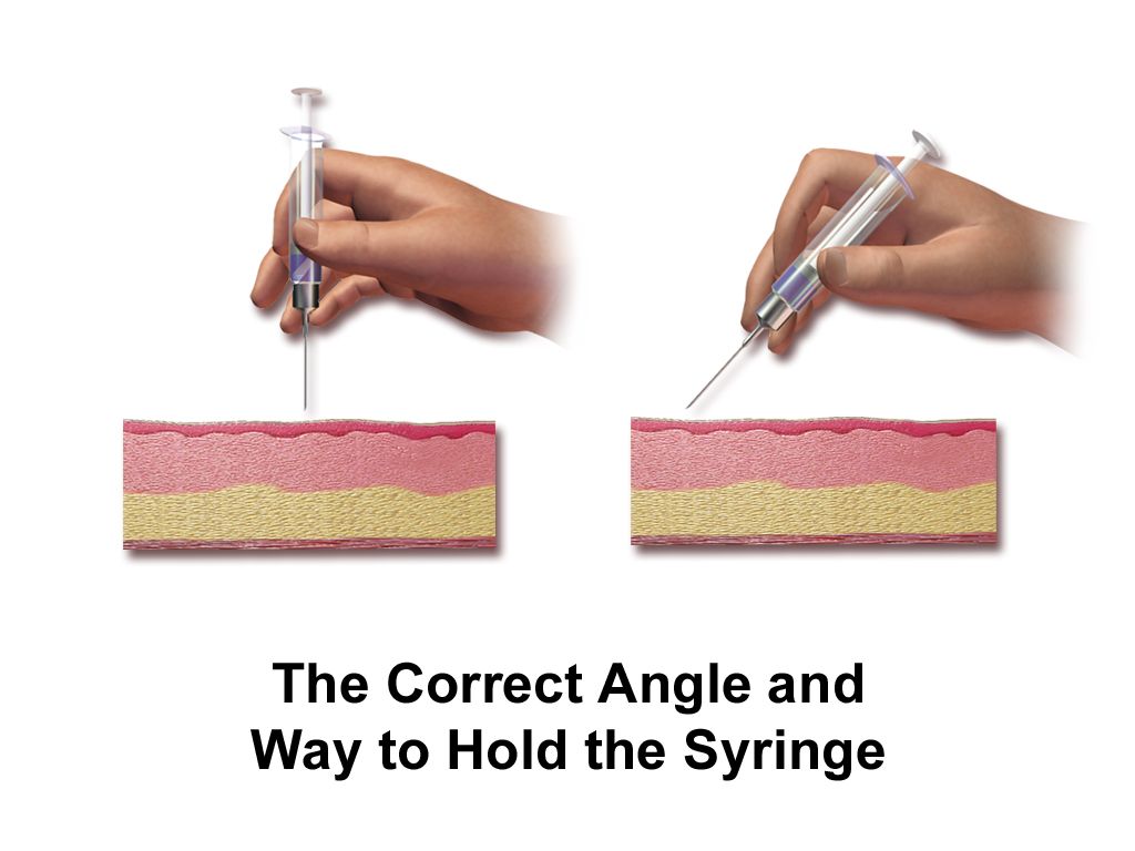
The epidermis is avascular. It is a stratified sheet of keratinocytes that migrate from the basal layer to the surface over roughly 28 days, seeded with melanocytes for pigment and Langerhans cells for immune surveillance. Because it has no vessels of its own, an injury confined to epidermis cannot bleed and cannot scar — it peels and resolves. That single fact is why a sunburn is painful and red but heals in a week without a mark, and why it is not counted in burn surface-area calculations.
The dermis is where the clinically interesting structures live: capillary loops, sensory nerve endings, sebaceous and sweat glands, and hair follicles. Those appendages dip down from the epidermis into the dermis, and they carry epithelial cells with them. When a wound destroys the surface but spares the follicles, new epithelium crawls out of each follicle like grass seeding a field, and the wound closes on its own. When the wound destroys the deep dermis, the seed is gone, and closure requires a graft. Every burn-depth decision you will make is really a question about follicles.
The hypodermis is subcutaneous fat — insulation, energy store and mechanical cushion over bony prominences. Collectively the integument provides barrier, thermoregulation, sensation, vitamin D synthesis, immune defence and fluid containment. Lose enough of it and the patient loses heat, loses plasma, loses the ability to keep organisms out, and loses the sensory feedback that would normally make them shift their weight off an ischaemic heel.
Looking, and looking in every skin tone
Describe what you see in the standard vocabulary, because that is how dermatology and the medical record talk. Primary lesions arise on previously normal skin; secondary lesions are what happens to a primary lesion after time, scratching, infection or treatment. Add configuration (annular, linear, grouped, dermatomal) and distribution (flexural, extensor, sun-exposed, dermatomal, intertriginous), because distribution is often more diagnostic than morphology.
| Lesion | Definition | Typical example |
|---|---|---|
| Macule | Flat, non-palpable colour change under 1 cm | Freckle, early drug rash |
| Papule | Raised, solid, under 1 cm | Acne, early varicella |
| Plaque | Raised, flat-topped, over 1 cm | Psoriasis |
| Nodule | Solid, deeper, 1–2 cm | Lipoma, erythema nodosum |
| Vesicle / bulla | Fluid-filled under 1 cm / over 1 cm | Herpes zoster / burn blister, TEN |
| Pustule | Pus-filled | Folliculitis, pustular psoriasis |
| Wheal | Transient oedematous plaque | Urticaria |
| Erosion / ulcer | Loss of epidermis / loss through dermis | Ruptured blister / pressure injury |
| Fissure, lichenification, excoriation | Linear crack, leathery thickening, scratch-induced loss | Chronic eczema |
Erythema in deeply pigmented skin does not look red. It looks violaceous, darker than the surrounding skin, or simply different in a way that is easy to miss under fluorescent light. This is not a cosmetic point — it is the single largest source of missed early pressure injury and missed cellulitis. Use natural or high-quality light, compare the area with adjacent skin and with the contralateral limb, and lead with your hands: warmth, coolness, boggy softness and induration are palpable regardless of pigment. Ask about pain and itch, because a patient will report tenderness over a stage 1 injury before you can see it. For pallor, cyanosis and jaundice, look at conjunctivae, hard palate, palms, soles and nail beds.
How wounds heal, and what stops them
Healing runs in overlapping phases. Haemostasis is immediate: vasoconstriction, platelet plug, fibrin. The inflammatory phase (roughly days 0–4) brings neutrophils then macrophages; the wound looks red, warm and swollen, and this is normal, not infection. The proliferative phase (days 4–21) builds beefy red granulation tissue, contracts the edges and re-epithelialises from the margins and remaining appendages. The maturation phase runs from three weeks to two years, remodelling collagen; a healed wound reaches only about 80 percent of original tensile strength, which is why you keep teaching lifting restrictions long after the incision looks fine. Wounds closed surgically heal by primary intention; open wounds granulating in heal by secondary intention; a contaminated wound left open and closed later heals by tertiary intention.
Healing fails for predictable reasons, and each one is a nursing intervention. Perfusion and oxygenation come first — tissue cannot build collagen without oxygen, which is why smoking, peripheral arterial disease and hypotension are wound problems. Nutrition is second: protein for collagen, vitamin C for cross-linking, zinc and arginine for proliferation. Hyperglycaemia impairs neutrophil function, so glycaemic control is wound care. Add corticosteroids and other immunosuppressants, chemotherapy, infection, repeated trauma, moisture imbalance (too wet macerates, too dry desiccates) and simple age. A moist, warm, protected wound bed heals faster than a dry scab — that is the entire rationale behind modern dressings.
Document so the next nurse can tell whether the wound is improving without ever having seen it: anatomical location; measurements in centimetres as length (head to toe) × width (side to side) × depth; undermining and tunnelling described in clock positions with 12 o’clock toward the head; percentage of the bed that is granulation, slough or eschar; exudate amount, colour and odour after cleansing; wound edges (attached, rolled/epibole, macerated); and the periwound skin. Photograph per policy. “Wound looks better” is not documentation.
Whether a wound can close itself depends on whether epithelial cells survive in the dermal appendages — and whether it will close depends on perfusion, protein and glucose.
Section 2Inflammatory, Infectious and Life-Threatening Skin Disorders
Bacterial infection: from cellulitis to a surgical emergency
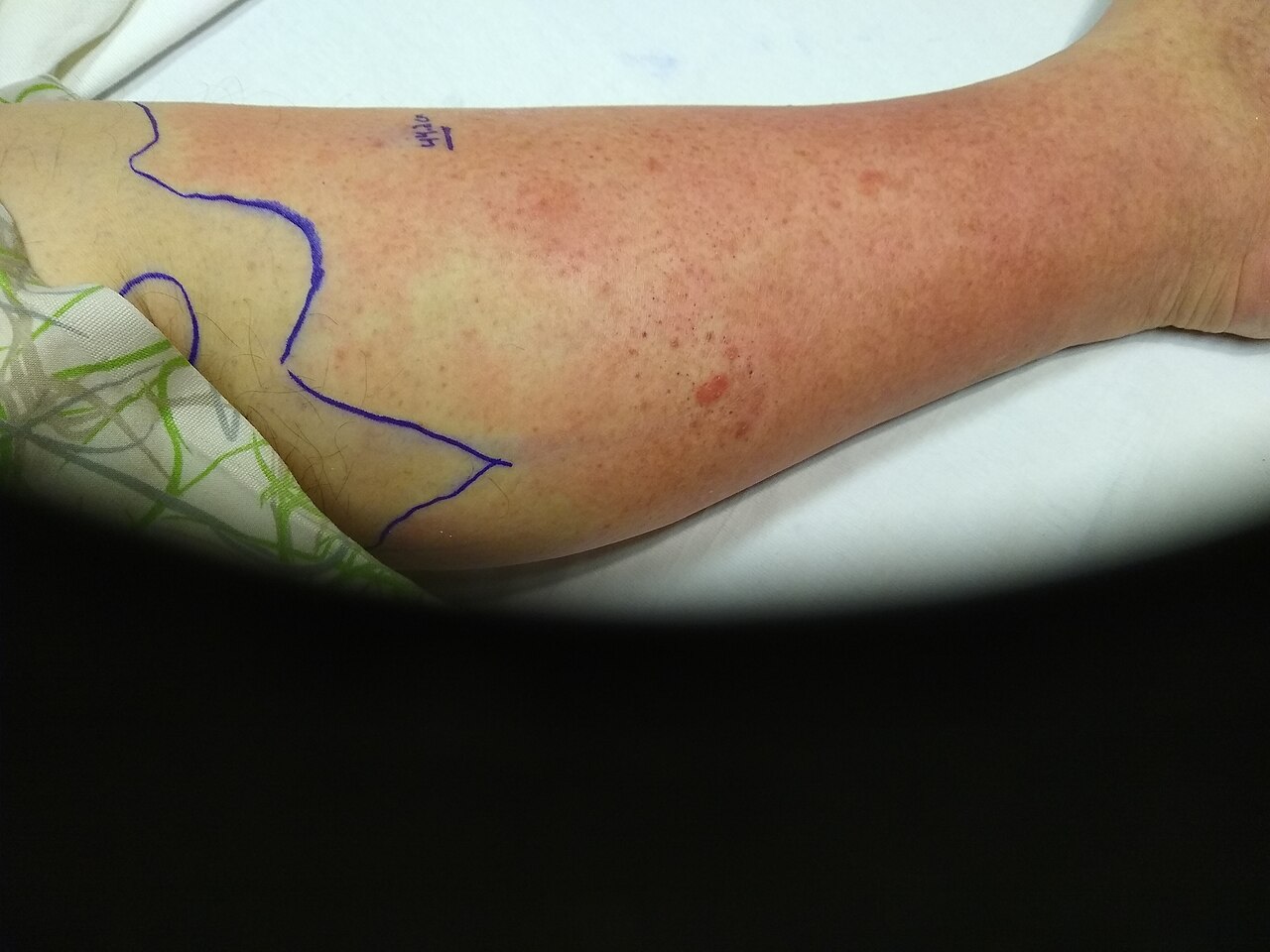
Cellulitis is infection of the deep dermis and subcutaneous tissue, usually Streptococcus pyogenes or Staphylococcus aureus, entering through a break you may have to hunt for — tinea pedis between the toes, a venous ulcer, an IV site, a shaving nick. It presents as warm, tender, oedematous erythema with indistinct borders. Erysipelas is the more superficial cousin: sharply demarcated, raised, fiery red, often on the face or shin, classically streptococcal and more likely to spike fever early. Risk is stacked in patients with diabetes, lymphoedema, chronic venous insufficiency, obesity and prior episodes.
Nursing management is unglamorous and effective. Outline the erythema with a skin marker and write the date and time on the skin, then reassess each shift — expansion beyond the mark on appropriate antibiotics is an escalation trigger. Elevate the limb above heart level, because gravity does more for the oedema than any drug. Warm compresses for comfort. Mark and monitor temperature, heart rate and white count. Treatment for non-purulent cellulitis targets streptococci: cephalexin orally, cefazolin intravenously. Purulent cellulitis with an abscess needs incision and drainage — drainage is the treatment, antibiotics are the adjunct — plus MRSA coverage with trimethoprim–sulfamethoxazole, doxycycline or clindamycin orally, or vancomycin intravenously with trough or AUC monitoring and attention to renal function. Send cultures before the first dose when you can.
Necrotising fasciitis is the diagnosis you must not miss. Infection tracks along fascial planes, thromboses the perforating vessels and kills the overlying skin from below, so the surface can look deceptively unimpressive while the patient is dying. The cardinal early sign is pain out of proportion to the visible findings. Then come rapid hourly progression, oedema extending past the erythema, dusky or grey mottled skin, haemorrhagic bullae, crepitus, foul “dishwater” drainage, and later anaesthesia over the area as the cutaneous nerves die — a numb, agonising limb is a catastrophic sign. Systemically the patient is toxic: fever, tachycardia, hypotension, altered mentation, rising lactate, marked leukocytosis. Management is emergent surgical debridement plus broad-spectrum antibiotics (typically vancomycin with piperacillin–tazobactam, plus clindamycin for its antitoxin effect on streptococcal exotoxin). Imaging must never delay the operating room.
Viral, fungal and parasitic disease
Herpes zoster is reactivation of varicella-zoster virus latent in a dorsal root ganglion. Patients report two to three days of burning, itching or stabbing pain in a band before anything appears; then grouped vesicles on an erythematous base in a unilateral dermatomal distribution that stops at the midline. Antivirals — acyclovir, valacyclovir or famciclovir — shorten the course and reduce postherpetic neuralgia, but only if started within 72 hours of rash onset, so a patient reporting a new one-sided rash needs to be seen today, not next week. Pain is neuropathic: gabapentin or pregabalin, tricyclics, lidocaine 5 percent patches, opioids as a bridge. Postherpetic neuralgia may persist for months and is far more common in older adults. Lesions are infectious to varicella-naive contacts until crusted: cover localised lesions and use standard plus contact precautions; disseminated zoster or zoster in an immunocompromised patient requires airborne plus contact precautions. Involvement of the ophthalmic branch of the trigeminal nerve — especially vesicles on the tip of the nose, the Hutchinson sign — is a same-day ophthalmology referral. Recombinant zoster vaccine, two doses, is recommended from age 50 and for immunocompromised adults from 19.
Fungal disease is common and usually simple. Tinea corporis is an annular plaque with a scaly, advancing, raised border and central clearing; tinea pedis macerates the web spaces and is a frequent portal for cellulitis; tinea capitis needs an oral agent because topicals cannot reach the follicle. Candidiasis favours warm moist folds — inframammary, panniculus, groin — and produces beefy red plaques with satellite lesions, the finding that distinguishes it from simple intertrigo. Treat with topical antifungals and, more importantly, keep the fold dry and separated; barrier products and moisture-wicking textiles do more than any cream.
Scabies is Sarcoptes scabiei burrowing in the stratum corneum, and it should be suspected in any patient with relentless nocturnal itch and burrows or papules in the finger web spaces, flexor wrists, axillae, waistline, umbilicus or genitalia. Treat with permethrin 5 percent cream applied from the neck down (including under nails), left on 8–14 hours, repeated in seven days; treat all household and intimate contacts simultaneously even if asymptomatic; launder linens and clothing in hot water and dry on high heat, or bag non-washables for 72 hours. Oral ivermectin is used for crusted (Norwegian) scabies, which is highly contagious and warrants contact precautions. Teach explicitly that itching commonly persists two to four weeks after successful treatment — that is a hypersensitivity reaction to dead mite antigen, not failure, and re-treating repeatedly causes dermatitis.
Chronic inflammatory disease and severe drug reactions
Psoriasis is T-cell–driven keratinocyte hyperproliferation: turnover drops from 28 days to about four, so cells pile up as well-demarcated erythematous plaques with adherent silvery scale over extensor surfaces, scalp, umbilicus and sacrum. Removing scale causes pinpoint bleeding (Auspitz sign); new plaques appear at sites of trauma (Koebner phenomenon); nails show pitting and onycholysis. Treat mild disease topically with corticosteroids, vitamin D analogues such as calcipotriene, tar and keratolytics; moderate-to-severe disease with narrowband UVB phototherapy, methotrexate, cyclosporine, apremilast or biologics targeting TNF-alpha, IL-17 and IL-23. Nursing implications matter here: methotrexate is weekly, not daily — a daily dosing error is lethal — requires folic acid supplementation, CBC and liver function monitoring, alcohol avoidance and contraception, since it is teratogenic. Biologics require screening for latent tuberculosis and hepatitis B before the first dose and holding for active infection. Psoriasis is not contagious and it is not a hygiene failure; it travels with psoriatic arthritis, metabolic syndrome, cardiovascular disease and depression, so screen for joint pain and mood.
Atopic dermatitis is a skin-barrier disease with an itch–scratch cycle on top. Teach “soak and seal”: short lukewarm bath, gentle non-soap cleanser, pat dry, then a thick ointment-based emollient within three minutes, with topical corticosteroid applied to active lesions first. Use the lowest effective steroid potency on the face, neck and skin folds, teach the fingertip-unit measure, and warn about atrophy, striae and telangiectasia with prolonged high-potency use. Topical calcineurin inhibitors (tacrolimus, pimecrolimus) are steroid-sparing for the face; dupilumab is available for moderate-to-severe disease. Sudden worsening with punched-out erosions and fever suggests eczema herpeticum — a same-day referral.
Stevens–Johnson syndrome and toxic epidermal necrolysis are a single spectrum of keratinocyte apoptosis, almost always drug-induced, separated by the percentage of body surface with epidermal detachment: SJS under 10 percent, overlap 10–30 percent, TEN over 30 percent. The usual culprits are allopurinol, sulfonamide antibiotics, aromatic anticonvulsants (lamotrigine, carbamazepine, phenytoin), nevirapine and oxicam NSAIDs, typically 4–28 days after starting. Suspect it when a flu-like prodrome is followed by painful skin, dusky targetoid macules, and erosions of at least two mucosal surfaces — eyes, mouth, genitalia. Lateral pressure sloughs the epidermis (Nikolsky sign positive). The single most important intervention is to stop the offending drug immediately; every hour of continued exposure worsens mortality. After that, care is burn care: transfer to a burn or intensive care unit, fluid and electrolyte replacement, meticulous non-adherent wound care with no tape on skin, temperature regulation, aggressive analgesia, nutrition, and an early ophthalmology consult, because ocular scarring and blindness are the leading long-term morbidity. Document the culprit drug as a true allergy in every system, and teach the patient and family to name that drug and its whole class for the rest of their life.
In skin infection, pain out of proportion to appearance means necrotising fasciitis until a surgeon says otherwise; in a drug rash, mucosal involvement plus a positive Nikolsky sign means stop the drug now and call the burn unit.
Section 3Pressure Injuries, Chronic Wounds and Skin Cancer
Pressure injury: mechanism, staging and prevention
Pressure injury develops when sustained pressure over a bony prominence exceeds capillary closing pressure and occludes perfusion; ischaemia, reperfusion injury and cell death follow. Shear makes it far worse by tearing the deeper tissue planes while the skin stays fixed — which is exactly what happens when the head of the bed is elevated and the patient slides down. Deep muscle over bone dies before the skin does, which is why a small surface lesion can sit over a large cavity, and why deep tissue pressure injury exists as its own category. Add moisture, malnutrition, impaired sensation, immobility, hypotension and friction and you have every risk factor on the Braden Scale.
| Category | Defining findings | Practice implication |
|---|---|---|
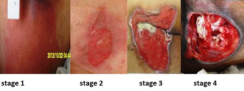
| Stage 1 | Intact skin, non-blanchable erythema; in darker tones may be purple or maroon, with warmth, firmness or pain | Off-load immediately; fully reversible |
| Stage 2 | Partial-thickness loss of dermis: shallow pink-red bed, or intact/ruptured serum-filled blister. No slough, granulation or eschar | Not a skin tear, moisture-associated damage or burn — those are staged separately |
| Stage 3 | Full-thickness loss; fat may be visible; slough or eschar may be present; undermining and tunnelling possible; no exposed bone, tendon or muscle | Depth varies by site — the ear and bridge of the nose have no fat, so stage 3 there is shallow |
| Stage 4 | Full-thickness with exposed or directly palpable bone, tendon, muscle, ligament or cartilage | Assume osteomyelitis risk; surgical consult |
| Unstageable | Base obscured by slough or eschar | Cannot stage until debrided — except stable, dry, intact eschar on an ischaemic heel, which is left alone as a biological cover |
| Deep tissue pressure injury | Persistent non-blanchable deep red, maroon or purple discoloration, or a blood-filled blister; skin may be intact | May declare itself as a stage 3 or 4 within days; document evolution |
| Medical device–related | Conforms to the pattern of the device — oxygen tubing, cervical collar, SCD, endotracheal tube tie | Assess under and around every device at least twice daily |
The Braden Scale scores six subscales — sensory perception, moisture, activity, mobility, nutrition, and friction/shear — for a total of 6 to 23. Lower is worse: 15–18 mild risk, 13–14 moderate, 10–12 high, 9 or below severe. Score on admission, at every change in condition and per unit policy, and treat the low subscale, not just the total: a patient scoring low only on moisture needs a barrier and a continence plan, not a specialty mattress.
Prevention is a bundle and every element is testable. Reposition on an individualised schedule, conventionally every two hours in bed and hourly in a chair — or teach the alert patient to do a 15-second weight shift every 15 minutes. Use the 30-degree lateral tilt rather than a full 90-degree side-lying position, which loads the trochanter. Keep the head of bed at or below 30 degrees unless contraindicated, and use the knee gatch or a lift sheet to stop sliding. Float the heels completely off the mattress with a pillow lengthwise under the calves or an offloading boot — the heel is the second most common site and the hardest to heal. Never massage over a bony prominence, never use ring or donut cushions, and never turn a patient onto an existing injury. Manage incontinence with a structured barrier regimen, keep linens dry and wrinkle-free, optimise protein and calorie intake, and choose a support surface matched to risk. And never reverse-stage: a healing stage 4 is a “healing stage 4,” not a stage 2, because muscle and fat are replaced by scar, not by the original tissue.
Lower-extremity ulcers: three diseases that look alike
A leg ulcer is not a diagnosis, it is a finding, and the treatment for one type actively harms another. Compression heals a venous ulcer and can infarct an arterial one, so you must know which you are looking at before the wrap goes on.
| Feature | Venous | Arterial | Neuropathic (diabetic) |
|---|---|---|---|
| Location | Medial malleolus, gaiter area | Lateral malleolus, toes, pressure points | Plantar surface, metatarsal heads, heel |
| Appearance | Shallow, irregular border, ruddy granulation | “Punched out,” pale or necrotic dry base | Round, callused rolled rim |
| Exudate | Moderate to heavy | Minimal | Variable; purulent if infected |
| Pain | Aching, better with elevation | Severe, worse with elevation, better dangling | Absent — that is the danger |
| Surrounding skin | Haemosiderin staining, oedema, lipodermatosclerosis, weeping | Thin, shiny, hairless, cool, pale on elevation | Dry, cracked, callused; deformity, Charcot changes |
| Pulses / ABI | Present; ABI usually normal | Diminished or absent; ABI under 0.9 | Often present but ABI may be falsely high (over 1.3) from calcification |
| Treatment core | Compression, elevation, moist dressing | Revascularisation; never compress | Offloading (total contact cast), debride callus, glycaemic control |
The ankle–brachial index is the gatekeeper: 0.91–1.30 is normal, 0.90 or below indicates arterial disease, 0.40 or below is severe ischaemia with rest pain, and above 1.30 means non-compressible calcified vessels that make the number unreliable. Full multilayer compression is generally reserved for an ABI above 0.8; modified or no compression below that, pending vascular assessment. Every diabetic patient gets daily foot inspection with a mirror, well-fitted closed shoes, no barefoot walking, no heating pads, straight-across nail trimming and an annual monofilament and pulse exam — teaching that prevents amputations far more effectively than any dressing.
Skin cancer and photoprotection
Basal cell carcinoma is the most common human cancer: a pearly, translucent papule with telangiectasias and a rolled border, often on the nose, ear or eyelid, sometimes with a central ulcer that scabs and re-bleeds. It invades locally and rarely metastasises. Squamous cell carcinoma is a firm, scaly, hyperkeratotic or ulcerated nodule on sun-exposed skin, arising from actinic keratoses; it can metastasise, and its risk is markedly higher in solid-organ transplant recipients and in chronic wounds and old burn scars — a non-healing ulcer within a mature burn scar is a Marjolin ulcer until biopsied. Melanoma is the one that kills. Teach ABCDE: Asymmetry, Border irregularity, Colour variegation, Diameter over 6 mm, and Evolving — plus the “ugly duckling” lesion that simply does not match the patient’s other moles. Diagnosis is by full-thickness biopsy; Breslow depth in millimetres is the dominant prognostic factor and drives sentinel lymph node biopsy. Prevention teaching is concrete: broad-spectrum SPF 30 or higher applied 15–30 minutes before exposure and reapplied every two hours and after swimming, protective clothing and wide-brimmed hats, shade between 10:00 and 16:00, no tanning beds at any age, and a monthly self-examination including scalp, soles and between the toes.
Pressure injuries are prevented by offloading and staged only when the base is visible; leg ulcers are treated by their cause — compression for venous, revascularisation for arterial, offloading for neuropathic — and any non-healing lesion or changing mole gets a biopsy, not another dressing.
Section 4Burn Injury: Depth, Extent and the First 24 Hours
Mechanism: why a skin injury becomes a whole-body problem
Heat coagulates protein. At the centre of the burn is the zone of coagulation — irreversibly dead tissue. Surrounding it is the zone of stasis, where perfusion is marginal and the tissue is alive but threatened; and outside that the zone of hyperaemia, which recovers. Everything you do in the first 24 hours — fluid, warmth, avoiding pressure and vasoconstrictors — exists to keep the zone of stasis from converting to necrosis. Under-resuscitate and the burn gets deeper. Over-resuscitate and you drown the patient. Both errors are nursing-visible before they are physician-visible.
Once a burn exceeds roughly 20–25 percent total body surface area (TBSA), the inflammatory response stops being local. Histamine, prostaglandins, bradykinin and thromboxane flood the circulation and make capillaries everywhere leak. Plasma, protein and electrolytes shift into the interstitium — massive third-spacing — producing generalised oedema and intravascular volume loss. This is burn shock: hypovolaemic and distributive at once, with an early depression of cardiac output out of proportion to the volume lost. Haematocrit rises from haemoconcentration even as the patient bleeds volume into the tissues. Potassium rises from cell lysis. Sodium falls as it follows water into the interstitium. Metabolic acidosis develops from hypoperfusion. Glucose rises with the catecholamine surge. The gut becomes ileus.
Then, at roughly 48–72 hours, capillary integrity is restored and the process reverses. Interstitial fluid remobilises into the vasculature, the patient diureses briskly, haematocrit falls by dilution, and potassium now drops — back into cells and out through the urine. The nurse who keeps running the resuscitation rate through this transition causes pulmonary oedema. The transition point is the boundary between the emergent and acute phases, and recognising it is a bedside judgement.
| Parameter | Emergent phase (0–48 h) | Acute phase (after fluid remobilisation) |
|---|---|---|
| Fluid shift | Intravascular to interstitial (third spacing) | Interstitial back to intravascular |
| Urine output | Low — the resuscitation target | Diuresis |
| Haematocrit | Elevated (haemoconcentration) | Falls (dilution, plus anaemia of injury) |
| Potassium | Hyperkalaemia from cell lysis | Hypokalaemia from shift and urinary loss |
| Sodium | Hyponatraemia (sodium follows water out) | Hyponatraemia (dilutional) or hypernatraemia with evaporative loss |
| Acid–base | Metabolic acidosis, base deficit, elevated lactate | Corrects with perfusion; watch for sepsis-driven acidosis |
| Dominant threat | Airway and hypovolaemic shock | Infection, hypermetabolism, contracture |
Depth and extent: naming the burn correctly
| Depth | Tissue involved | Appearance and sensation | Healing |
|---|---|---|---|
| Superficial (first-degree) | Epidermis only | Dry, red, blanches, no blisters, painful | 3–7 days, no scar. Not counted in TBSA |
| Superficial partial-thickness | Epidermis + papillary dermis | Moist, weeping, blisters, blanches with brisk refill, very painful | 7–21 days, minimal scarring |
| Deep partial-thickness | Into reticular dermis | Mottled red and waxy white, drier, sluggish or absent blanching, decreased light touch but pressure is painful | Over 21 days; hypertrophic scarring; usually excised and grafted |
| Full-thickness (third-degree) | Entire dermis | Dry, leathery eschar — waxy white, tan, brown or charred; does not blanch; insensate; thrombosed vessels visible | Cannot re-epithelialise; requires grafting |
| Fourth-degree | Fat, fascia, muscle, tendon, bone | Charred, exposed deep structures | Debridement, flaps, often amputation |
Two cautions students get wrong. First, a full-thickness burn is painless in its centre, but the patient still hurts badly, because full-thickness areas are ringed by partial-thickness tissue with intact nerves — never withhold analgesia because “third-degree burns do not hurt.” Second, burn depth evolves over 48–72 hours; the wound you chart on admission may be deeper on day three, and that is expected, not a documentation error.
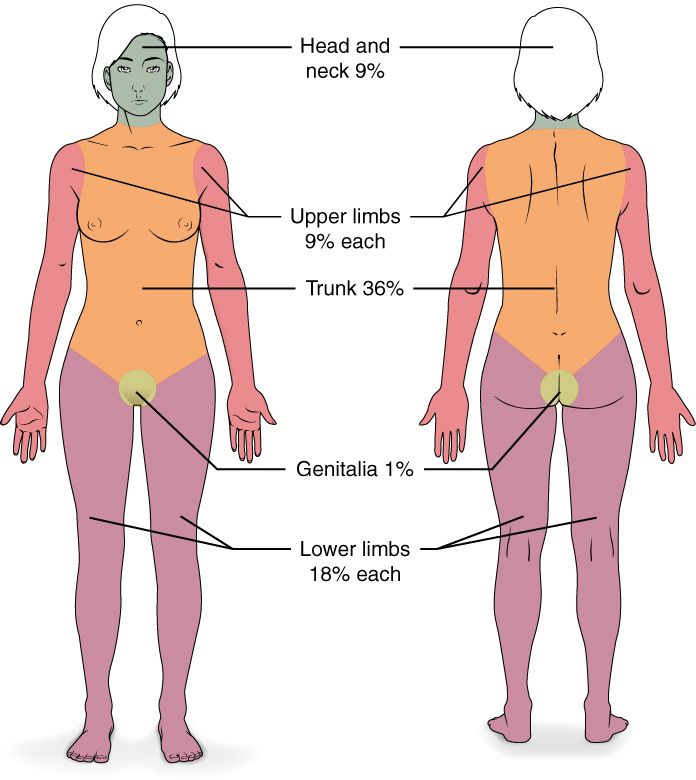
For extent, the Rule of Nines gives a fast field estimate in adults: head and neck 9 percent; each arm 9; anterior trunk 18; posterior trunk 18; each leg 18; perineum 1. In infants and small children the head is proportionally much larger — about 18 percent — and each leg smaller, about 13.5 percent, so the adult rule overestimates the legs and underestimates the head. The Lund and Browder chart adjusts by age and is the accurate tool used in burn centres. For scattered or irregular burns, use the rule of palm: the patient’s own palm including the fingers is approximately 1 percent TBSA. Count only partial- and full-thickness burns.
The American Burn Association transfer criteria decide destination: partial-thickness burns over 10 percent TBSA; any full-thickness burn; burns of the face, hands, feet, genitalia, perineum or over major joints; electrical injury including lightning; chemical burns; inhalation injury; burns in patients with significant comorbidity; burns with concomitant trauma; and children in facilities without paediatric burn capability.
What you do first, in order
Stop the burning process — remove smouldering clothing and all jewellery and constricting items before oedema develops, brush off dry chemical powder before irrigating, and irrigate chemical burns with copious room-temperature water for at least 20 minutes (longer for alkalis, which cause deeper liquefactive necrosis; never attempt to neutralise). Cool small burns briefly with room-temperature water, but never apply ice — it vasoconstricts the zone of stasis and deepens the injury — and never cool a large burn, because hypothermia kills burn patients. Cover with a clean dry sheet and keep the patient warm.
Then run ABC. The airway is the first killer and the one with a closing window. Two lists, and students collapse them into one. Suspect inhalation injury with any of: burns sustained in an enclosed space; loss of consciousness at the scene; facial burns; singed nasal hairs, eyebrows or eyelashes; soot in the mouth or nose; or carbonaceous sputum. Those are exposure findings, and they are poor standalone predictors of obstruction — what they mandate is close serial airway assessment, humidified 100 percent oxygen and a carboxyhaemoglobin level, not a reflex tube. Do not intubate on exposure history alone; an unnecessary tube in a burn patient costs ventilator days, pneumonia, sedation and unplanned extubations. Escalate for immediate airway control when the dynamic signs appear: new or worsening hoarseness or voice change; a brassy cough; stridor; drooling or difficulty swallowing; wheezing; respiratory distress; deep circumferential facial or neck burns; or altered mentation. Those mean intubate now, before the oedema closes the airway. Upper-airway swelling builds over the first 12 hours and peaks at roughly 24 to 48 hours, and the resuscitation fluid you are about to give makes it worse — so vigilance does not relax at the end of your shift. A patient talking comfortably now can be unintubatable in ninety minutes; do not wait for hypoxia to declare itself.
Remember two poisons that pulse oximetry will not show you. Carbon monoxide binds haemoglobin with roughly 200 times the affinity of oxygen, and standard pulse oximetry cannot distinguish carboxyhaemoglobin from oxyhaemoglobin — the SpO2 reads normal while the patient is starving. Diagnose with a carboxyhaemoglobin level by co-oximetry: under 5 percent is normal in a non-smoker, 15–20 percent causes headache and confusion, 20–40 percent nausea and disorientation, 40–60 percent coma and seizures. Treat with 100 percent oxygen by non-rebreather, which shortens the carboxyhaemoglobin half-life from about 4–5 hours on room air to roughly 60–90 minutes; hyperbaric therapy is considered for severe poisoning. Cyanide, released when plastics, wool and silk burn, blocks cellular oxygen use; suspect it with profound persistent lactic acidosis and altered mentation despite adequate oxygen, and treat with intravenous hydroxocobalamin, warning the team that it turns skin, urine and serum deep red and interferes with colorimetric laboratory assays and pulse oximetry.
Fluid resuscitation, worked through
Two large-bore IVs go through unburned skin if possible, through burned skin if not — access matters more than aesthetics. Lactated Ringer’s is the crystalloid of choice, because it is isotonic and its lactate is metabolised to bicarbonate, helping the acidosis. Fluid is required for burns of roughly 20 percent TBSA or more in adults (10 percent in children and older adults).
The classic Parkland formula is 4 mL × body weight in kg × %TBSA of lactated Ringer’s over the first 24 hours, with half given in the first 8 hours and the remaining half over the next 16 hours. The critical detail: the clock starts at the time of injury, not the time of arrival. Current American Burn Association consensus starts lower — 2 mL/kg/%TBSA for thermal injury in adults, 3 mL for high-voltage electrical injury — and titrates upward, because decades of over-resuscitation (“fluid creep”) produced compartment syndromes and pulmonary oedema. Know both; the formula is a starting point, not a prescription.
Worked example. An 80 kg man sustains 45 percent TBSA partial- and full-thickness flame burns at 20:00. He reaches your emergency department at 22:00.
Parkland total: 4 mL × 80 kg × 45 = 14,400 mL lactated Ringer’s in the first 24 hours. First half = 7,200 mL, due in the first 8 hours from injury — that is by 04:00. But two hours have already elapsed, so those 7,200 mL must run over the remaining 6 hours: 7,200 ÷ 6 = 1,200 mL/hr. The second half, 7,200 mL, runs over the following 16 hours (04:00 to 20:00): 7,200 ÷ 16 = 450 mL/hr. Had he arrived immediately, the first rate would have been 7,200 ÷ 8 = 900 mL/hr. Using the ABA 2 mL starting point instead: 2 × 80 × 45 = 7,200 mL total, 3,600 mL in the first 8 hours = 450 mL/hr, then 225 mL/hr.
Now the part that actually matters at the bedside: the formula is titrated to urine output every hour. Insert an indwelling catheter and target 0.5 mL/kg/hr in adults — about 30–50 mL/hr for most adults, so roughly 40 mL/hr for this 80 kg man — and 1 mL/kg/hr in children under 30 kg. In high-voltage electrical injury with tea-coloured or port-wine urine from myoglobin, target 75–100 mL/hr until the urine clears, because myoglobin precipitating in tubules causes acute kidney injury. Falling urine output, rising heart rate, worsening base deficit and rising lactate say increase the rate. Urine output well above target, ballooning oedema, rising peak airway pressures and a tense abdomen say back off — over-resuscitation causes abdominal compartment syndrome, extremity compartment syndrome, ocular compartment syndrome and pulmonary oedema. Colloid, typically 5 percent albumin, is commonly added after the first 12–24 hours once capillaries begin to seal.
Meanwhile: keep the room hot (about 30–33 °C) and use warmed fluids and blankets, because a burn patient cannot thermoregulate and hypothermia worsens coagulopathy and acidosis. Give analgesia as intravenous opioid only — intramuscular and subcutaneous absorption is unreliable during third spacing and will dump a bolus into the circulation when perfusion returns. Insert a nasogastric tube for burns over 20 percent TBSA because ileus is near-universal, and start stress-ulcer prophylaxis to prevent a Curling ulcer. Update tetanus immunisation. Monitor circumferential burns hourly with a Doppler: loss of pulse, deep unrelenting pain, pallor, paraesthesia and delayed capillary refill in an extremity, or rising peak airway pressures with restricted chest excursion, mean the eschar is acting as a tourniquet and an escharotomy is needed — a bedside incision through the insensate eschar; if the deeper compartments are involved, a fasciotomy in theatre.
Estimate depth and TBSA accurately, protect the airway before oedema closes it, start Parkland from the time of injury — and then let hourly urine output, not the formula, decide the rate.
Section 5Acute and Rehabilitative Burn Care: Infection, Nutrition and Function
Infection: the threat after 48 to 72 hours
Once diuresis begins, the emergent phase ends and the acute phase runs until every wound is closed. The burn wound is a warm, moist, protein-rich, avascular surface with no epidermal barrier and no white cells able to reach it — a perfect culture medium. Infection and sepsis are now the leading cause of death. Early colonisation is with gram-positive skin flora; by the end of the first week gram-negatives dominate, especially Pseudomonas aeruginosa, followed by fungi such as Candida in patients on prolonged antibiotics.
Prophylactic systemic antibiotics are not given — they select resistant organisms without reducing wound infection. Control is topical, plus early excision of dead tissue. Recognising infection in a burn patient is a nursing skill, because the usual signs are unreliable. Watch the wound for a change in colour (green or blue-green discoloration suggests Pseudomonas), for rapid or premature separation of eschar, for focal dark brown or black spots in previously clean tissue, for conversion of a partial-thickness wound to full-thickness, and for a new odour. Watch the patient for the burn-specific sepsis picture: hypothermia or hyperthermia, new confusion or agitation, return of ileus or feeding intolerance, unexplained hyperglycaemia and rising insulin requirement, thrombocytopenia, tachypnoea and falling urine output. A burn patient who suddenly stops tolerating tube feeds is septic until proved otherwise.
| Topical agent | How it works / penetration | Nursing implications |
|---|---|---|
| Silver sulfadiazine 1 percent | Broad-spectrum; poor eschar penetration | Contraindicated in sulfa allergy, late pregnancy and infants under 2 months (kernicterus risk); may cause transient leukopenia — monitor CBC; forms a pseudo-eschar that must be cleansed off; painless to apply |
| Mafenide acetate | Penetrates eschar and cartilage well — agent of choice for ear and nose burns | Painful and burning on application — premedicate; carbonic anhydrase inhibitor, so it causes metabolic acidosis — monitor ABG and respiratory rate |
| Silver nitrate 0.5 percent solution | Surface antisepsis; does not penetrate | Stains everything black; dressings must be kept wet; causes hyponatraemia, hypochloraemia and hypokalaemia — monitor electrolytes |
| Silver-impregnated dressings | Sustained silver release | May stay in place 3–7 days — fewer painful changes; do not use with oil-based products |
| Bacitracin / plain petrolatum gauze | Low-toxicity coverage | Common on facial and superficial partial-thickness burns; frequent reapplication |
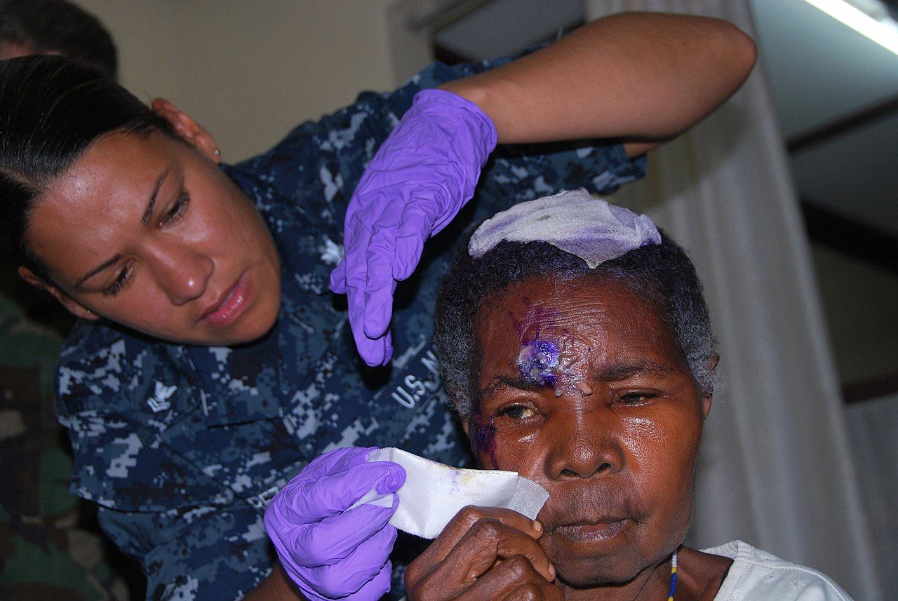
Wound care itself is hydrotherapy or bedside cleansing, debridement and re-dressing, usually once or twice daily and always after adequate analgesia. Debridement may be mechanical (careful cleansing, forceps), enzymatic (collagenase applied to slough), autolytic (occlusive dressing) or surgical. Early excision and grafting — taking full-thickness eschar to a viable bed within the first several days — is the single biggest advance in burn survival, because it removes the culture medium. Use strict infection-prevention practice: hand hygiene, gowns and gloves for wound contact, dedicated equipment, no fresh flowers or standing water, and cohorting per policy.
Hypermetabolism, nutrition and pain
A major burn produces the most extreme hypermetabolic state in medicine: resting energy expenditure can approach twice normal, peaking around days 5–7 and persisting for months after wound closure. Catecholamines, cortisol and glucagon drive gluconeogenesis, insulin resistance and relentless skeletal-muscle catabolism. If you do not feed the patient, they will burn their own diaphragm and their wounds will not close.
Start enteral nutrition early — within 24 hours, and often within 4–6 hours in major burns — because early feeding preserves gut mucosal integrity and reduces bacterial translocation. Targets are high-calorie and high-protein: commonly 1.5–2 g/kg/day of protein and 25–35 kcal/kg/day plus a burn factor (the older Curreri formula, 25 kcal/kg plus 40 kcal per %TBSA, still illustrates the magnitude). Supplement vitamin C, vitamin A, zinc and often glutamine. Nursing monitoring is daily weights on the same scale at the same time, strict intake and output, calorie counts, glucose control with insulin infusions as needed, and tracking of nitrogen balance and prealbumin with the understanding that prealbumin falls in inflammation regardless of intake. Hold nothing for the sake of routine: burn patients are chronically underfed by NPO orders that nobody cancels.
Pain has two components and both need a plan. Background pain is continuous and is best covered by scheduled or continuous opioid plus adjuvants; procedural pain during dressing changes and physiotherapy is severe, predictable and requires premedication timed to peak effect, often with ketamine or short-acting agents, plus non-pharmacological methods — virtual reality distraction, music, controlled breathing and giving the patient a say in pausing. Anxiety amplifies pain; treat both. Later, pruritus in healing burns is intense and under-treated: use emollients, cool compresses, antihistamines, and gabapentin or pregabalin for the neuropathic component, and keep nails short.
Grafting, contracture prevention and going home
Deep partial- and full-thickness burns are closed with a split-thickness autograft harvested from the patient’s own unburned skin. Meshing the graft expands coverage and lets exudate escape but leaves a permanent lattice pattern. Cultured epithelial autograft is used when donor sites are scarce. Cadaver allograft and porcine xenograft are temporary biological dressings only — they will be rejected, and teaching families otherwise causes real distress. After grafting, immobilise and elevate the site for three to five days, protect absolutely against shear, monitor for haematoma or seroma beneath the graft (which lifts it off its blood supply and is the commonest cause of failure), and keep the dressing intact until the surgeon says otherwise. The donor site is a fresh superficial partial-thickness wound: it is often more painful than the graft, heals in 10–14 days, and may be reharvested.
Rehabilitation begins on day one, not at discharge. The rule to remember is that the position of comfort is the position of contracture: burned tissue contracts into flexion, and a patient left comfortable will fuse into a useless posture. Anterior neck burns are positioned in slight hyperextension with no pillow; axillae are abducted 90–110 degrees on airplane splints; elbows and knees are splinted in extension; hips stay extended and neutrally rotated with no pillow under the knees; hands are splinted in the intrinsic-plus position (wrist slightly extended, MCP joints flexed 70–90 degrees, IP joints straight, thumb abducted). Range-of-motion exercise happens daily, and ambulation starts as soon as haemodynamics allow. Once wounds are closed, custom pressure garments worn 23 hours a day for 12–18 months flatten hypertrophic scar and are the treatment patients most often abandon — make adherence an explicit teaching goal.
Discharge teaching covers wound and graft care with return demonstration; signs of infection to report; the pressure-garment and splinting schedule; daily moisturising of healed skin, which has no sebaceous glands and cracks; strict sun avoidance and SPF 30 or higher for at least a year because new skin hyperpigments permanently; itch management; a high-protein diet with weight monitoring; and realistic expectations about fatigue, cold intolerance and the two-year timeline of scar maturation. Screen for post-traumatic stress, depression and altered body image, and connect the patient with burn survivor peer support — psychosocial recovery is slower than wound closure and predicts return to work better than TBSA does.
After the first 48–72 hours the burn patient no longer dies of shock — they die of infection and catabolism, so the acute-phase priorities are topical antimicrobials with early excision, aggressive enteral protein, and splinting that protects function before the scar decides for you.
The chapter in one breath
Skin is a barrier, a thermostat and a fluid container, and every integumentary disorder is a version of one of those failing. Assess it in every skin tone using light, comparison and your hands, name lesions in standard morphology, and document wounds in centimetres with clock-position tunnelling. In infection, the finding that changes everything is pain out of proportion to appearance — that is necrotising fasciitis and it goes to theatre, not to a dressing cart. In drug eruptions, mucosal involvement with a positive Nikolsky sign is Stevens–Johnson syndrome or toxic epidermal necrolysis, and stopping the drug is the intervention that saves the patient. Pressure injuries are prevented by offloading, floated heels, 30-degree tilts and moisture control, staged only when the base is visible, and never reverse-staged. Leg ulcers are treated by cause: compress venous, revascularise arterial, offload neuropathic, and check the ankle–brachial index first. In burns, classify depth by whether dermal appendages survive, estimate extent with the Rule of Nines, protect the airway before oedema closes it, and start Parkland from the time of injury while titrating hourly to a urine output of 0.5 mL/kg/hr. Past 48–72 hours, the enemy changes from shock to infection and catabolism — topical antimicrobials, early excision and grafting, high-protein enteral feeding, and splinting that preserves the function the scar would otherwise take.
Words worth knowing
| Term | What it means |
|---|---|
| Eschar | Dry, leathery, devitalised tissue over a full-thickness burn or wound; acts as a tourniquet when circumferential and as a culture medium when retained |
| Escharotomy | Bedside incision through full-thickness eschar to relieve constriction of a limb or the chest; the eschar itself is insensate |
| Zone of stasis | The salvageable ring of marginally perfused tissue around a burn; adequate resuscitation and warmth prevent it converting to necrosis |
| Parkland formula | 4 mL × kg × %TBSA of lactated Ringer’s in 24 hours, half in the first 8 hours from the time of injury; ABA consensus starts at 2 mL and titrates |
| Rule of Nines | Rapid adult TBSA estimate in multiples of 9 percent; the paediatric head is 18 percent and each leg 13.5 percent |
| Rule of palm | The patient’s palm including fingers approximates 1 percent TBSA; used for scattered burns |
| Carboxyhaemoglobin | Haemoglobin bound to carbon monoxide; requires co-oximetry because pulse oximetry reads it as saturated |
| Curling ulcer | Stress-related gastroduodenal ulceration following major burns; the reason for early acid suppression |
| Autograft / allograft / xenograft | Permanent graft from the patient’s own skin / temporary cadaveric skin / temporary porcine skin |
| Intrinsic-plus position | Anti-contracture hand splinting: wrist extended, MCP joints flexed 70–90 degrees, IP joints straight, thumb abducted |
| Nikolsky sign | Epidermis shears off with gentle lateral pressure; positive in TEN and pemphigus |
| Deep tissue pressure injury | Persistent non-blanchable deep red, maroon or purple discoloration, or a blood-filled blister, over intact or non-intact skin |
| Braden Scale | Six-subscale pressure-injury risk tool scored 6–23; 18 or below indicates risk, 9 or below severe risk |
| Ankle–brachial index (ABI) | Ankle-to-arm systolic pressure ratio; 0.91–1.30 normal, 0.90 or less arterial disease, above 1.30 non-compressible calcified vessels |
| Marjolin ulcer | Squamous cell carcinoma arising in a chronic wound or old burn scar; any non-healing scar ulcer needs biopsy |
| Auspitz sign | Pinpoint bleeding after removing psoriatic scale, from the dilated capillaries of elongated dermal papillae |
| Erysipelas | Superficial dermal and lymphatic streptococcal infection with a sharply demarcated raised border, unlike the indistinct margin of cellulitis |
| Hypermetabolic response | The post-burn catabolic surge that can double resting energy expenditure for months; managed with early high-protein enteral feeding |
| Undermining / tunnelling | Tissue destruction under intact wound edges / a narrow sinus tract; both charted in clock positions with 12 o’clock toward the head |
| Pressure garment | Custom compression worn 23 hours daily for 12–18 months to flatten hypertrophic burn scar |