Sensory Disorders: Eye and Ear
Sensory nursing is quiet work until it suddenly is not. Most of what you do for eyes and ears is patient teaching, drop technique, fall prevention and the unglamorous business of making sure a frightened person can hear the plan of care — and then, once or twice a year, a patient tells you their vision went dark in one eye an hour ago, or that a splash of drain cleaner hit their face, or that they woke up deaf on one side, and the difference between a good outcome and permanent loss is measured in minutes. This chapter teaches the mechanism first, because the mechanism predicts the finding: pressure that cannot escape gives you a rock-hard painful eye with a fixed mid-dilated pupil, a retina starved of blood gives you painless blackout, endolymph under pressure gives you vertigo with fluctuating hearing loss. Learn the mechanism and you will never have to memorise a list of symptoms again.
By the end of this chapter you can
- Perform and document a focused bedside eye and ear assessment including visual acuity, confrontation fields, pupil testing, the whisper test and the Weber and Rinne tuning-fork tests.
- Explain aqueous humour dynamics and distinguish primary open-angle glaucoma from acute angle-closure glaucoma, including which one is a same-hour emergency and why.
- Compare the drug classes that lower intraocular pressure by mechanism, and state the systemic monitoring each one demands.
- Triage sudden painless vision loss among central retinal artery occlusion, retinal detachment, vitreous haemorrhage and giant cell arteritis, and act on the correct time window.
- Manage diabetic retinopathy and age-related macular degeneration, including anti-VEGF therapy, the Amsler grid and AREDS2 supplementation.
- Differentiate the red eye and initiate immediate irrigation for a chemical ocular burn before any other assessment.
- Distinguish conductive from sensorineural hearing loss, recognise sudden sensorineural hearing loss as an emergency, and identify common ototoxic drugs.
- Differentiate benign paroxysmal positional vertigo, vestibular neuritis, labyrinthitis and Ménière disease, and teach safety for each.
- Escalate ocular and otologic red flags using SBAR and delegate appropriately, keeping assessment, teaching, evaluation and unstable patients with the registered nurse.
Section 1Assessing Vision, Hearing and the Safety They Buy
The mechanism you need before anything else
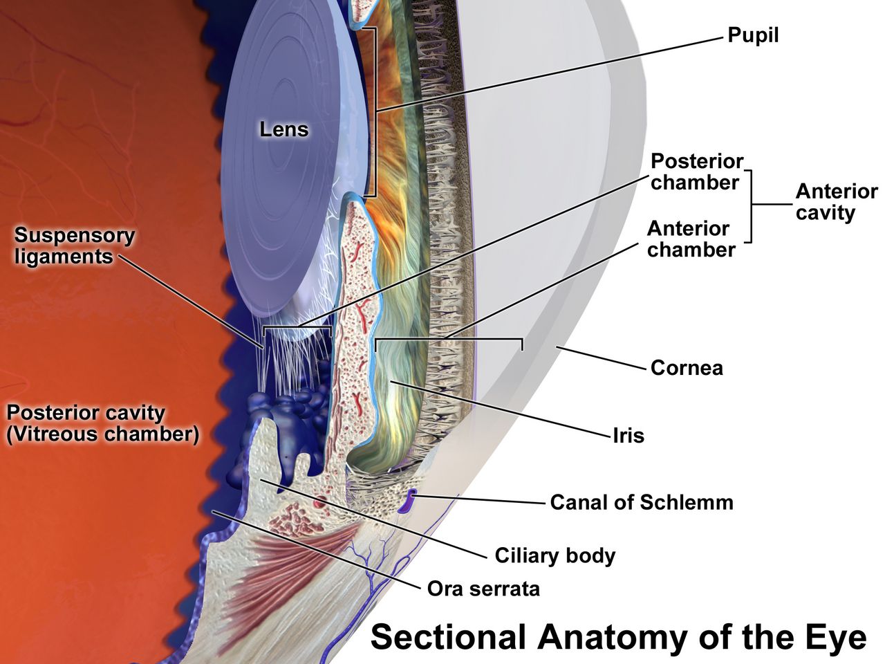
The eye is a pressurised sphere with a one-way plumbing problem. The ciliary body, sitting behind the iris, secretes aqueous humour continuously at roughly 2 to 3 microlitres per minute. That fluid flows from the posterior chamber, through the pupil, into the anterior chamber, and drains out through the trabecular meshwork at the angle where the iris meets the cornea. Production never stops. If drainage slows or the angle closes, pressure rises, and pressure is transmitted to the one structure that cannot tolerate it: the axons of the optic nerve as they turn and exit at the optic disc. Everything you will learn about glaucoma is a variation on that sentence.
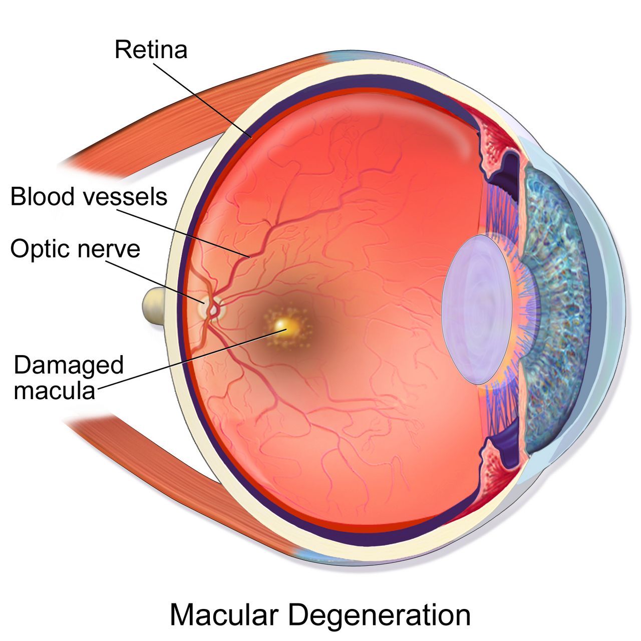
Light that survives the trip lands on the retina, and the retina is metabolically the hungriest tissue in the body per gram. It is fed by two independent systems — the central retinal artery for the inner layers and the choroidal circulation for the outer layers — and neither has meaningful collateral supply. That is why retinal ischaemia behaves like brain ischaemia rather than like a bruised muscle: the tissue dies on a clock measured in minutes to a couple of hours, not days. The macula, the small central zone packed with cones, provides reading vision and face recognition; lose the macula and you keep peripheral navigation but lose the ability to live independently.
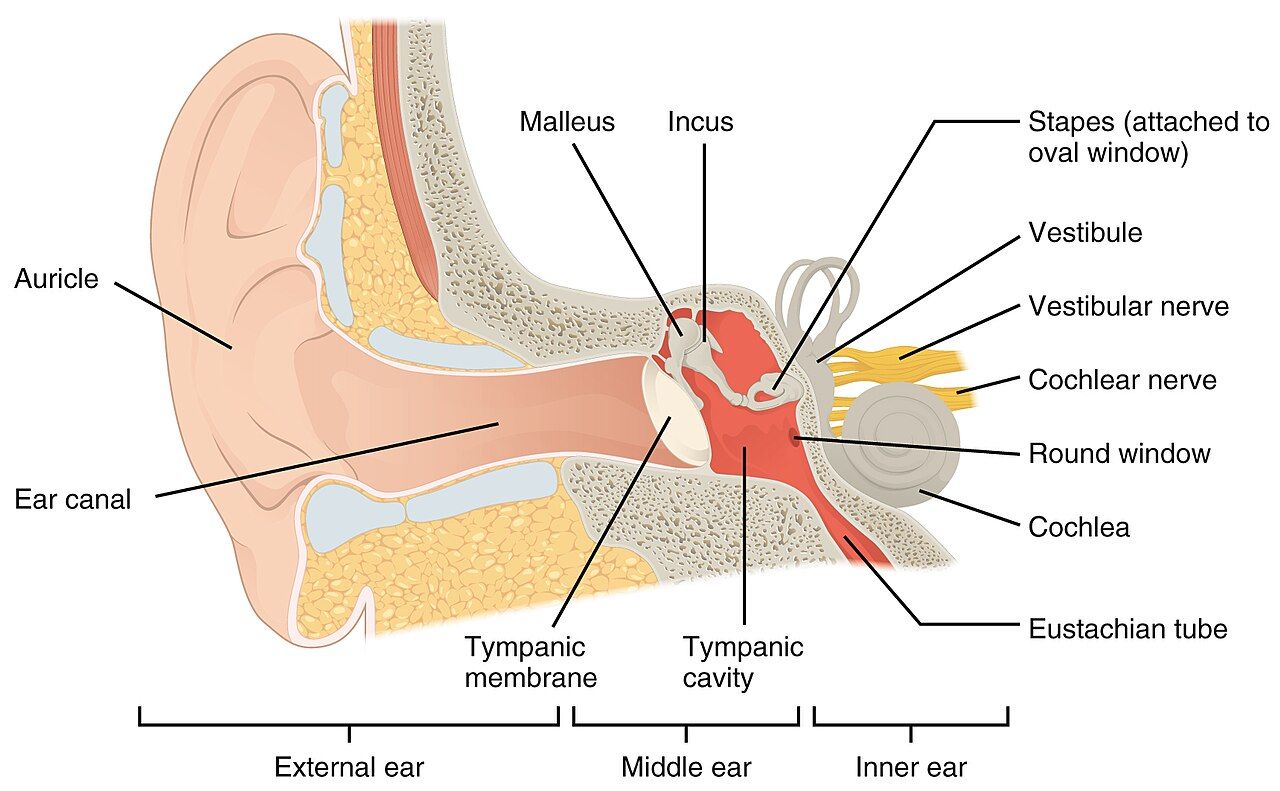
The ear divides the same way into a mechanical half and a neural half, and that division is the whole of hearing-loss diagnosis. The external canal and the middle ear ossicles are a mechanical amplifier: anything that blocks or stiffens them — cerumen, fluid, a perforated drum, a fixed stapes — produces conductive loss. The cochlea and the eighth cranial nerve are the transducer and the cable: anything that damages hair cells or the nerve — noise, ageing, aminoglycosides, a schwannoma — produces sensorineural loss. Sitting alongside the cochlea, sharing its fluid, is the vestibular labyrinth, which is why so many patients arrive with hearing and balance complaints in the same breath.
The vision assessment you can do at the bedside
Always test and document visual acuity before you do anything else to an eye, with the single exception of a chemical splash. Acuity is the vital sign of the eye. Use a Snellen chart at 20 feet or a handheld near card at 14 inches, test each eye separately with the other occluded, and let the patient wear their usual glasses or contacts — you want corrected acuity, not a refraction study. Record it exactly as measured: 20/40 means the patient sees at 20 feet what a normal eye sees at 40. Normal is 20/20; legal blindness in the United States is 20/200 or worse in the better eye with correction, or a visual field no wider than 20 degrees. If a patient cannot read the largest letter, step down through counting fingers, hand motion, and light perception, and document which.
Then check the pupils, the fields and the external eye. Test direct and consensual reaction and swing the light rapidly between eyes: if the pupil dilates when the light arrives on it, that is a relative afferent pupillary defect and it means the optic nerve or a large area of retina on that side is not conducting. That single finding separates true optic-nerve or retinal disease from cataract, refractive error and functional complaints. Confrontation fields are crude but free — sit at eye level, cover the opposite eyes, and bring a wiggling finger in from each quadrant. Inspect for redness, discharge, lid position, proptosis and corneal clarity, and test extraocular movements in the six cardinal directions, noting pain on movement.
Diagnostics you will see ordered follow logically. Tonometry measures intraocular pressure; normal is roughly 10 to 21 mm Hg. Slit-lamp examination with fluorescein stain shows corneal abrasions and ulcers as green uptake under cobalt-blue light. Gonioscopy visualises the drainage angle and tells the ophthalmologist whether a glaucoma is open-angle or closed. Automated perimetry maps visual fields and is how open-angle glaucoma is followed over years. Optical coherence tomography images retinal and optic-nerve layers in cross-section, and fluorescein angiography demonstrates leaking or non-perfused vessels in diabetic retinopathy and wet macular degeneration.
The hearing and balance assessment you can do at the bedside
Start with the otoscope and with observation. Pull the pinna up and back in an adult to straighten the canal — down and back in a child under three — and inspect the canal for cerumen, oedema, discharge and foreign bodies before judging the drum. A normal tympanic membrane is pearly grey, translucent, and shows a cone of light; a bulging erythematous drum with loss of landmarks means acute otitis media, an amber immobile drum means effusion, and a retraction pocket packed with white debris suggests cholesteatoma. Note whether pulling the pinna or pressing the tragus hurts — that pain is external otitis, not middle-ear disease.
The whisper test is a legitimate screen: stand about two feet behind the patient, occlude and rub the tragus of the non-tested ear, exhale fully, and whisper a combination of three numbers and letters. Inability to repeat at least three of six items is a positive screen. Then use a 512 Hz tuning fork. In the Weber test you place the fork on the midline forehead or vertex; sound should be heard equally in both ears. In the Rinne test you compare air conduction at the meatus with bone conduction on the mastoid; normally air conduction lasts about twice as long as bone conduction. The pattern of the two tests, not either alone, tells you which half of the ear is broken.
| Test | Normal | Conductive loss (left ear) | Sensorineural loss (left ear) |
|---|---|---|---|
| Weber (fork on midline) | Heard equally, midline | Lateralises TO the left (bad) ear — blocked ear is shielded from room noise | Lateralises AWAY, to the right (good) ear — left cochlea cannot transduce |
| Rinne (air vs bone) | Air conduction greater than bone conduction, both ears | Bone conduction greater than air conduction on the left | Air conduction still greater than bone conduction, but both are reduced on the left |
| Typical causes | — | Cerumen, effusion, perforation, otosclerosis, cholesteatoma | Presbycusis, noise, ototoxicity, Ménière, acoustic neuroma |
Formal audiometry quantifies what the fork suggests. Normal adult thresholds are 0 to 25 decibels; 26 to 40 dB is mild loss, 41 to 55 dB moderate, 56 to 70 dB moderately severe, 71 to 90 dB severe, and above 90 dB profound. Tympanometry measures drum mobility — a type A tracing is normal, a flat type B curve means effusion or perforation, and a type C curve means negative middle-ear pressure from eustachian tube dysfunction. Otoacoustic emissions and auditory brainstem response testing are used when the patient cannot cooperate, including newborn screening.
Sensory loss is a safety diagnosis
A patient who cannot see the call light or hear the alarm is a fall risk, an aspiration risk, a medication-error risk and a delirium risk, and none of that shows up on a lab report. Older adults arrive with layered baseline changes: presbyopia from a stiffened lens, slowed dark adaptation, glare sensitivity, reduced tear production, arcus senilis, lens yellowing that dulls blues and greens, drier and more impacted cerumen, and presbycusis that eats the high frequencies first. Because consonants live in the high frequencies, presbycusis does not make speech quieter so much as mushy — which is why shouting fails and why the patient is so often mislabelled confused when they are simply not receiving the sentence.
Communicate the way the deficit requires. Face the patient at eye level in good light without covering your mouth, lower your pitch rather than raising your volume, rephrase instead of repeating the identical sentence, reduce background noise by turning off the television, and confirm hearing aids are in and switched on with working batteries before you conclude anything about cognition. For low vision, announce yourself on entering and before touching, keep the room arrangement constant, orient the patient to the tray using clock positions, and use the sighted-guide technique — the patient holds your elbow and walks half a step behind rather than being pushed from in front.
Delegation follows the usual rule with no sensory exception. Unlicensed assistive personnel may reinforce teaching you have already delivered, position the patient, assist with ambulation and hygiene, clean eyeglasses and hearing aids, gather irrigation supplies, and report what they observe. They do not perform the initial or ongoing assessment, do not test acuity as a nursing judgement, do not teach a newly diagnosed patient, do not evaluate response to therapy, and do not take the unstable patient. Instillation of eye and ear medication is medication administration — it belongs to the licensed nurse within scope and facility policy, and a licensed practical nurse giving drops still reports abnormal findings to the registered nurse.
Document visual acuity and a hearing screen on every patient with an eye or ear complaint, because acuity is the eye’s vital sign and unrecognised sensory loss masquerades as confusion, non-adherence and falls.
Section 2Pressure and the Lens: Glaucoma and Cataract
Two glaucomas, two tempos
Primary open-angle glaucoma is the common form and accounts for roughly nine in ten cases in the United States. The drainage angle is anatomically open, but the trabecular meshwork offers increasing outflow resistance, so pressure creeps up over years. The optic nerve dies from the outside in: peripheral vision is lost first, in arcuate patterns the patient does not notice because the brain fills in the gaps and the other eye compensates. By the time a patient reports tunnel vision, a large fraction of the retinal ganglion cells are gone and cannot be recovered. Glaucomatous vision loss is permanent, which reframes the entire nursing goal — you are not restoring sight, you are protecting what remains.
Risk factors matter because screening depends on them: age over 40, African ancestry (earlier onset and more aggressive course), Hispanic ancestry in older age groups, family history, high myopia, diabetes, chronic corticosteroid use in any form including inhaled and topical, and prior ocular trauma. The disease is defined by optic-nerve damage and field loss, not by pressure alone — normal-tension glaucoma damages the nerve at pressures under 21 mm Hg, and ocular hypertension raises pressure without damaging the nerve. Screening is a dilated examination with tonometry, optic-disc assessment for an enlarged cup-to-disc ratio, and perimetry.
Because the disease is silent, adherence is the entire nursing intervention. Patients stop drops because the drops sting, because nothing feels different when they skip them, because a bottle costs money, and because arthritic hands cannot squeeze a small bottle over an eye they cannot aim at. Assess each of those explicitly. Teach the drop schedule attached to existing routines, demonstrate and then watch a return demonstration, offer drop-guide devices, and warn that missing doses causes no symptom at all until the field is gone. Tell the patient to inform every clinician they see that they have glaucoma, especially before any procedure involving anticholinergic or sympathomimetic drugs.
Acute angle-closure glaucoma: the one that cannot wait
In a predisposed eye — short axial length, hyperopia, shallow anterior chamber, thick or forward-sitting lens, often an older adult of East Asian or Inuit ancestry — the peripheral iris can bunch forward and occlude the trabecular meshwork completely. Production continues into a closed compartment, and intraocular pressure rockets from a normal 10 to 21 mm Hg to 40, 60, even 80 mm Hg within hours. The trigger is anything that mid-dilates the pupil: walking into a dark room or cinema, emotional stress, and above all drugs — anticholinergics, antihistamines, tricyclic antidepressants, some antipsychotics, scopolamine, topical mydriatics, high-dose sympathomimetics, and even nebulised ipratropium that leaks around a loose mask.
The presentation is dramatic and easy to miss because the loudest complaints are systemic. The patient reports sudden severe unilateral eye pain and a frontal headache, blurred vision with coloured haloes around lights, and nausea and vomiting that can be severe enough for them to land on a medical floor with a presumed gastrointestinal problem or migraine. On examination the eye is injected with a ciliary flush, the cornea is hazy or steamy from oedema, the pupil is mid-dilated and fixed or minimally reactive, and the globe feels rock-hard compared with the other eye. Vision is markedly reduced. This is an emergency — untreated, optic-nerve damage begins within hours and can be irreversible within a day or two.
What you do first: call the ophthalmologist immediately and do not wait for rounds, keep the patient supine because that lets the lens settle backward and deepens the anterior chamber, keep the room quiet but normally lit rather than darkened — darkness mid-dilates the pupil and worsens the block, which is the same reason a dark cinema can trigger an attack — hold every anticholinergic and every mydriatic drop, give the prescribed analgesic and antiemetic because vomiting and Valsalva raise pressure further, and start nothing by mouth in case an intravenous osmotic or a procedure is coming. Medical therapy layers agents that cut production and pull fluid out: a topical beta-blocker, a topical alpha-2 agonist, a topical or oral carbonic anhydrase inhibitor, and intravenous mannitol as an osmotic. Once pressure begins to fall, pilocarpine constricts the pupil and pulls the peripheral iris off the meshwork — it does not work at extremely high pressure because the sphincter is ischaemic. Definitive treatment is laser peripheral iridotomy, and the fellow eye is treated prophylactically because it shares the anatomy.
Drugs that lower intraocular pressure
Every agent works by one of two levers: make less aqueous or drain more of it. Knowing which lever a drug pulls tells you its systemic risk. Anything that suppresses production by blocking beta receptors or carbonic anhydrase carries systemic cardiopulmonary or metabolic consequences; anything that improves outflow tends to cause local cosmetic and comfort effects. Teach punctal occlusion with every drop — press the inner canthus for one to two minutes after instillation — because it both increases ocular contact time and dramatically reduces systemic absorption through the nasolacrimal duct.
| Class and example | Mechanism | What the nurse watches and teaches |
|---|---|---|
| Prostaglandin analogue — latanoprost, bimatoprost, travoprost | Increases uveoscleral outflow | First-line; once daily at bedtime. Permanent brown iris darkening, darker and longer lashes, periorbital fat atrophy, conjunctival hyperaemia. Refrigerate latanoprost until opened. Warn about the cosmetic changes before the patient discovers them. |
| Beta-blocker — timolol, betaxolol | Decreases aqueous production | Systemic absorption is real: bradycardia, hypotension, bronchospasm, fatigue, masked hypoglycaemia. Contraindicated or used with great caution in asthma, chronic obstructive pulmonary disease, heart block and decompensated heart failure. Check apical pulse before instilling. Punctal occlusion is essential. |
| Alpha-2 agonist — brimonidine, apraclonidine | Decreases production and increases uveoscleral outflow | Dry mouth, allergic conjunctivitis, fatigue, hypotension. Contraindicated in infants and young children and in patients on monoamine oxidase inhibitors; can cause central nervous system depression and apnoea in the very young. |
| Topical carbonic anhydrase inhibitor — dorzolamide, brinzolamide | Decreases production | Bitter taste, stinging, blurred vision. Sulfonamide-derived — ask about sulfa allergy. |
| Systemic carbonic anhydrase inhibitor — acetazolamide | Decreases production sharply | Reserved for acute angle closure and refractory cases. Monitor potassium, sodium bicarbonate and creatinine; expect paraesthesias, polyuria, metabolic acidosis, malaise. Avoid in sulfa allergy, renal impairment and marked electrolyte derangement. |
| Rho-kinase inhibitor — netarsudil | Increases trabecular outflow | Conjunctival hyperaemia is very common, plus corneal verticillata and small conjunctival haemorrhages. Reassure but report pain or vision change. |
| Cholinergic miotic — pilocarpine | Constricts pupil, pulls iris off the meshwork, increases trabecular outflow | Mainly used in angle closure. Brow ache, dim vision especially at night, accommodative spasm. Teach fall precautions and no night driving. |
When drops fail, laser trabeculoplasty is offered for open-angle disease and surgical drainage follows: trabeculectomy creates a filtering bleb, and tube-shunt devices divert aqueous to an external plate. Minimally invasive glaucoma surgery stents are increasingly combined with cataract extraction. After any filtering procedure, teach the patient to avoid bending below the waist, heavy lifting, straining at stool, vigorous coughing and sexual activity for the period the surgeon specifies, to sleep with an eye shield, and to report immediately any increase in pain, decrease in vision, purulent discharge or flashes and floaters — a bleb infection can seed the inside of the eye.
Cataract and the post-operative eye
A cataract is opacification of the crystalline lens, and it is the leading treatable cause of blindness worldwide. Age is the dominant cause, with lens proteins denaturing and aggregating over decades, but the accelerators are worth teaching: ultraviolet exposure, smoking, diabetes, chronic corticosteroids, blunt or penetrating trauma, and prior intraocular surgery or radiation. Symptoms are painless and gradual — progressive blurring, glare and haloes around headlights that make night driving frightening, colours that look faded or yellowed, and, characteristically, improved near vision in some patients as the lens becomes more refractive, the so-called second sight. The red reflex dims and the pupil may look cloudy or white in advanced disease.
There is no drug that clears a cataract. Surgery is elective and driven by function, not by an appearance on examination: when the patient can no longer read, drive safely or work, phacoemulsification with intraocular lens implantation is offered. It is usually a same-day procedure under local or topical anaesthesia with light sedation, one eye at a time. Preoperatively you confirm the correct eye is marked, review anticoagulants and alpha-blockers such as tamsulosin with the surgical team because the latter causes intraoperative floppy iris syndrome, instil the ordered dilating and anti-inflammatory drops on schedule, and confirm the patient has a ride home and someone to help with drops.
Postoperative teaching is where nurses prevent the real complications. The patient wears a protective shield at night and sunglasses in daylight, sleeps on the non-operative side, and does not rub or press the eye. They avoid bending at the waist, lifting more than about ten pounds, straining at stool, and vigorous coughing or vomiting for the period ordered — a stool softener is a legitimate nursing suggestion. Mild scratchiness, itching and a bloodshot appearance are expected; what is not expected is increasing pain, decreasing vision, a sudden shower of floaters or flashing lights, or purulent discharge. Those mean endophthalmitis or retinal detachment and require the surgeon the same hour, not at the next scheduled visit.
Open-angle glaucoma steals vision silently over years and is a war of adherence, while acute angle-closure glaucoma produces a painful red eye with a mid-dilated fixed pupil and rock-hard globe and must be treated within hours — and any anticholinergic can precipitate it.
Section 3The Retina: Sudden Loss and Slow Loss
Sudden painless vision loss: sorting it in five minutes
Sudden vision loss is one of the few complaints where the correct nursing action changes depending on which of four diagnoses it is, so learn to sort them by pain, pattern and preceding symptoms. Central retinal artery occlusion is the retinal stroke: profound painless loss over seconds in one eye, an afferent pupillary defect, a pale retina with a cherry-red spot at the fovea on fundoscopy. The retina tolerates roughly ninety minutes to a few hours of complete ischaemia before infarction, so this is a hyperacute emergency handled like a cerebrovascular event — activate the stroke pathway, obtain urgent imaging, and expect a carotid and cardiac embolic workup, because the next embolus may go to the brain. Amaurosis fugax, a transient monocular blackout that resolves, is the same warning in a reversible package.
Retinal detachment announces itself differently. The patient describes a sudden shower of new floaters, flashes of light called photopsia, and then a curtain, shadow or veil advancing across the field from one side. There is no pain and no redness. Risk factors are high myopia, prior cataract or vitreoretinal surgery, blunt trauma, proliferative diabetic retinopathy and a previous detachment in either eye. It is urgent rather than hyperacute, but the clock still matters because a detachment that reaches the macula converts a good visual prognosis into a poor one. Keep the patient at rest, restrict activity and head movement, avoid Valsalva, keep them nothing by mouth pending surgery, and position as the ophthalmologist directs so that gravity holds the detached area against the wall of the eye.
Two more belong on the list. Vitreous haemorrhage — common in proliferative diabetic retinopathy and in anticoagulated patients — produces sudden floaters, cobwebs or a red or black haze, with a lost red reflex; it is managed with head elevation, activity restriction and observation, or vitrectomy if it does not clear. Giant cell arteritis is the one you must not miss in anyone over 50: monocular vision loss with a new temporal headache, scalp tenderness, jaw claudication when chewing, fever, weight loss and proximal muscle aching from polymyalgia rheumatica. Erythrocyte sedimentation rate is typically above 50 mm per hour and C-reactive protein is elevated. High-dose corticosteroid is started immediately on suspicion, before the temporal artery biopsy, because the whole purpose of treatment is to save the second eye.
| Presentation | Likely cause | First nursing action |
|---|---|---|
| Instant, total, painless monocular loss; afferent pupillary defect | Central retinal artery occlusion | Emergency — treat as a stroke, notify ophthalmology and the stroke team now, note the exact time of onset |
| Flashes, new floaters, then an advancing curtain; painless | Retinal detachment | Urgent ophthalmology, bed rest, restrict head movement, nothing by mouth, position as directed |
| Sudden floaters or red haze, lost red reflex, diabetic or anticoagulated | Vitreous haemorrhage | Head of bed elevated, limit activity, ophthalmology referral, review anticoagulation |
| Vision loss plus temporal headache, jaw claudication, age over 50 | Giant cell arteritis | Draw sedimentation rate and C-reactive protein, expect high-dose steroids before biopsy — do not delay the steroid |
| Severe pain, haloes, vomiting, hard eye, fixed mid-dilated pupil | Acute angle-closure glaucoma | Ophthalmology now, supine in a quiet room with the lights left on (darkness dilates the pupil), hold anticholinergics, antiemetic and analgesia |
Diabetic retinopathy
Chronic hyperglycaemia damages the retinal microvasculature through glycation, pericyte loss and capillary basement-membrane thickening. The result is leaky, weak capillaries and, eventually, capillaries that close entirely. Non-proliferative retinopathy shows microaneurysms, dot-and-blot haemorrhages, hard exudates and cotton-wool spots, and it becomes vision-threatening when fluid collects at the macula — diabetic macular oedema is the most common cause of vision loss in diabetes. When enough capillary bed has closed, the ischaemic retina releases vascular endothelial growth factor and grows fragile new vessels: this is proliferative retinopathy, and those vessels bleed into the vitreous and contract into fibrous bands that pull the retina off, causing tractional detachment.
Screening schedules are examination questions and clinic reality. A patient with type 1 diabetes needs a dilated eye examination within five years of diagnosis, then annually. A patient with type 2 diabetes needs one at the time of diagnosis, because they have often been hyperglycaemic for years already, then annually. Pregnancy accelerates retinopathy, so a woman with pre-existing diabetes should be examined before conception or in the first trimester and followed closely. The most powerful interventions remain metabolic: glycaemic control with an individualised haemoglobin A1c target commonly around less than 7 percent, blood pressure control generally below 130/80 mm Hg, lipid management and smoking cessation.
Treatment of established disease is intravitreal anti-VEGF injection — ranibizumab, aflibercept, or bevacizumab used off-label — given at intervals of roughly four to eight weeks initially, which is a substantial adherence burden and a common reason patients drop out of care. Panretinal photocoagulation ablates ischaemic peripheral retina to remove the VEGF stimulus, at the cost of some peripheral and night vision, and vitrectomy clears non-resolving haemorrhage and relieves traction. After any intravitreal injection, teach the patient that mild grittiness and a subconjunctival red patch are normal but that increasing pain, worsening vision, a shower of floaters or discharge over the next several days means endophthalmitis and requires the retina service immediately.
Age-related macular degeneration
Macular degeneration is the leading cause of irreversible central vision loss in adults over 60 in high-income countries. The dry, or non-exudative, form accounts for roughly 85 to 90 percent of cases: metabolic waste accumulates as drusen beneath the retinal pigment epithelium, cells atrophy, and central vision fades gradually over years, ending in geographic atrophy. The wet, or exudative, form is less common but far more aggressive: abnormal choroidal vessels grow up under the macula and leak blood and fluid, and central vision can distort or drop out over days to weeks. The hallmark early symptom is metamorphopsia — straight lines such as door frames and window blinds appear wavy or bent — followed by a central grey or blank spot.
Teach every at-risk patient to use an Amsler grid daily: tape it at reading distance, wear reading glasses, cover one eye, fix on the central dot, and report on the same day any new wavy, blurred, missing or dark area. That single home test is what converts wet macular degeneration from a catastrophe into a treatable problem, because anti-VEGF injections preserve and often improve vision when started early. Modifiable risk factors are smoking, which roughly doubles risk and is the strongest one you can change, hypertension, obesity and low dietary intake of leafy greens and fish.
For intermediate or advanced dry disease, the AREDS2 formulation slows progression: vitamin C 500 mg, vitamin E 400 international units, zinc oxide 80 mg, cupric oxide 2 mg, lutein 10 mg and zeaxanthin 2 mg. The critical safety point is that beta-carotene was removed because it increased lung-cancer risk in current and former smokers — never hand a smoker an older beta-carotene formulation. AREDS2 does not prevent macular degeneration in people who do not have it and does not help early disease. Beyond drugs, nursing care is low-vision rehabilitation: magnifiers, high-contrast large-print materials, task lighting, screen-reader and text-to-speech settings on the phone the patient already owns, referral to occupational therapy and a state low-vision service, and an honest conversation about driving.
Sudden painless monocular vision loss is a retinal emergency until proved otherwise — note the exact time of onset, and remember that a curtain means detachment, instant blackout means arterial occlusion, and vision loss with a temporal headache over age 50 means steroids before biopsy.
Section 4The Red Eye: Infection, Inflammation and Trauma
Sorting the red eye
Most red eyes are conjunctivitis and most conjunctivitis is self-limited, so the clinical skill is finding the minority that will blind the patient. Four findings should stop you every time: significant pain rather than irritation, reduced visual acuity, photophobia, and a ciliary flush — redness concentrated in a ring around the cornea rather than spread diffusely toward the lids. Add a hazy cornea, a fixed or irregular pupil, or a history of contact-lens wear or trauma, and the eye needs to be seen the same day. Simple conjunctivitis itches or feels gritty, spares acuity, and does not hurt in the deep aching way that uveitis and keratitis do.
Bacterial conjunctivitis produces thick purulent discharge with lids crusted shut on waking, usually starting in one eye; it is treated with topical antibiotic drops and scrupulous hand hygiene. Viral conjunctivitis, usually adenoviral, produces watery discharge, a gritty foreign-body sensation, a tender preauricular lymph node and frequent bilateral spread; it is exquisitely contagious for up to two weeks, so teach separate towels, no shared pillows and no eye makeup, and it needs supportive care rather than antibiotics. Allergic conjunctivitis is bilateral, intensely itchy, with stringy mucus and often rhinitis; it responds to antihistamine or mast-cell-stabiliser drops and cool compresses.
The dangerous inflammations look different. Bacterial keratitis, most often in a contact-lens wearer who slept in lenses or used tap water, gives pain, photophobia, a white corneal infiltrate and reduced acuity, and Pseudomonas can perforate a cornea in a day or two — fortified antibiotic drops are given as often as hourly around the clock, and the eye is never patched. Herpes simplex keratitis produces a branching dendritic ulcer on fluorescein staining and is treated with antivirals; topical corticosteroid alone can melt that cornea, which is why unlabelled steroid drops must never be borrowed from a family member. Anterior uveitis gives deep aching pain, photophobia, a small or irregular pupil and ciliary flush, and is treated with steroid and cycloplegic drops while the team looks for an associated systemic inflammatory disease.
Chemical splash and ocular trauma
A chemical ocular burn is the single ophthalmic situation where you act before you assess. Begin copious irrigation immediately with normal saline or lactated Ringer solution — or with plain water if that is what is within reach at the moment of injury — and irrigate continuously for at least fifteen to thirty minutes, holding the lids open, sweeping the fornices, and directing flow from the inner canthus outward so runoff does not cross the other eye. Remove contact lenses. Only after irrigation do you test pH with litmus paper in the lower fornix, aiming for a normal 7.0 to 7.4, and if pH remains abnormal you keep irrigating and recheck every twenty to thirty minutes. Do not neutralise an acid with a base or a base with an acid; the exothermic reaction adds a thermal burn.
Alkali burns — drain cleaner, oven cleaner, lye, ammonia, wet cement and plaster — are far worse than acid burns because alkali causes liquefactive necrosis and penetrates deep into the anterior chamber, while acid causes coagulative necrosis that partly seals the surface. A deceptively white, quiet-looking eye after an alkali splash is an ominous sign of limbal ischaemia, not a reassuring one. All chemical burns need urgent ophthalmology, and cement burns need particulate lime removed mechanically from the fornices.
Blunt and penetrating trauma follow different rules. A hyphema is blood layered in the anterior chamber after blunt injury: elevate the head of the bed 30 to 45 degrees so blood settles inferiorly and clears the visual axis, enforce rest, shield the eye, avoid aspirin and other antiplatelet or anticoagulant agents when possible, and watch for a pressure spike and for rebleeding around days two to five. A suspected globe rupture or penetrating foreign body demands the opposite of instinct: do not remove an impaled object, do not irrigate, do not instil drops, do not measure pressure, and do not press on the eye. Place a rigid shield — not a pressure patch — over the eye, keep the patient nothing by mouth with the head elevated, control pain and nausea aggressively because vomiting can extrude intraocular contents, give tetanus prophylaxis and antibiotics as ordered, and get the patient to the operating room. A corneal abrasion, by contrast, is common and benign: fluorescein confirms it, topical antibiotic prophylaxis and oral analgesia manage it, and topical anaesthetics are never sent home because repeated use prevents epithelial healing.
Periocular infection, drops, and the technique that matters
Distinguishing preseptal from orbital cellulitis is a high-yield bedside judgement. Preseptal (periorbital) cellulitis involves only the tissue anterior to the orbital septum: the lid is red and swollen, but the eye moves fully and painlessly, acuity is normal and there is no proptosis, and many patients are treated with oral antibiotics. Orbital cellulitis involves the orbit itself and typically follows sinusitis: it adds pain with eye movement, restricted or painful extraocular motion, proptosis, reduced acuity, an afferent pupillary defect and systemic toxicity. That patient is admitted for intravenous antibiotics with computed tomography of the orbits and sinuses, and can progress to abscess, cavernous sinus thrombosis or intracranial extension. Herpes zoster ophthalmicus — a vesicular dermatomal rash in the first trigeminal division, especially with vesicles on the tip of the nose, the Hutchinson sign — needs systemic antiviral therapy and an ophthalmology examination because corneal involvement is common.
Endophthalmitis is infection inside the eye, usually after intraocular surgery or an intravitreal injection, occasionally from a penetrating injury or haematogenous seeding. It presents within days as increasing pain, rapidly worsening vision, marked redness, a hypopyon of layered white cells in the anterior chamber, and lid oedema. It is a same-hour emergency requiring vitreous sampling and intravitreal antibiotics. This is precisely why every post-operative and post-injection teaching sheet says the same three things: increasing pain, decreasing vision, and increasing redness or discharge mean call today.
Drop technique is a nursing skill that changes drug delivery more than most dose adjustments do. Perform hand hygiene, verify the correct eye, have the patient tilt the head back and look up, pull the lower lid down to form a conjunctival sac, and drop the medication into that sac without letting the bottle tip touch lashes, lid or eye. One drop is the full dose — the conjunctival sac holds far less than a drop, and a second drop mostly washes the first away. Have the patient close the eye gently, without squeezing, and occlude the punctum at the inner canthus for one to two minutes. Wait at least five minutes between different drops so the second does not rinse out the first, give all drops before any ointment because ointment blocks absorption, and if a suspension such as a steroid or brinzolamide is ordered, shake it first. For ear drops the parallel rules are: warm the bottle to body temperature in your hands to avoid provoking vertigo, position the affected ear up, straighten the canal, instil along the canal wall rather than onto the drum, and keep the patient on their side for about five minutes.
For a chemical splash, irrigation begins before assessment, before history and before the ophthalmology call — and for any other red eye, pain, reduced acuity, photophobia or a ciliary flush moves the patient from routine to same-day.
Section 5The Ear: Infection, Hearing Loss and Vertigo
External and middle ear disease
Otitis externa is infection of the canal skin, classically after swimming or after a cotton swab has stripped the protective cerumen layer. The distinguishing sign is pain on traction of the pinna or pressure on the tragus, with a swollen weeping canal and often debris that obscures the drum. Treatment is topical antibiotic drops with or without a steroid — ofloxacin or ciprofloxacin with dexamethasone — sometimes with a wick placed to carry drops down a swollen canal. Teach the patient to keep the ear dry, to stop using swabs entirely, and to use a drying agent after swimming once the infection clears. The dangerous variant is necrotising (malignant) otitis externa in an older adult with diabetes or immunocompromise: it is a Pseudomonas osteomyelitis of the skull base with severe deep pain, granulation tissue in the canal and possible cranial nerve palsies, and it needs admission and prolonged systemic antipseudomonal therapy.
Acute otitis media is a middle-ear infection behind an intact drum, driven by eustachian tube dysfunction after a viral upper respiratory infection. Adults present with ear pain, muffled hearing, fever and a bulging erythematous immobile drum; sudden relief of pain with purulent drainage means the drum has perforated. Amoxicillin remains first-line, with amoxicillin-clavulanate for treatment failure or recent antibiotic exposure. Otitis media with effusion is fluid without acute infection: muffled hearing, an amber or retracted drum, a flat type B tympanogram, and usually watchful waiting. Persistent unilateral effusion in an adult is not casual — it can signal a nasopharyngeal mass obstructing the eustachian tube and warrants referral.
Two chronic middle-ear problems deserve recognition. Mastoiditis follows untreated or inadequately treated otitis media: postauricular pain, swelling and erythema with the pinna pushed outward and downward, fever and toxicity, requiring intravenous antibiotics and sometimes mastoidectomy. Cholesteatoma is a keratin-filled sac that erodes bone: chronic painless foul-smelling drainage, progressive conductive hearing loss and, if it erodes further, facial nerve palsy or vertigo — surgical removal is the only cure. Finally, cerumen impaction is the most common and most easily fixed cause of conductive loss in an older adult. Irrigate only after confirming there is no perforation, no tympanostomy tube, no previous mastoid surgery and no active infection; use body-temperature solution and direct the stream along the canal wall, and stop for pain or vertigo.
Hearing loss, including the emergency version
Sort every hearing complaint into conductive or sensorineural first, because the pathways diverge completely. Conductive loss is mechanical and usually fixable — cerumen, effusion, perforation, ossicular disruption, otosclerosis where the stapes footplate fixes to the oval window and which is corrected by stapedectomy. Sensorineural loss is cochlear or neural: presbycusis from cumulative hair-cell loss, noise-induced loss that classically notches around 4000 Hz, ototoxic injury, Ménière disease, and vestibular schwannoma (acoustic neuroma). Unilateral sensorineural hearing loss with tinnitus and imbalance is a schwannoma until magnetic resonance imaging with gadolinium says otherwise — asymmetry is the finding that must never be shrugged off as ageing, because ageing is symmetric.
Sudden sensorineural hearing loss is the otologic emergency students routinely miss. It is defined as a loss of 30 decibels or more across three contiguous frequencies developing within 72 hours, usually in one ear, often noticed on waking, and often accompanied by aural fullness and tinnitus. Patients and even clinicians attribute it to wax or fluid and wait a week — and the treatment window closes. If the Weber lateralises away from the affected ear and the Rinne shows air conduction still greater than bone conduction, the loss is sensorineural and the patient needs urgent audiometry and ear-nose-throat evaluation. High-dose oral corticosteroid, sometimes with intratympanic steroid injection, gives the best chance of recovery when started within 72 hours and diminishing benefit after two weeks.
| Ototoxic agent | Injury pattern | Monitoring and mitigation |
|---|---|---|
| Aminoglycosides — gentamicin, tobramycin, amikacin | Cochlear and vestibular hair cells; often permanent, may progress after the drug stops | Trough levels and renal function, shortest effective course, baseline and serial audiometry, ask daily about tinnitus, fullness and unsteadiness |
| Loop diuretics — furosemide, bumetanide | Stria vascularis; usually transient but permanent with rapid high-dose intravenous administration | Do not exceed the recommended intravenous push rate (furosemide about 4 mg per minute in adults); risk rises sharply in renal failure and when combined with aminoglycosides |
| Cisplatin and other platinum agents | Outer hair cells, dose-cumulative, high-frequency first, usually permanent | Baseline and pre-cycle audiometry, report new tinnitus promptly, dose modification decisions belong to oncology |
| High-dose salicylates and quinine | Tinnitus and reversible high-frequency loss | Tinnitus is an early warning of salicylate toxicity — report it rather than dismissing it |
| Vancomycin, macrolides at high dose | Uncommon, usually reversible; risk increases with concurrent ototoxins | Levels, renal function, and audiometric surveillance in prolonged therapy |
Vertigo: telling the peripheral causes apart
Start by clarifying what the patient means. Vertigo is the illusion of movement — the room spins, or the patient spins — and it points to the vestibular system. Lightheadedness or near-syncope points to perfusion: orthostatic hypotension, arrhythmia, anaemia, hypoglycaemia, medication. Disequilibrium without spinning points to the legs, the joints, the eyes and the cerebellum. Once true vertigo is established, the useful questions are duration of each episode, what triggers it, and whether hearing is involved. Then screen for central causes, which are the ones that kill: sudden severe headache, diplopia, dysarthria, dysphagia, focal weakness or numbness, ataxia out of proportion to the vertigo, or vertical or direction-changing nystagmus all mean posterior-circulation stroke until imaging says otherwise.
| Disorder | Episode length and trigger | Hearing | Management |
|---|---|---|---|
| Benign paroxysmal positional vertigo | Seconds to under a minute, triggered by rolling over, lying down, looking up | Normal | Dix-Hallpike confirms; Epley canalith-repositioning manoeuvre treats it, often in one visit; fall precautions |
| Ménière disease | 20 minutes to 12 hours, spontaneous, clustered then quiescent | Fluctuating low-frequency sensorineural loss, tinnitus, aural fullness | Sodium restriction 1500 to 2000 mg daily, thiazide or thiazide-plus-potassium-sparing diuretic, limit caffeine, alcohol and nicotine; acute attacks treated with meclizine, a benzodiazepine and an antiemetic; refractory cases get intratympanic steroid or gentamicin, endolymphatic sac surgery or labyrinthectomy |
| Vestibular neuritis | Days of continuous vertigo, worse with head motion, after a viral illness | Normal | Short-course vestibular suppressants and antiemetics, then early vestibular rehabilitation; prolonged suppressant use delays compensation |
| Labyrinthitis | Days of continuous vertigo | Hearing loss and tinnitus present — this is what separates it from neuritis | As for neuritis, plus urgent audiology; consider steroids and evaluate for a suppurative source |
| Central (stroke, tumour, multiple sclerosis) | Variable, often with headache or focal signs | Usually spared | Emergency neuroimaging and stroke pathway — do not treat as peripheral vertigo |
Ménière disease deserves its mechanism spelled out because the teaching follows from it. Excess endolymph distends the membranous labyrinth — endolymphatic hydrops — and when the membrane ruptures, potassium-rich endolymph floods the perilymphatic space and paralyses the hair cells, producing the attack. That is why sodium and fluid load matter, why diuretics help some patients, and why hearing fluctuates early and becomes permanently worse over years. Practically, teach the patient to sit or lie down at the first hint of aural fullness rather than trying to walk it off, to keep the environment free of clutter, to avoid ladders, swimming alone and driving during clusters, to keep a rescue antiemetic accessible, and to keep a symptom and salt diary to find their own triggers.
Otologic surgery, devices and the older adult
After tympanoplasty, stapedectomy or mastoidectomy, teaching prevents the complications. The patient does not blow the nose, sneezes with the mouth open, avoids heavy lifting, straining and vigorous coughing, keeps water out of the ear, and does not fly until cleared — all because pressure changes displace a graft or a prosthesis. Warn them that hearing in the operated ear will be worse initially because of packing and oedema, and that popping and crackling are expected. What they must report is vertigo that is new or worsening, facial droop or weakness on the operated side because the facial nerve runs through the temporal bone, clear watery drainage suggesting cerebrospinal fluid leak, fever, or a sudden increase in pain. Keep the operated ear up when lying down unless the surgeon says otherwise, and use antiemetics liberally since vomiting raises middle-ear pressure.
Hearing devices are nursing territory because patients are discharged with them and readmitted without them. For hearing aids, teach daily cleaning of the earmould, opening the battery door at night to extend battery life and dry the compartment, keeping them out of the bathroom and away from hairspray, checking for feedback whistle that usually means poor fit or wax, and always labelling and storing them in a container rather than a napkin on a meal tray. Cochlear implants bypass dead hair cells and stimulate the cochlear nerve directly and are indicated for severe to profound sensorineural loss with poor benefit from hearing aids; recipients need pneumococcal vaccination because of meningitis risk, must have the external processor removed for certain imaging, and require months of auditory rehabilitation to interpret the new signal. An implant does not restore normal hearing; set that expectation before surgery.
Older adults concentrate all of this. Presbycusis plus impacted cerumen plus a noisy unit plus a mask over your mouth equals a patient who looks confused and is not. Vestibular function declines with age, so vertigo in an older adult is a fall risk before it is anything else — and meclizine and benzodiazepines, the very drugs that relieve it, are on the Beers criteria for causing sedation, anticholinergic effects and falls, so use the smallest effective dose for the shortest time and pair it with a bed alarm, non-skid footwear, a clear path to the bathroom and a physical-therapy referral. Older adults with an ear infection may present with confusion and anorexia rather than fever and pain. And the same anticholinergic burden that we so casually add for nausea, sleep or bladder spasm is what precipitates acute angle-closure glaucoma, so review the medication list of every older adult with a red painful eye or a new balance complaint.
Match the tempo to the diagnosis — seconds of positional spinning is benign paroxysmal positional vertigo, hours with fluctuating hearing loss and fullness is Ménière, days without hearing loss is vestibular neuritis, and a 30-decibel drop within 72 hours is a steroid emergency.
The chapter in one breath
Eyes and ears reward mechanism-based thinking. Aqueous humour is made continuously behind the iris and must drain through the angle in front of it, so open-angle glaucoma silently strangles the optic nerve over years while acute angle-closure glaucoma slams the door in hours and produces a painful red eye, a steamy cornea, a mid-dilated fixed pupil and a rock-hard globe — commonly after an anticholinergic. The retina has no collateral supply, so sudden painless monocular loss is an emergency: instant blackout is arterial occlusion handled as a stroke, flashes and an advancing curtain are detachment, and vision loss with a temporal headache after 50 is giant cell arteritis that gets steroids before the biopsy. Diabetic retinopathy and macular degeneration are slower losses managed with glycaemic and blood-pressure control, dilated screening, anti-VEGF injections, an Amsler grid and AREDS2 without beta-carotene for smokers. A red eye with pain, reduced acuity, photophobia or ciliary flush is seen the same day, and a chemical splash is irrigated before anything else is done. In the ear, conductive loss is mechanical and usually fixable, sensorineural loss is cochlear or neural, sudden sensorineural loss is a 72-hour steroid emergency, and vertigo is sorted by how long each episode lasts and whether hearing is involved. Throughout, undetected sensory loss looks like confusion, non-adherence and falls.
Words worth knowing
| Term | What it means |
|---|---|
| Aqueous humour | Clear fluid made continuously by the ciliary body that nourishes the lens and cornea and must drain through the trabecular meshwork; its outflow determines intraocular pressure. |
| Tonometry | Measurement of intraocular pressure; normal is roughly 10 to 21 mm Hg. |
| Ciliary flush | Redness concentrated in a ring at the limbus around the cornea, suggesting keratitis, uveitis or acute glaucoma rather than simple conjunctivitis. |
| Miotic | A drug such as pilocarpine that constricts the pupil, pulling the peripheral iris away from the drainage angle. |
| Mydriatic and cycloplegic | Drugs that dilate the pupil and paralyse accommodation; they relieve uveitis pain but can precipitate angle closure in a susceptible eye. |
| Afferent pupillary defect | Pupil dilates rather than constricts when light swings to it, indicating optic-nerve or extensive retinal dysfunction on that side. |
| Photopsia | Perceived flashes of light, classically from vitreoretinal traction preceding a retinal detachment. |
| Metamorphopsia | Straight lines appear wavy or bent; the earliest symptom of wet macular degeneration, detected on the Amsler grid. |
| Drusen | Yellow deposits of metabolic waste beneath the retinal pigment epithelium, the hallmark of dry macular degeneration. |
| Anti-VEGF therapy | Intravitreal injection of ranibizumab, aflibercept or bevacizumab that blocks vascular endothelial growth factor to stop abnormal vessel growth and leakage. |
| Hyphema | Blood layered in the anterior chamber after blunt trauma; managed with head elevation, rest, a shield and pressure monitoring. |
| Endophthalmitis | Infection inside the globe after surgery, injection or penetrating injury; increasing pain with decreasing vision is the warning pair. |
| Hutchinson sign | Herpes zoster vesicles on the tip of the nose, predicting corneal involvement in herpes zoster ophthalmicus. |
| Presbyopia | Age-related stiffening of the lens causing loss of near focus, usually noticed after age 40. |
| Presbycusis | Age-related, symmetric, high-frequency sensorineural hearing loss that blurs consonants and makes speech sound mumbled. |
| Otosclerosis | Fixation of the stapes footplate producing progressive conductive hearing loss, correctable by stapedectomy. |
| Cholesteatoma | Keratin-filled sac in the middle ear that erodes bone, causing chronic foul painless drainage and conductive loss; requires surgery. |
| Endolymphatic hydrops | Excess endolymph distending the membranous labyrinth, the mechanism underlying Ménière disease. |
| Nystagmus | Involuntary rhythmic eye movement; vertical or direction-changing nystagmus suggests a central rather than peripheral cause of vertigo. |
| Tympanometry | Test of tympanic membrane mobility; type A is normal, flat type B suggests effusion or perforation, type C indicates negative middle-ear pressure. |