Musculoskeletal Disorders and Orthopaedic Trauma
Bone is living tissue with an arterial supply, a nerve supply and a metabolic life, and almost everything that goes wrong with it goes wrong through one of those three channels. A fracture is not simply a broken stick: it is a haemorrhaging, swelling, inflamed compartment of tissue wrapped in fascia that does not stretch, threaded with nerves that die after four to six hours of ischaemia, sitting on a patient who is now immobile and therefore at risk of clot, pneumonia, pressure injury, delirium and deconditioning. The orthopaedic patient rarely dies of the broken bone. They die of the pulmonary embolus, the fat embolism, the sepsis from an infected implant, or the cascade that begins when an eighty-four-year-old breaks a hip and never walks again. This chapter teaches you the assessment that catches those complications early, the priorities that keep a limb attached, and the teaching that keeps the next fracture from happening.
By the end of this chapter you can
- Perform and document a structured neurovascular assessment using the six Ps, and state which finding is earliest and which is latest.
- Interpret common musculoskeletal diagnostics — radiograph, CT, MRI, bone scan, DEXA T-score, alkaline phosphatase, calcium, ESR and CRP — against real reference ranges.
- Classify fractures, describe the four stages of bone healing, and provide safe nursing care for casts, skin traction, skeletal traction, external fixation and ORIF.
- Recognise acute compartment syndrome, fat embolism syndrome, venous thromboembolism, rhabdomyolysis and osteomyelitis, and take the correct first action for each.
- Escalate limb-threatening and life-threatening orthopaedic findings using SBAR within a defined time frame.
- Deliver pre-operative and post-operative care for total hip arthroplasty, total knee arthroplasty and amputation, including approach-specific precautions and prevention of flexion contracture.
- Differentiate osteoarthritis, osteoporosis, osteomalacia and Paget disease by mechanism, laboratory pattern and treatment.
- Teach bisphosphonate administration, fall prevention and assistive-device use, and identify which orthopaedic tasks may and may not be delegated.
- Describe how fracture, infection and bone disease present differently in the older adult, including atypical pain and delirium.
Section 1Reading the Musculoskeletal System: Assessment and Diagnostics
Why bone hurts, swells and stops working
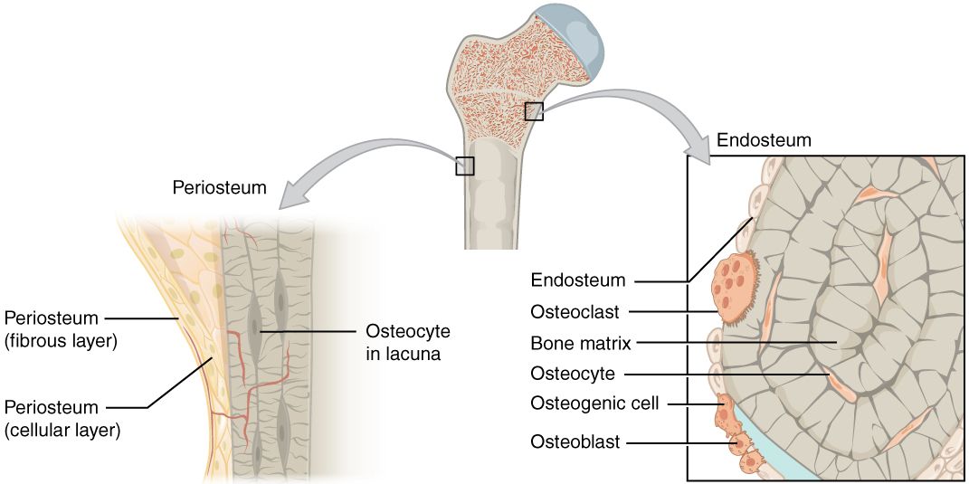
Three structures generate almost every musculoskeletal symptom you will assess. The periosteum is the fibrous sleeve over the bone surface and it is densely innervated — which is why a fracture, a bone tumour or an expanding infection under the periosteum produces pain that is deep, boring, unrelieved by position and often worse at night. The cortex and marrow hold a rich blood supply, so a fractured femur can sequester one to two litres of blood into the thigh without a drop of it appearing on the sheets. And the fascial compartments that bundle muscle, nerve and vessel are effectively inelastic; once bleeding and oedema raise the pressure inside one of them, the small vessels collapse and the tissue inside starts to die while the pulse distal to it is still perfectly palpable.
Function loss follows mechanism. A torn ligament (a sprain) makes a joint unstable and painful with stress in one direction. A torn muscle or tendon (a strain) makes a movement weak and painful with resisted contraction. A fracture produces pain, deformity, crepitus, loss of function, shortening and ecchymosis that often blooms twelve to twenty-four hours later, far from the injury, as blood tracks along fascial planes. A compressed nerve root produces pain in a dermatome, weakness in a myotome and a diminished deep tendon reflex; the pain radiates in a stripe rather than spreading in a blob.
Your general survey therefore has a shape: look at the limb (deformity, swelling, colour, wounds, shortening, rotation), touch it (temperature, capillary refill, pulses, tenderness), move it or watch the patient move it (active before passive; never move an obviously deformed limb before imaging unless the limb is pulseless), and always compare with the other side. The uninjured extremity is your control, and no textbook range of motion beats it.
The neurovascular assessment: the skill that saves the limb
The neurovascular check — also charted as circulation, motion and sensation (CMS) — is the single most important repeated assessment in orthopaedic nursing. Perform it on admission, after any manipulation, cast, splint or surgery, then hourly for the first twenty-four hours (many units use every hour for four hours, then every two hours for eight, then every four), and any time the patient complains. Compare it to the other limb and to the previous entry. A trend is worth more than a snapshot.
| The six Ps | What you actually do | What it means |
|---|---|---|
| Pain | Rate at rest and on passive stretch of the muscles in the compartment (extend the toes or fingers) | Pain out of proportion to injury, escalating despite opioids, and severe pain on passive stretch is the earliest sign of compartment syndrome |
| Paraesthesia | Light touch and two-point discrimination in each nerve distribution | Numbness or tingling is the earliest reliable neurological sign; sensory fibres die before motor fibres |
| Pallor | Colour and capillary refill (normal under 3 seconds) | Late arterial finding; a dusky or mottled limb is worse than a pale one |
| Pulselessness | Palpate, then Doppler if not palpable; mark the spot | Latest and least reliable sign in compartment syndrome — never wait for it; in true arterial injury it is decisive |
| Paralysis | Active wiggle of fingers or toes, then strength against resistance | Late; motor loss suggests irreversible damage is starting |
| Poikilothermia | Back of your hand on the limb, compared to the other side | The limb takes on room temperature when perfusion fails |
Two nerve checks are worth memorising because they are commonly missed. After an upper extremity injury or cast, ask the patient to make the “OK” sign (anterior interosseous or median), spread the fingers against resistance (ulnar), and extend the wrist and thumb (radial); check sensation in the web space between thumb and index finger (radial), the tip of the index finger (median) and the tip of the little finger (ulnar). After a lower extremity injury, cast or knee surgery, check dorsiflexion of the ankle and extension of the great toe and sensation in the first web space of the foot — that is the peroneal nerve, the nerve most often injured by a tight cast, a poorly padded knee immobiliser, a fibular neck fracture or prolonged leg-crossed positioning.
Diagnostics and the numbers that matter
Plain radiograph is the first study for suspected fracture; two views at ninety degrees to each other, plus the joint above and below. Remember that a normal X-ray does not exclude a fracture. Occult hip fractures, scaphoid fractures and stress fractures are commonly invisible for seven to ten days; if the older adult cannot bear weight and the hip film is clean, the next step is MRI (most sensitive) or CT, not reassurance. CT defines complex intra-articular and pelvic fractures and is the study of choice for operative planning. MRI is the study for soft tissue — disc herniation, meniscus, rotator cuff, ligament, spinal cord compression and early osteomyelitis. A bone scan (technetium-99m) lights up anywhere osteoblasts are busy, which makes it sensitive but non-specific: it detects metastases, osteomyelitis and stress fractures but cannot tell them apart. DEXA measures bone mineral density. Arthrocentesis samples joint fluid; bone biopsy with culture is the gold standard for osteomyelitis and for tumour diagnosis.
| Test | Adult reference | Orthopaedic meaning |
|---|---|---|
| Total serum calcium | 8.6–10.2 mg/dL | Normal in osteoporosis and Paget disease; low in osteomalacia; high in hyperparathyroidism and malignant bone disease |
| Ionised calcium | 4.6–5.3 mg/dL | The physiologically active fraction; check before denosumab and IV bisphosphonate |
| Phosphate | 2.5–4.5 mg/dL | Low in osteomalacia and vitamin D deficiency |
| Alkaline phosphatase (ALP) | 30–120 U/L | Marker of osteoblast activity; markedly elevated in Paget disease and osteosarcoma; modestly elevated in healing fracture and osteomalacia |
| 25-hydroxy vitamin D | Sufficient 30–100 ng/mL; deficient under 20 ng/mL | Correct before starting any antiresorptive drug |
| ESR | Roughly under 20 mm/h (men), under 30 mm/h (women) | Non-specific inflammation; used to trend osteomyelitis and prosthetic joint infection |
| CRP | Under 1.0 mg/dL (10 mg/L) | Rises and falls faster than ESR; the better day-to-day infection trend |
| Creatine kinase (CK) | 30–170 U/L | Rhabdomyolysis after crush or prolonged compression is CK above roughly five times the upper limit of normal — about 1,000 U/L against this range; above 5,000 U/L marks severe muscle injury and a high risk of acute kidney injury |
| Synovial fluid WBC | Under 200/mm³ normal | 2,000–50,000/mm³ inflammatory; over 50,000/mm³ with high neutrophils suggests septic arthritis — a surgical emergency |
| DEXA T-score | Normal above −1.0 | −1.0 to −2.5 is low bone mass (osteopenia); −2.5 or lower is osteoporosis |
Two cautions. First, ESR and CRP are elevated for days to weeks after any surgery, so a single raised value on post-operative day two means nothing; the alarming pattern is a value that was falling and starts to climb again. Second, D-dimer is essentially useless after orthopaedic surgery for the same reason — it is elevated in nearly everyone, so a suspected post-operative pulmonary embolism goes to CT pulmonary angiography, not to a D-dimer.
The neurovascular check — performed on a schedule, compared to the other limb and to the last entry, and charted in findings rather than adjectives — is the assessment that separates a limb that is saved from a limb that is lost.
Section 2Fractures and Soft-Tissue Injury: From Field to Fixation
Patterns, healing and the timeline you will be asked about
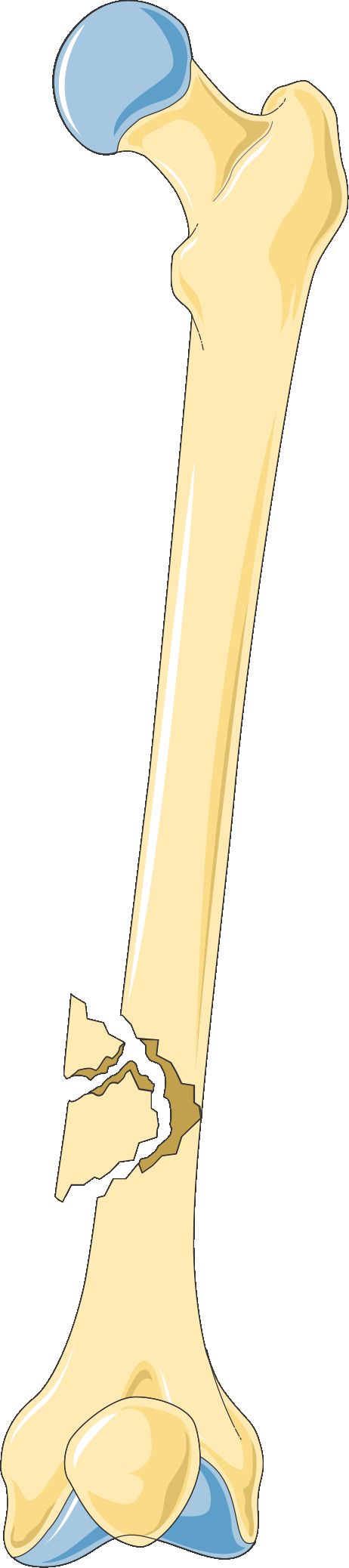
A fracture is complete or incomplete, displaced or non-displaced, and — the distinction that changes everything — closed or open. An open (compound) fracture communicates with the skin surface and is contaminated by definition. It needs intravenous antibiotics within one hour of arrival (typically cefazolin, with gram-negative coverage added for larger wounds), tetanus prophylaxis, sterile moist covering of exposed bone, and urgent operative irrigation and debridement. Patterns tell you mechanism: transverse from direct blow, oblique and spiral from twisting (a spiral fracture in a non-ambulatory infant is a child-abuse red flag), comminuted (more than two fragments) from high energy or from weak bone, impacted from axial load, greenstick in children, compression in the osteoporotic vertebra, and pathologic through a bone already weakened by tumour, infection or osteoporosis.
Healing proceeds in four overlapping stages: haematoma formation in the first hours to days, when bleeding at the fracture site forms the scaffold; fibrocartilaginous (soft) callus over roughly days three to fourteen; bony callus from about week three to week twelve, when woven bone bridges the gap and the fracture becomes clinically stable; and remodelling, which continues for months to years as osteoclasts and osteoblasts convert woven bone to lamellar bone along lines of stress. Practical timelines: upper extremity fractures generally unite in six to eight weeks, lower extremity in eight to twelve or more, and femur or tibia in an older adult with diabetes considerably longer. Smoking, corticosteroids, poor protein intake, diabetes, infection, inadequate immobilisation and distraction at the fracture site all delay healing. Delayed union is healing that is slower than expected; non-union is healing that has stopped; malunion is healing in the wrong position.
Nutrition is nursing therapy here, not a footnote. Fracture healing needs adequate protein (roughly 1.2 to 1.5 g/kg/day in an older adult unless renal disease dictates otherwise), calcium, vitamin D, vitamin C for collagen cross-linking, and enough total calories that the patient is not catabolising the muscle they will need for rehabilitation. The frail hip-fracture patient who eats twenty per cent of their tray needs a dietitian referral and oral nutrition supplements, and that is a nursing judgement you make without an order.
Immobilisation: casts, traction and fixation
Casts. Plaster is heavy, moulds beautifully, and takes twenty-four to seventy-two hours to dry fully; fibreglass is lighter, water-resistant and sets in about thirty minutes. Handle a wet cast with the palms only — fingertips leave indentations that become pressure points. Leave the cast uncovered to air-dry, support it on a cloth-covered pillow, and elevate the limb above heart level for the first twenty-four to forty-eight hours with ice over (not under) the fracture site for twenty minutes at a time. Teach the patient never to insert anything under a cast to scratch: use cool air from a hairdryer instead. Report immediately any drainage staining through, foul odour, a warm spot on the cast surface, increasing pain, numbness, tingling, or an inability to move the fingers or toes. If circulation is compromised the cast is bivalved or a window is cut — and a nurse never cuts a cast without an order and the correct tool.
Traction. Skin traction (Buck extension, Russell traction) applies pull through a boot or adhesive strapping and is limited to about 5 to 10 pounds (2.3 to 4.5 kg) because more will shear the skin; it is used chiefly for comfort and muscle-spasm control before hip-fracture surgery. Skeletal traction pulls through a pin or wire in the bone and tolerates 15 to 30 pounds (7 to 13 kg); it is continuous and the weights are never removed. For any traction: weights hang freely and never rest on the floor or the bed frame, ropes stay in the pulley grooves and are not frayed, knots are clear of the pulleys, the patient stays aligned in the middle of the bed so that body weight provides counter-traction, and the pull remains in the correct line. Assess pin sites at least every shift for the expected serous crusting versus the abnormal purulent drainage, erythema spreading beyond the pin, tenderness, loosening or fever. Standard pin care is once-daily cleansing with chlorhexidine solution after the first forty-eight to seventy-two hours, following the facility protocol.
Surgical fixation. ORIF (open reduction with internal fixation) uses plates, screws, rods or intramedullary nails and permits earlier mobilisation — the reason it is now standard for the older adult with a hip fracture. External fixation uses pins connected to an outside frame, chosen for open fractures with soft-tissue loss, comminuted fractures or when the patient is too unstable for definitive surgery. Its advantage is access to the wound; its price is pin-tract infection and a device that looks alarming to the patient and family, so build reassurance and body-image support into your care plan. Never use the external fixator frame as a handle to lift or turn the limb.
Soft-tissue injury and repetitive strain
Sprains (ligament) and strains (muscle or tendon) are managed acutely with rest, ice for twenty minutes several times daily for the first twenty-four to forty-eight hours, compression and elevation, followed by early protected motion — prolonged immobilisation produces stiffness and weakness, not healing. A grade III sprain is a complete tear with instability and may require surgery. Teach patients that the inability to bear weight for four steps immediately and in the emergency department, or bony tenderness over the malleoli, is what prompts an ankle radiograph.
Rotator cuff tear usually involves the supraspinatus; the patient reports pain with overhead reach, night pain lying on the shoulder, and weakness in abduction, and a positive drop-arm test (the arm falls when lowered from ninety degrees of abduction) suggests a full-thickness tear. Meniscal injury presents with a twisting mechanism, joint-line tenderness, clicking and locking. Carpal tunnel syndrome is median nerve compression at the wrist producing numbness and burning in the thumb, index, middle and radial half of the ring finger, worse at night, with thenar wasting late; Tinel and Phalen signs support the diagnosis, and first-line treatment is a neutral-position wrist splint worn at night plus ergonomic modification. Epicondylitis, bursitis and plantar fasciitis follow the same pattern: overuse, localised tenderness, pain reproduced by loading the involved structure, treated with relative rest, ice, NSAIDs, stretching and correction of the offending activity.
The older adult and the fragility fracture
A fragility fracture is a fracture from a fall at standing height or less — the definition matters because it establishes osteoporosis clinically regardless of the DEXA result, and it obliges you to think about the next fracture before this one has healed. The classic presentation of an extracapsular hip fracture is a shortened, externally rotated, adducted leg with severe groin pain and inability to bear weight. But do not anchor on that picture: an impacted femoral neck fracture can leave the patient able to walk with a limp, and a vertebral compression fracture may present only as sudden mid-back pain when lifting a laundry basket, or as progressive height loss and kyphosis with no remembered injury at all.
Older adults also present atypically in ways that catch students out. Pain may be reported as vague or denied entirely in the patient with dementia, in whom the fracture announces itself as agitation, refusal to transfer, or new delirium. Untreated pain is itself a leading cause of post-operative delirium, so the answer is scheduled analgesia and a validated observational pain scale (PAINAD) rather than withholding opioids out of fear. Best practice for hip fracture is surgical repair within twenty-four to forty-eight hours, because delay increases mortality, pneumonia, pressure injury and delirium; one-year mortality after hip fracture remains roughly twenty to thirty per cent, and only about half of survivors regain their previous level of function. Regional nerve blocks (fascia iliaca) reduce opioid requirement and delirium. Screen and treat for the reason they fell: orthostatic hypotension, polypharmacy, sedative-hypnotics, vision, footwear, home hazards and vitamin D deficiency.
Immobilisation devices are only as safe as the assessment that surrounds them: elevate and ice a new cast, never remove skeletal traction weights, keep the patient in line with the pull, and re-check circulation, motion and sensation after every device change.
Section 3When a Fracture Turns Dangerous
Acute compartment syndrome: the six-hour clock
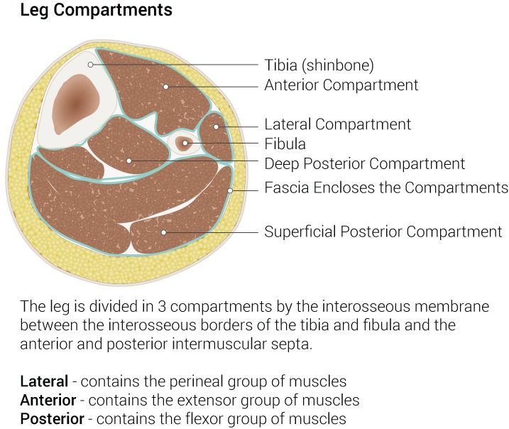
Pressure inside a closed fascial compartment rises above the pressure in the capillaries that feed it, perfusion stops, muscle and nerve become ischaemic, and after roughly four to six hours the damage is permanent. Causes divide into things that increase the contents (bleeding, oedema after fracture, reperfusion after vascular repair, infiltrated intravenous fluid, burn oedema) and things that decrease the space (a tight cast, a circumferential dressing or burn eschar, a poorly applied splint). The tibia is the most common site, followed by the forearm; a supracondylar humerus fracture in a child is the classic precursor of Volkmann ischaemic contracture, the permanent claw deformity that follows untreated forearm compartment syndrome.
The presentation is deceptive because the limb often looks fine. The cardinal findings are pain out of proportion to the injury and escalating despite escalating opioid doses, pain on passive stretch of the compartment muscles, and a compartment that feels tense and woody. Paraesthesia follows. Pallor, pulselessness and paralysis are late; the artery is a large high-pressure vessel and it keeps flowing long after the capillaries have collapsed, so a palpable pulse never rules this out. Diagnosis is clinical, supported by compartment pressure measurement: normal is 0 to 8 mmHg, and either an absolute pressure above about 30 mmHg or a delta pressure (diastolic blood pressure minus compartment pressure) below 30 mmHg indicates fasciotomy.
Your first actions, in order: stop and do a focused neurovascular assessment; loosen or remove anything constricting — unwrap the elastic bandage, cut the cast padding after the cast is bivalved by the provider (bivalving a cast without removing the underlying padding relieves only about half the pressure); place the limb at heart level; remove ice; and notify the surgeon immediately. Do not wait for the next scheduled check, do not medicate and reassess in an hour, and do not elevate. Fasciotomy wounds are left open and later closed or grafted. Follow-up laboratory studies matter because devitalised muscle releases myoglobin and potassium: watch CK, potassium, creatinine and urine colour for rhabdomyolysis and give aggressive intravenous crystalloid to protect the kidneys.
Fat embolism, venous thromboembolism and haemorrhage
Fat embolism syndrome (FES) is a distinct entity from pulmonary embolism, and the timing is the giveaway. Marrow fat globules enter the venous circulation after long-bone or pelvic fracture and produce inflammation in the lung and brain, typically twenty-four to seventy-two hours after injury — often in a young adult with a femur fracture who was fine yesterday. The classic triad is hypoxaemia (dyspnoea, tachypnoea, PaO₂ below 60 mmHg on room air), neurological change (restlessness, confusion, agitation, sometimes the very first sign) and a petechial rash across the chest, axillae, neck and conjunctivae, which appears in only a minority of patients but is close to diagnostic when it does. Laboratory findings include falling platelets and haematocrit. There is no specific treatment: management is oxygen, sometimes mechanical ventilation, and supportive care. Prevention is what nursing controls — early immobilisation of the fracture, early operative fixation within twenty-four hours where possible, and careful handling of the fractured limb.
Venous thromboembolism is the leading preventable cause of death after major orthopaedic surgery. Hip fracture, hip arthroplasty and knee arthroplasty carry among the highest venous thromboembolism risk of any surgery, because the triad is complete: stasis from immobility, endothelial injury from the operation, and a hypercoagulable post-operative state. Prophylaxis combines mechanical measures (sequential compression devices worn whenever the patient is in bed, early ambulation, ankle pumps) with pharmacologic prophylaxis for ten to thirty-five days — commonly enoxaparin 40 mg subcutaneously daily (or 30 mg every twelve hours), a direct oral anticoagulant such as rivaroxaban 10 mg daily, or aspirin in selected low-risk arthroplasty protocols. Assess daily for unilateral calf swelling, warmth, tenderness and a calf circumference difference; never massage a suspicious calf. Sudden dyspnoea, pleuritic chest pain, tachycardia, anxiety or unexplained hypoxaemia is a pulmonary embolism until proven otherwise: oxygen, rapid response, ECG and CT pulmonary angiography.
Haemorrhage and hypovolaemia are quiet in orthopaedics because the blood hides inside the limb. A closed femur fracture can hold one to two litres; a pelvic fracture can hold far more and can exsanguinate a patient into the retroperitoneum with an unremarkable abdomen. Monitor haemoglobin, heart rate, blood pressure trend and urine output rather than the dressing. Post-operative transfusion thresholds are restrictive — generally a haemoglobin below 7 g/dL, or below 8 g/dL in patients with cardiovascular disease or symptomatic anaemia.
Infection, and the complications that show up late
Osteomyelitis reaches bone by direct inoculation (open fracture, surgery, pin tract), by contiguous spread (diabetic foot ulcer, pressure injury) or haematogenously. Staphylococcus aureus is the leading organism. Acute presentation is localised bone pain, warmth, swelling, fever, malaise and elevated WBC, ESR and CRP. Plain films lag by ten to fourteen days; MRI detects it earliest, and bone biopsy with culture is the gold standard and should ideally be obtained before antibiotics are started. Treatment is four to six weeks of intravenous antibiotics, often through a PICC line at home, plus surgical debridement when there is dead bone. Chronic osteomyelitis produces a sequestrum (a segment of dead, avascular bone that antibiotics cannot reach), an involucrum of new bone around it, and draining sinus tracts — and it is why debridement rather than antibiotics alone is curative.
Septic arthritis destroys articular cartilage within days and is a surgical emergency: a hot, swollen, exquisitely painful joint the patient will not move, with fever, requires urgent arthrocentesis (synovial WBC often over 50,000/mm³) and washout. Avascular necrosis follows disruption of the blood supply — classically the femoral head after a displaced femoral neck fracture, the scaphoid proximal pole, and the talus — presenting weeks to months later as insidious groin or joint pain worsening with weight bearing. Complex regional pain syndrome presents after an often minor injury with burning pain far out of proportion, allodynia, swelling, colour and temperature change and sweating asymmetry; early aggressive mobilisation and physical therapy, with gabapentinoids and sympathetic block, is treatment. Contracture, muscle atrophy, pressure injury, constipation and hospital-acquired pneumonia are all complications of immobility, which is why turning, incentive spirometry, bowel regimen and getting the patient out of bed on the day of surgery are not comfort measures — they are the treatment.
| Complication | Typical timing | Hallmark | Your first action |
|---|---|---|---|
| Acute compartment syndrome | 6–48 h | Pain out of proportion; pain on passive stretch; tense compartment | Release constriction, limb to heart level, remove ice, call surgeon STAT |
| Fat embolism syndrome | 24–72 h | Hypoxaemia, confusion, petechiae on chest and axillae | Oxygen, high Fowler if tolerated, rapid response, ABG and pulse oximetry |
| Deep vein thrombosis | Days 2–14 and beyond | Unilateral calf swelling, warmth, tenderness | Do not massage; notify provider; anticipate duplex ultrasound |
| Pulmonary embolism | Any time post-op | Sudden dyspnoea, pleuritic pain, tachycardia, hypoxaemia | Oxygen, sit upright, rapid response, CT pulmonary angiography |
| Rhabdomyolysis | Hours after crush or prolonged compression | Tea-coloured urine, CK above five times normal (roughly over 1,000 U/L), hyperkalaemia | Aggressive isotonic IV fluids at once; trend potassium, creatinine and urine output, ECG — do not wait for CK to reach a set number |
| Osteomyelitis or prosthetic joint infection | Days to months | Fever, increasing pain, wound drainage after day 5, rising CRP | Culture before antibiotics, notify surgeon, do not simply redress |
In orthopaedic trauma the deadly and limb-threatening complications each have a signature time window — compartment syndrome in the first two days, fat embolism at one to three days, thromboembolism from day two onward, and infection from day five onward — so assess for the complication the calendar is pointing at.
Section 4Degenerative Joints, Arthroplasty, Amputation and the Painful Back
Osteoarthritis: mechanical failure, not systemic inflammation
Osteoarthritis (OA) is progressive loss of articular cartilage with reactive change in the underlying bone. Cartilage thins and fissures, the joint space narrows, subchondral bone becomes sclerotic and forms cysts, and the joint margins throw out osteophytes. The result is pain that is worse with use and better with rest, morning stiffness that resolves in under thirty minutes, crepitus, bony rather than boggy enlargement, and an asymmetric distribution in the weight-bearing joints — knees, hips, lumbar and cervical spine — plus the hands, where Heberden nodes at the distal interphalangeal joints and Bouchard nodes at the proximal interphalangeal joints are characteristic. Systemic features are absent, and ESR and CRP are normal. That last point is the examination discriminator: rheumatoid arthritis is symmetric, involves the metacarpophalangeal and proximal interphalangeal joints while sparing the distal interphalangeal joints, produces morning stiffness lasting longer than an hour with fatigue and low-grade fever, and raises inflammatory markers. Rheumatoid and other inflammatory arthritides are covered in Chapter 9.
Management is a ladder. Non-pharmacologic first and always: weight loss (each pound lost removes roughly four pounds of force from the knee), low-impact exercise and quadriceps strengthening, physical and occupational therapy, assistive devices, heat before activity and cold after. Pharmacologic: topical NSAIDs are first-line for knee and hand OA in older adults because systemic absorption is minimal; oral acetaminophen up to 3 g/day (and no more than 2 g/day in the older adult, in liver disease or with regular alcohol use); oral NSAIDs at the lowest effective dose with attention to gastrointestinal bleeding, hypertension, kidney injury and heart failure — a genuinely risky combination in the frail patient already taking an anticoagulant and an ACE inhibitor. Intra-articular corticosteroid injections give weeks to a few months of relief and are limited to about three or four per joint per year because repeated injection damages cartilage. Opioids are not a maintenance therapy for osteoarthritis. When pain limits sleep and daily function despite the ladder, joint replacement is offered.
Total hip and total knee arthroplasty
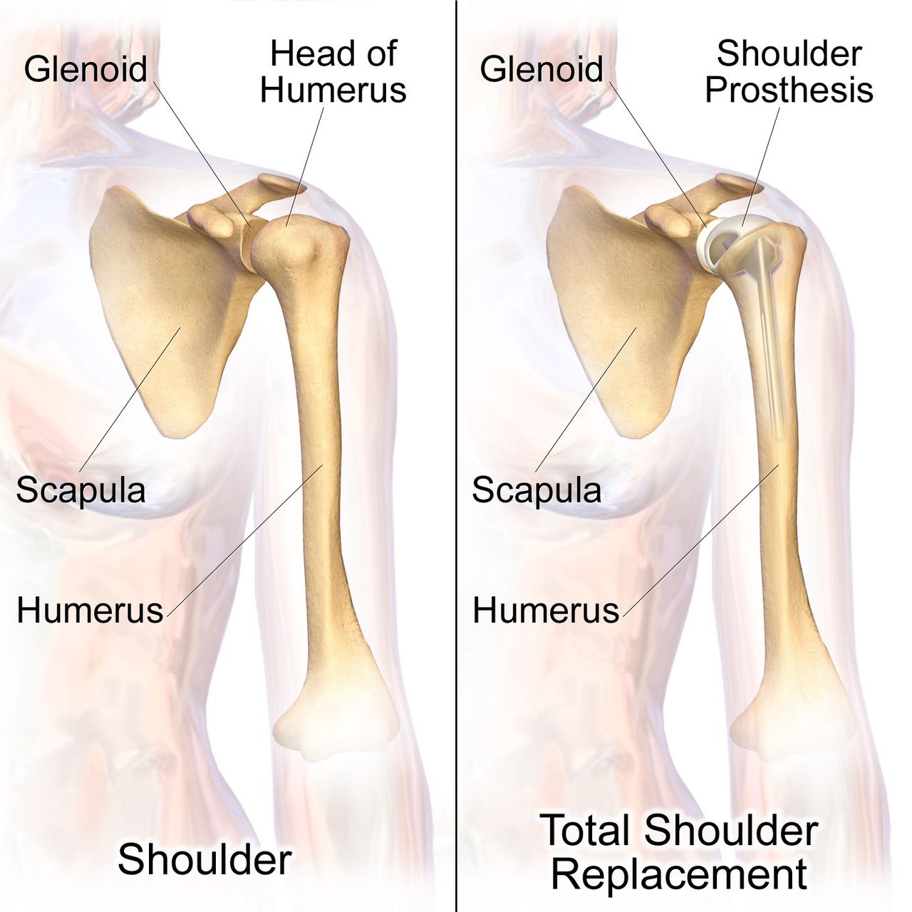
Pre-operatively, teach before the anaesthetic rather than after it: incentive spirometry, ankle pumps, the transfer technique, the pain plan and the precautions. Screen for infection anywhere in the body, since bacteraemia seeds a prosthesis; confirm that the surgical-site antibiotic (usually cefazolin, vancomycin for MRSA colonisation) is given within sixty minutes of incision — vancomycin is the exception and is started within one hundred and twenty minutes because it must be infused slowly; and verify the surgical site marking with the patient awake. Post-operatively the priorities are airway and haemodynamics first, then neurovascular checks distal to the joint, then pain control adequate to permit the physiotherapist to work, then VTE prophylaxis, then mobilisation — usually on the day of surgery or the first post-operative morning.
Hip precautions depend on the surgical approach and you must know which one your patient had. After a posterior approach (the most common), the prohibited movements are the ones that lever the head out the back of the socket: no hip flexion beyond ninety degrees, no adduction past midline (no crossing legs or ankles), and no internal rotation of the operative leg. After an anterior approach, the risk is the opposite: avoid hyperextension, external rotation and bridging. Precautions typically last six to twelve weeks. Equip the home accordingly: elevated toilet seat, chairs with arms and a firm high seat, a reacher, a long-handled shoe horn and sponge, sock aid, and no low soft couches or deep recliners. An abduction pillow or wedge is used in bed after posterior approaches, and the patient is turned toward the unoperated side with the pillow between the legs unless the surgeon specifies otherwise.
Prosthetic hip dislocation is an emergency you must recognise: the patient reports a pop and sudden severe pain, cannot move the leg, and the leg is shortened and internally rotated (posterior dislocation, the usual direction). Keep the patient still and supine, support the limb, do not attempt to reposition or ambulate them, notify the surgeon and keep them nil by mouth for closed reduction under sedation. After total knee arthroplasty, the priorities differ: the enemy is a flexion contracture, so the knee is kept in extension at rest and a pillow is never placed under the knee — elevate the whole leg on a pillow under the calf instead. Goals are typically ninety degrees of flexion by discharge and full extension, progressing toward one hundred and ten to one hundred and twenty degrees. Expect knee swelling, use cold therapy, and remember that new fever with wound drainage after post-operative day five, escalating rather than improving pain, or drainage that persists beyond seventy-two hours suggests a prosthetic joint infection and must be reported rather than simply redressed.
| Task on the orthopaedic unit | RN | LPN/LVN | UAP |
|---|---|---|---|
| Initial and post-operative neurovascular assessment; any change in status | Yes — RN only | No | No |
| Teaching hip precautions, crutch gait, bisphosphonate administration | Yes — RN only | Reinforce established teaching | No |
| Evaluating response to analgesia or to a new mobility plan | Yes — RN only | No | No |
| First post-operative ambulation; ambulating an unstable patient | Yes — RN (with therapy) | Per policy once stable | No |
| Pin-site care and dressing changes | Yes | Yes, per protocol | No |
| Applying sequential compression devices, ice packs, repositioning, assisting a stable patient to ambulate with a taught device, intake and output, vital signs | Supervises | Yes | Yes — report findings to the RN |
Amputation and the painful back
Amputation is most often performed for peripheral arterial disease and diabetic limb infection, and less often for trauma, tumour or congenital deformity. Immediately post-operatively the priorities are haemorrhage and pain: keep a large tourniquet at the bedside for the first twenty-four to forty-eight hours, monitor the dressing and the drain, and record the amount of drainage rather than describing it. Elevate the residual limb on a pillow for the first twenty-four hours only to control oedema; after that, elevation risks a hip or knee flexion contracture that will make prosthetic fitting impossible. From day two, position the patient prone for twenty to thirty minutes three to four times daily after a lower-limb amputation, keep the residual limb flat when supine, avoid sitting in a chair for prolonged periods with the knee flexed, and never place a pillow between the thighs (that promotes abduction contracture). Teach residual-limb wrapping in a figure-of-eight pattern with more pressure distally to shape the limb into a cone.
Phantom limb sensation — the feeling that the limb is still present — is nearly universal and is normal. Phantom limb pain is burning, cramping or shooting pain perceived in the missing limb, is real neuropathic pain, and is treated with gabapentin or pregabalin, tricyclic antidepressants such as amitriptyline, mirror therapy, desensitisation, transcutaneous electrical nerve stimulation and early prosthetic use. Never tell a patient the pain cannot be real. Grief, altered body image and depression are expected; ask directly, involve peer support and rehabilitation early, and remember that the contralateral limb must be protected obsessively in the diabetic patient because it is now the only one.
Low back pain is the most common musculoskeletal complaint you will meet. Most acute mechanical back pain resolves in four to six weeks with continued activity (bed rest actually worsens outcomes), heat, NSAIDs, and reassurance; imaging is not indicated in the first six weeks unless red flags exist. A herniated intervertebral disc, usually at L4–L5 or L5–S1, compresses a nerve root and produces radiating pain below the knee, dermatomal numbness, a positive straight-leg raise and myotomal weakness — foot dorsiflexion for L5, plantar flexion and a reduced ankle reflex for S1. The red flags that must never be missed are: cauda equina syndrome (new urinary retention or incontinence, faecal incontinence, saddle anaesthesia, bilateral leg weakness) which is a surgical emergency requiring decompression within hours; fever with back pain and intravenous drug use (spinal epidural abscess or vertebral osteomyelitis); a history of cancer with new unrelenting night pain (metastasis with cord compression); and significant trauma or unexplained weight loss. After lumbar fusion, teach the “no BLT” rule — no bending, no lifting more than five to ten pounds, no twisting — and log-roll the patient in bed with the shoulders and hips moving as a unit.
After joint replacement and amputation, most of what protects the patient is positioning — approach-specific hip precautions, the knee kept in extension, and the residual limb kept flat with scheduled prone lying — and positioning is teaching, which belongs to the RN.
Section 5Bone Integrity: Osteoporosis, Metabolic Bone Disease and Bone Tumours
Osteoporosis: the silent disease until it is not
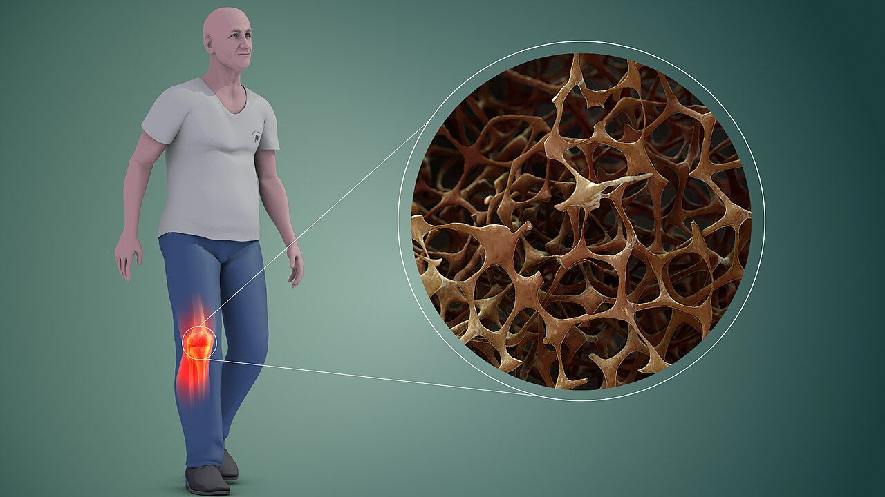
Bone is continuously remodelled: osteoclasts resorb, osteoblasts rebuild. Osteoporosis is the state in which resorption outpaces formation, so bone mass falls and microarchitecture deteriorates while the bone that remains is chemically normal — which is why serum calcium, phosphate and ALP are all typically normal in uncomplicated osteoporosis. Oestrogen restrains osteoclasts, so bone loss accelerates sharply for five to ten years after menopause. Other major risks: age, low body weight, white or Asian ancestry, family history of hip fracture, smoking, more than two or three alcoholic drinks daily, sedentary lifestyle, low calcium and vitamin D intake, hyperthyroidism and hyperparathyroidism, malabsorption, chronic kidney disease, and drugs — particularly glucocorticoids (prednisone 5 mg daily or more for three months or longer), anticonvulsants, proton pump inhibitors, aromatase inhibitors, androgen deprivation therapy and long-term heparin.
Osteoporosis produces no symptoms until it produces a fracture. The three classic sites are the vertebrae (compression fractures causing acute back pain, progressive height loss of more than 1.5 inches and thoracic kyphosis, the “dowager hump”), the hip and the distal radius (Colles fracture from a fall on an outstretched hand). Screening with DEXA of the hip and lumbar spine is recommended for all women aged 65 and older, men aged 70 and older, and younger post-menopausal women or men over 50 with risk factors or a fragility fracture. The T-score compares the patient to a healthy young adult: above −1.0 is normal, −1.0 to −2.5 is low bone mass (osteopenia), and −2.5 or below is osteoporosis; a T-score of −2.5 or below plus a fragility fracture is severe osteoporosis. The Z-score compares to age-matched peers and is the score used in premenopausal women, men under 50 and children; a Z-score of −2.0 or lower prompts a search for a secondary cause. FRAX estimates ten-year fracture probability and helps decide whether to treat osteopenia.
Every patient gets the foundation: 1,000 to 1,200 mg of elemental calcium daily from diet where possible (dairy, fortified plant milks, sardines with bones, kale, broccoli, tofu set with calcium) and 800 to 1,000 international units of vitamin D daily; weight-bearing and resistance exercise; smoking cessation; alcohol moderation; and rigorous fall prevention. Calcium supplements are absorbed only about 500 to 600 mg at a time, so divide the dose. Calcium carbonate is cheap but needs gastric acid, so take it with food and expect constipation; calcium citrate is absorbed without acid and is the correct choice for a patient on a proton pump inhibitor or with achlorhydria. Warn about constipation and, in the patient with a history of calcium stones or hypercalciuria, keep supplementation conservative and push fluids.
The drugs, and the teaching that makes them safe
| Drug class | Mechanism | Representative agent | What the nurse monitors and teaches |
|---|---|---|---|
| Bisphosphonate (oral) | Binds hydroxyapatite and inhibits osteoclast resorption | Alendronate 70 mg weekly; risedronate; ibandronate | Take first thing in the morning on an empty stomach with a full 6 to 8 oz glass of plain water; remain upright for 30 minutes (60 for ibandronate) and take nothing else by mouth in that time; report new dysphagia, heartburn or retrosternal pain (oesophagitis and ulceration); dental examination before starting (osteonecrosis of the jaw); report new thigh or groin pain (atypical femoral fracture); avoid if creatinine clearance is under about 30–35 mL/min; correct hypocalcaemia and vitamin D deficiency first |
| Bisphosphonate (intravenous) | Same | Zoledronic acid 5 mg IV once yearly | Hydrate well before and after; expect a flu-like acute-phase reaction for 24–72 hours after the first dose, treated with acetaminophen; check calcium and renal function first |
| RANK ligand inhibitor | Monoclonal antibody that blocks osteoclast formation and survival | Denosumab 60 mg subcutaneously every 6 months | Usable in renal impairment but check calcium first — hypocalcaemia risk is real, especially in CKD; osteonecrosis of the jaw; never simply stop it without transitioning to another agent, because rebound vertebral fractures follow discontinuation; must be given on schedule |
| Selective oestrogen receptor modulator | Oestrogen agonist at bone, antagonist at breast | Raloxifene 60 mg daily | Reduces vertebral fracture and breast cancer risk; increases VTE risk — stop before prolonged immobility or surgery; causes hot flashes and leg cramps |
| Anabolic PTH analogue | Intermittent dosing stimulates osteoblasts — builds new bone | Teriparatide, abaloparatide, daily subcutaneous | For severe osteoporosis; generally limited to about 2 years of lifetime use; teach self-injection and pen refrigeration; watch for transient hypercalcaemia and orthostatic dizziness after the first doses (sit or lie down) |
| Sclerostin inhibitor | Increases formation and decreases resorption | Romosozumab monthly for 12 months | Boxed warning for myocardial infarction, stroke and cardiovascular death — avoid within one year of MI or stroke; follow with an antiresorptive agent |
Two safety themes cut across the whole table. First, correct the calcium and vitamin D before the antiresorptive, or you will cause symptomatic hypocalcaemia — perioral numbness, tingling fingers, cramps, Trousseau and Chvostek signs, and in severe cases tetany, laryngospasm and a prolonged QT interval. Second, every antiresorptive carries a small risk of osteonecrosis of the jaw and atypical femoral fracture, so a dental examination before starting and a low threshold for investigating new thigh, groin or jaw pain are standing nursing responsibilities, not optional extras.
Osteomalacia, Paget disease and tumours of bone
Osteomalacia is defective mineralisation of the bone matrix in adults, almost always from vitamin D deficiency (poor intake, minimal sun exposure, dark skin, malabsorption after bariatric surgery or in coeliac disease, chronic kidney disease or certain anticonvulsants). Unlike osteoporosis, the mineral chemistry is abnormal: calcium is low or low-normal, phosphate is low, ALP is elevated, PTH rises in compensation, and 25-hydroxy vitamin D is low. The patient reports diffuse aching bone pain and tenderness, proximal muscle weakness and a waddling gait, and radiographs may show Looser zones (pseudofractures). Treatment is vitamin D repletion with calcium, and the symptoms improve within weeks. The paediatric equivalent is rickets, with bowed legs and growth-plate widening.
Paget disease of bone is a focal disorder of wildly accelerated, disorganised remodelling: osteoclasts resorb at high speed and osteoblasts lay down structurally chaotic bone that is larger but weaker. Calcium and phosphate are normal and ALP is markedly elevated — often two to several times the upper limit — which is the classic laboratory fingerprint. Many patients are asymptomatic and are found on an incidental radiograph or ALP; others report deep bone pain, bowing of the tibia or femur, skull enlargement (a hat that no longer fits), hearing loss from compression of the eighth cranial nerve, headache, pathologic fracture, secondary osteoarthritis of adjacent joints, and rarely high-output heart failure from hypervascular bone or malignant transformation to osteosarcoma. Treatment is a bisphosphonate, most often intravenous zoledronic acid, with ALP monitored as the marker of response, plus calcium and vitamin D, analgesia and fall precautions.
Bone tumours. Benign lesions (osteochondroma, osteoid osteoma, giant cell tumour) are far more common than malignant ones and are often incidental. Among primary malignancies, osteosarcoma is the most common, occurs mostly in adolescents and young adults around the knee (distal femur, proximal tibia) and in older adults with Paget disease, and presents with deep pain worse at night, a palpable mass and elevated ALP and LDH; radiographs show a “sunburst” periosteal reaction and Codman triangle. Ewing sarcoma occurs in children and adolescents and may mimic infection with fever and elevated inflammatory markers, with an “onion-skin” periosteal reaction. Chondrosarcoma occurs in middle-aged and older adults. Treatment is neoadjuvant chemotherapy, limb-salvage resection or amputation, and further chemotherapy. But by far the commonest malignant bone lesion is metastasis — breast, prostate, lung, kidney and thyroid primaries spread to the vertebrae, pelvis, ribs, proximal femur and skull. Watch for pathologic fracture from trivial force, and for hypercalcaemia of malignancy presenting as the classic quartet of bones, stones, abdominal groans and psychiatric overtones: nausea, constipation, polyuria, dehydration, confusion and lethargy. Treatment is aggressive isotonic saline followed by calcitonin and an intravenous bisphosphonate or denosumab. New back pain with leg weakness in a patient with known cancer is spinal cord compression until proven otherwise — MRI, high-dose corticosteroids and urgent radiation oncology or neurosurgery consultation.
Osteoporosis is normal bone in too small a quantity and its labs are normal, whereas osteomalacia and Paget disease announce themselves in the chemistry — and in all three the nursing win is the fracture that never happens.
The chapter in one breath
Musculoskeletal nursing rests on one repeated assessment and a handful of unforgiving timelines. The neurovascular check — pain, paraesthesia, pallor, pulselessness, paralysis, poikilothermia, compared side to side and entry to entry — is what protects a limb, and its most important finding is pain out of proportion with pain on passive stretch, because that is acute compartment syndrome and the tissue has four to six hours. Fractures heal through haematoma, soft callus, hard callus and remodelling, supported by casts that must be elevated and iced early and watched for constriction, by traction whose weights hang free and are never removed, and increasingly by early surgical fixation because early fixation reduces the risk of fat embolism and lets the older adult stand up. The complications keep a calendar: compartment syndrome in the first two days, fat embolism at one to three days with hypoxaemia, confusion and petechiae, thromboembolism from day two onward, infection from day five. Joint replacement care is largely positioning — approach-specific hip precautions, the knee in extension — and amputation care is contracture prevention with scheduled prone lying and a tourniquet at the bedside. Finally, bone integrity: normal labs and a T-score of −2.5 or lower mean osteoporosis, high ALP with normal calcium means Paget disease, low phosphate with high ALP means osteomalacia. The best orthopaedic outcome is always the fracture that never happens.
Words worth knowing
| Term | What it means |
|---|---|
| Acute compartment syndrome | Pressure within a closed fascial compartment exceeds capillary perfusion pressure, causing ischaemia of muscle and nerve; irreversible after roughly 4 to 6 hours; treated by fasciotomy |
| Delta pressure | Diastolic blood pressure minus measured compartment pressure; a value below 30 mmHg indicates fasciotomy |
| Fat embolism syndrome | Marrow fat entering the circulation 24 to 72 hours after long-bone or pelvic fracture, producing hypoxaemia, neurological change and a petechial rash over the chest and axillae |
| Volkmann ischaemic contracture | Permanent flexion deformity of the hand and wrist following untreated forearm compartment syndrome, classically after a supracondylar humerus fracture |
| Fragility fracture | A fracture caused by a fall from standing height or less; clinically establishes osteoporosis regardless of DEXA result |
| Bivalve | Cutting a cast lengthwise into two halves to relieve pressure; the underlying padding must also be cut to relieve pressure fully |
| Counter-traction | The opposing force — usually the patient’s body weight and bed position — that allows traction to pull effectively |
| External fixation | Pins driven into bone and connected to an external frame; used for open, comminuted or contaminated fractures and unstable patients |
| ORIF | Open reduction with internal fixation — surgical realignment with plates, screws, rods or intramedullary nails, allowing earlier mobilisation |
| Sequestrum | A fragment of dead, avascular bone in chronic osteomyelitis that antibiotics cannot penetrate; requires surgical debridement |
| Involucrum | New bone laid down around a sequestrum in chronic osteomyelitis |
| Avascular necrosis | Bone death from loss of blood supply, classically the femoral head after displaced femoral neck fracture, the scaphoid and the talus |
| Heberden and Bouchard nodes | Bony enlargements of the distal (Heberden) and proximal (Bouchard) interphalangeal joints characteristic of osteoarthritis |
| Osteophyte | Bony outgrowth at joint margins in osteoarthritis, visible on radiograph along with joint-space narrowing and subchondral sclerosis |
| Flexion contracture | Permanent shortening of soft tissue holding a joint in flexion; prevented after knee replacement by resting in extension and after amputation by prone positioning |
| Phantom limb pain | Genuine neuropathic pain perceived in an amputated limb, treated with gabapentinoids, tricyclics, mirror therapy and early prosthetic use |
| Cauda equina syndrome | Compression of the lumbosacral nerve roots causing saddle anaesthesia, urinary retention or incontinence and bilateral leg weakness; a surgical emergency |
| T-score | DEXA comparison with healthy young adults: above −1.0 normal, −1.0 to −2.5 low bone mass, −2.5 or lower osteoporosis |
| Looser zones | Radiolucent pseudofracture lines seen in osteomalacia |
| Rhabdomyolysis | Breakdown of skeletal muscle releasing myoglobin and potassium after crush or prolonged compression; tea-coloured urine, CK above five times the upper limit of normal (roughly over 1,000 U/L), hyperkalaemia, and a high risk of acute kidney injury once CK exceeds about 5,000 U/L |