Endocrine Disorders: Thyroid, Parathyroid, Adrenal and Pituitary
Every hormone in this chapter is a message, and every disorder is either too much message, too little message, or a message that never left the building. Learn the loop and the diseases stop being a list to memorise: the pituitary shouts at a gland, the gland answers with hormone, and the hormone tells the pituitary to be quiet. That single sentence explains why a high TSH means a lazy thyroid, why a suppressed TSH with a high free T4 means the gland has gone rogue, and why a patient on long-term prednisone cannot make cortisol on the day she gets pneumonia. Endocrine emergencies are quiet until they are not. Myxedema coma, thyroid storm, hypocalcaemic tetany, adrenal crisis and severe hyponatraemia all present first as vital-sign drift and a change in mentation, and all of them kill through airway, circulation or seizure. Your job on a busy floor is to notice the drift, name the pattern and escalate before the crash.
By the end of this chapter you can
- Explain negative feedback in the hypothalamic—pituitary—target axis and use paired hormone levels to distinguish primary from secondary (central) endocrine disease.
- Interpret TSH, free T4, serum calcium, PTH, cortisol, serum and urine osmolality against real reference ranges and state what the nurse does with each result.
- Prioritise nursing care in myxedema coma and thyroid storm, including why a thionamide must precede iodine and why aspirin is withheld.
- Deliver post-thyroidectomy care that anticipates airway obstruction from haematoma, recurrent laryngeal nerve injury and hypocalcaemia.
- Distinguish hyperparathyroidism from hypoparathyroidism by symptom cluster, ECG change and emergency drug, and demonstrate Chvostek and Trousseau signs.
- Compare Cushing syndrome and adrenal insufficiency, and manage adrenal crisis with fluids and IV hydrocortisone while teaching sick-day rules.
- Differentiate SIADH from diabetes insipidus using volume status, urine specific gravity and osmolality, and correct sodium at a safe rate.
- Recognise older-adult presentations such as apathetic thyrotoxicosis and drug-induced hyponatraemia, and state which tasks may and may not be delegated to assistive personnel.
- Compose an SBAR escalation for each endocrine red flag in this chapter.
Section 1Reading the Axes: The Logic That Makes Endocrine Make Sense
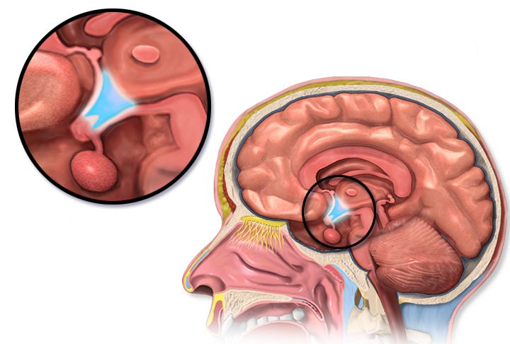
Feedback loops, and why the trophic hormone tells you where the lesion is
The hypothalamus secretes releasing hormones into a private portal circulation that drains straight into the anterior pituitary. The pituitary answers with trophic hormones — thyroid-stimulating hormone (TSH), adrenocorticotropic hormone (ACTH), growth hormone (GH), follicle-stimulating and luteinising hormone, prolactin. Those trophic hormones travel in the systemic blood to a target gland, and the target gland releases the hormone that actually does the work: thyroxine, cortisol, oestrogen, testosterone. The finished hormone then circles back and tells both the hypothalamus and the pituitary to stop shouting. That is negative feedback, and it is the whole game.
Here is the payoff for the bedside. If the target gland is the broken part — a thyroid destroyed by autoimmune thyroiditis, an adrenal cortex wrecked by autoimmune adrenalitis — then the target hormone is low and the pituitary, hearing no feedback, screams louder. So you see a low target hormone with a high trophic hormone. That is primary disease. If the pituitary or hypothalamus is the broken part, it cannot shout at all, so both hormones are low: low target hormone with a low or inappropriately normal trophic hormone. That is secondary, or central, disease. The same two-line rule works for excess. A rogue gland making too much hormone will suppress the trophic hormone to nearly nothing; a pituitary tumour making too much trophic hormone will drive the target hormone up while the trophic hormone stays high, which is biochemically impossible unless something upstream is autonomous.
Students lose points on this because they memorise diseases instead of patterns. Read the pair. A TSH of 18 mIU/L with a free T4 of 0.4 ng/dL is a failing thyroid. A TSH of 0.9 mIU/L with a free T4 of 0.4 ng/dL is a failing pituitary and a much more dangerous patient, because a pituitary that cannot make TSH usually cannot make ACTH either, and cortisol deficiency kills faster than hypothyroidism.
| Pattern | Trophic hormone | Target hormone | Where the lesion is | Classic example |
|---|---|---|---|---|
| Primary deficiency | High | Low | Target gland | Hashimoto hypothyroidism; Addison disease |
| Secondary (central) deficiency | Low or normal | Low | Pituitary or hypothalamus | Pituitary apoplexy; long-term exogenous steroids |
| Primary excess | Suppressed | High | Target gland | Graves disease; adrenal cortisol-secreting adenoma |
| Secondary excess | High or normal | High | Pituitary or ectopic tumour | Cushing disease; ectopic ACTH from small-cell lung cancer |
Stimulate when you suspect too little, suppress when you suspect too much
Random hormone levels are noisy. Cortisol swings with the clock, GH is secreted in bursts, and a single value can be normal in a patient who has almost no reserve. So endocrinology provokes the gland. When you suspect deficiency, you stimulate: give synthetic ACTH (cosyntropin 250 mcg IV or IM) and measure cortisol at 30 and 60 minutes. A healthy adrenal cortex jumps above roughly 18 to 20 mcg/dL. A failing one does not move. When you suspect excess, you suppress: give 1 mg of dexamethasone at 11 pm and draw cortisol at 8 am. A normal axis obediently shuts down to below 1.8 mcg/dL; an autonomous cortisol source ignores the instruction. The same grammar governs the water-deprivation test for diabetes insipidus and the oral glucose tolerance test for acromegaly.
Your nursing role in provocative testing is not glamorous and it is entirely about the validity of the result. Confirm the exact draw times and write them on the tubes; a cortisol drawn at 45 minutes instead of 30 is uninterpretable. Hold or document interfering drugs. Biotin supplements, which patients take for hair and nails and never think to report, distort many immunoassays including TSH and free T4 and should be stopped several days before. Keep the patient supine and calm if the test involves catecholamines. And for a water-deprivation test, the patient must not sneak fluids — which means the RN, not the assistive personnel, owns the observation, because the test is a live risk of hypernatraemia.
The labs that lie, and the assessment that does not
Acute illness perturbs every axis. In non-thyroidal illness syndrome (formerly euthyroid sick syndrome) a critically ill patient shows a low T3, sometimes a low free T4 and a TSH that is low during illness and rebounds high during recovery — none of it meaning thyroid disease. Steroids, dopamine, amiodarone, lithium, opioids and heparin all bend results. This is why a good endocrine assessment leans on the physical exam and the trend: weight over months, resting heart rate, skin and hair texture, deep-tendon-reflex relaxation phase, orthostatic vital signs, and the family’s report of how the person has changed. A daughter saying “she used to do the crossword” is data.
Older adults deserve a standing warning. Endocrine disease in a person over seventy-five rarely announces itself with textbook signs. Hypothyroidism looks like dementia or depression. Hyperthyroidism looks like new atrial fibrillation and weight loss with no tremor or agitation at all — apathetic thyrotoxicosis. Hypercalcaemia looks like constipation and confusion. Adrenal insufficiency looks like recurrent falls. If an older patient has an unexplained change in cognition, function or sodium, an endocrine cause belongs on your differential.
Interpret every endocrine result as a pair — trophic hormone with target hormone — because the pair, not the single value, tells you whether the gland, the pituitary or a drug is the problem.
Section 2The Thyroid: From Sluggish to Storm
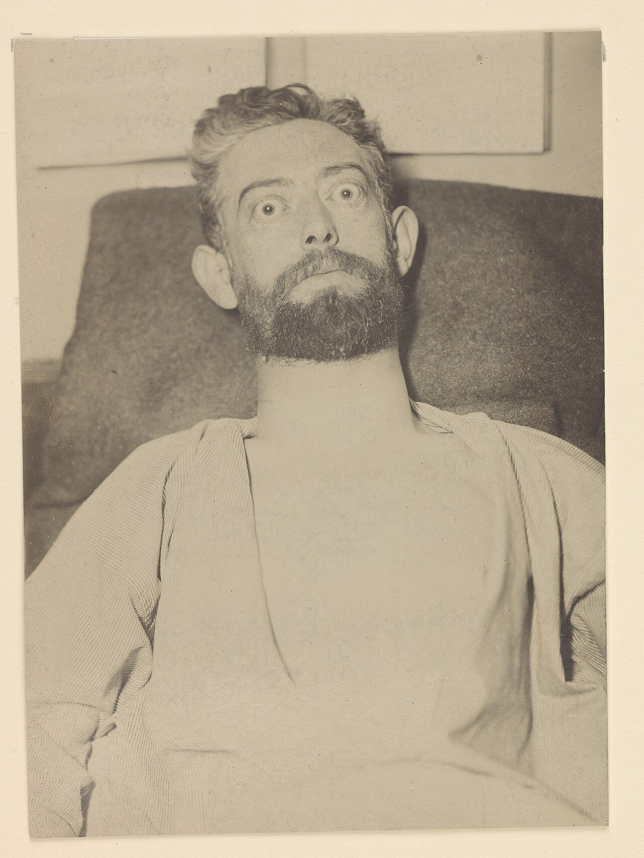
Hypothyroidism: everything slows, including the diaphragm
Thyroid hormone sets the basal metabolic rate of essentially every cell, so deficiency slows everything at once. Heat production falls, giving cold intolerance and a low-normal temperature. Gut motility falls, giving constipation. Cardiac output falls through bradycardia and reduced contractility, and diastolic hypertension appears because vessels stiffen. Cholesterol clearance falls, so the lipid panel worsens. Glycosaminoglycans accumulate in the dermis and draw water in, producing the non-pitting periorbital and facial puffiness called myxedema, along with generalised non-pitting swelling of the hands and feet, the coarse skin, the hoarse voice and the enlarged tongue. The separate sign called pretibial myxedema is not part of this picture — it is Graves dermopathy over the shins and belongs to the hyperthyroid patient. Reflexes show a characteristically slow relaxation phase. The most common cause in iodine-sufficient countries is Hashimoto thyroiditis, an autoimmune destruction marked by anti-thyroid peroxidase antibodies; other causes include prior radioactive iodine, thyroidectomy, amiodarone, lithium and, in the newborn, congenital absence — which is why every state screens.
Diagnosis is a TSH first. Reference range is roughly 0.4 to 4.0 mIU/L. An elevated TSH with a low free T4 (normal roughly 0.8 to 1.8 ng/dL) is overt primary hypothyroidism. An elevated TSH with a normal free T4 is subclinical disease, which may be treated if the TSH exceeds 10 mIU/L or the patient is pregnant or symptomatic. Treatment is levothyroxine, a synthetic T4 that the body converts to active T3. A healthy adult starts near 1.6 mcg/kg per day. An adult over sixty, or anyone with coronary disease, starts at 12.5 to 25 mcg daily and titrates slowly, because thyroid hormone raises myocardial oxygen demand and a full replacement dose can precipitate angina, infarction or a tachyarrhythmia. Recheck TSH six to eight weeks after any dose change — not sooner, because the axis takes that long to re-equilibrate.
Teaching levothyroxine is a nursing skill in itself. Take it on an empty stomach with water, thirty to sixty minutes before breakfast, at the same time daily. Separate it by at least four hours from calcium carbonate, iron, magnesium, aluminium antacids, sucralfate, cholestyramine and proton-pump inhibitors, all of which block absorption. Do not switch brands casually. It is lifelong; feeling well is a sign the dose is right, not a reason to stop. Report palpitations, chest pain, insomnia, tremor or unintended weight loss — those are over-replacement, and in an older adult chronic over-replacement causes atrial fibrillation and accelerates osteoporosis.
Myxedema coma: the hypothermic, hypercapnic, hyponatraemic patient
Myxedema coma is decompensated hypothyroidism, usually in an older woman in winter, precipitated by infection, cold exposure, surgery, sedatives, opioids or stopping levothyroxine. The picture is hypothermia often below 35 degrees Celsius, bradycardia, hypotension, hypoventilation with carbon dioxide retention, non-pitting oedema, hyponatraemia, hypoglycaemia and a depressed level of consciousness that progresses to coma. Mortality remains substantial even when treated, and the killer is respiratory failure.
Priorities are airway and ventilation first — anticipate intubation, monitor capnography and arterial blood gases, and hold every sedative and opioid you reasonably can, since these patients clear drugs at a crawl. Then IV levothyroxine, typically a loading dose of 200 to 400 mcg followed by 50 to 100 mcg daily, with liothyronine added in some protocols. Critically, give IV hydrocortisone 100 mg every 8 hours before or with the thyroid hormone, because coexisting adrenal insufficiency is common and giving thyroid hormone first accelerates cortisol metabolism and can tip the patient into adrenal crisis. Rewarm passively with blankets; active external rewarming vasodilates a patient with no cardiac reserve and drops the blood pressure. Treat hyponatraemia with fluid restriction rather than aggressive saline unless the sodium is dangerously low, monitor glucose, and hunt the precipitant — culture, chest film, urinalysis.
Hyperthyroidism, Graves disease and thyroid storm
Excess thyroid hormone speeds everything and up-regulates beta-adrenergic receptors, which is why the patient looks adrenergic: tachycardia, palpitations, atrial fibrillation, systolic hypertension with a widened pulse pressure, fine tremor, warm moist skin, heat intolerance, weight loss with a ravenous appetite, frequent stools, anxiety, insomnia and proximal muscle weakness. Graves disease, an autoimmune condition driven by thyroid-stimulating immunoglobulin, adds a diffuse goitre, ophthalmopathy with exophthalmos and lid lag, and rarely pretibial myxedema. Toxic multinodular goitre and a toxic adenoma cause hyperthyroidism without eye disease. Subacute thyroiditis causes a painful gland with transient hormone leak and a low radioactive iodine uptake — an important distinction, because thionamides do not help a gland that is leaking rather than manufacturing.
Labs show a suppressed TSH, often below 0.1 mIU/L, with a high free T4 or, in about five percent, an isolated high T3. A radioactive iodine uptake and scan sorts overproduction (high uptake) from leak (low uptake). Medical treatment has three arms. Beta blockers — propranolol, atenolol, or esmolol in the unstable patient — control symptoms within hours and are the first thing the patient actually feels. Thionamides block hormone synthesis: methimazole is preferred for most adults because it is once daily and less hepatotoxic; propylthiouracil (PTU) is reserved for the first trimester of pregnancy and for thyroid storm, because it also blocks peripheral conversion of T4 to T3. Radioactive iodine ablation is the common definitive therapy in the United States and usually leads to permanent hypothyroidism, which is an accepted trade. It is absolutely contraindicated in pregnancy and breastfeeding, because iodine-131 crosses the placenta and ablates the fetal thyroid and is excreted in breast milk: document a negative pregnancy test before dosing, confirm lactation has stopped, and teach that conception is deferred for roughly six months afterwards. Teach the radiation precautions for the first several days as well — sleep alone, limit close contact with pregnant women and young children, sit to void and flush the toilet twice, wash hands thoroughly, and keep laundry, towels and eating utensils separate. Surgery is chosen for large compressive goitres, suspected malignancy, pregnancy when drugs fail, or severe ophthalmopathy.
| Drug | Mechanism | Onset | Nursing implications |
|---|---|---|---|
| Propranolol | Non-selective beta blockade; high doses also reduce T4-to-T3 conversion | Hours | Hold and call for heart rate below 60 or systolic below 90; caution in asthma and decompensated heart failure; masks hypoglycaemia in diabetes |
| Methimazole | Blocks thyroid peroxidase, halting hormone synthesis | 2 to 6 weeks for full effect | Teach the agranulocytosis warning: sore throat, mouth ulcers or fever means stop and get a CBC with differential the same day; teratogenic in the first trimester |
| Propylthiouracil | Blocks synthesis and peripheral T4-to-T3 conversion | Days for conversion effect | Preferred in storm and in the first trimester; monitor for hepatotoxicity — dark urine, jaundice, right-upper-quadrant pain; check LFTs |
| Iodine (SSKI, Lugol solution) | Acutely blocks hormone release and, by the Wolff–Chaikoff effect, blocks iodide organification and new synthesis | Hours | Give at least one hour AFTER a thionamide; dilute in juice and use a straw to protect enamel; effect wanes after 10 to 14 days |
| Hydrocortisone | Reduces T4-to-T3 conversion; treats relative adrenal insufficiency | Hours | Monitor glucose and potassium; do not omit in storm |
| Cholestyramine | Binds hormone in the enterohepatic circulation | Days | Adjunct only; separate from all other oral drugs by four hours |
Thyroid storm is hyperthyroidism plus decompensation, usually triggered by infection, surgery, trauma, iodinated contrast, childbirth or abrupt withdrawal of antithyroid drugs. Expect fever above 38.5 and often above 40 degrees Celsius, heart rate above 140 with atrial fibrillation, agitation progressing to delirium and coma, vomiting and diarrhoea with dehydration, and high-output heart failure. Treat in an order that matters: beta blocker first for the cardiovascular emergency, thionamide (PTU) second to stop synthesis, iodine at least one hour after the thionamide — give iodine first and you simply hand the gland raw material — then hydrocortisone, cooling blankets and acetaminophen. Never give aspirin; salicylates displace thyroid hormone from thyroxine-binding globulin and raise the free fraction. Continuous cardiac monitoring, cooling, aggressive IV fluid with glucose, and a relentless search for the trigger complete the picture.
Nodules, cancer, and the neck after thyroidectomy
Thyroid nodules are common and mostly benign. Ultrasound characterises them and fine-needle aspiration samples the suspicious ones. Papillary carcinoma is the most frequent and has an excellent prognosis; follicular, medullary (associated with multiple endocrine neoplasia type 2 and elevated calcitonin) and anaplastic follow in decreasing frequency and increasing aggression. Treatment is usually total thyroidectomy, often with radioactive iodine ablation, then lifelong levothyroxine at a dose deliberately high enough to suppress TSH in higher-risk disease.
Post-thyroidectomy nursing is a masterclass in anticipating three complications. First, airway. A haematoma in a closed neck compartment compresses the trachea within hours; so does laryngeal oedema. Keep the head of bed elevated 30 to 45 degrees, support the head and neck with pillows when the patient moves, and check the dressing — including behind the neck, where blood tracks by gravity — every fifteen minutes initially. A complaint of neck tightness, pressure, or difficulty swallowing is an emergency, not a comfort issue. Keep suction, oxygen and an emergency tracheostomy tray at the bedside. Second, the recurrent laryngeal nerve. Ask the patient to speak every hour or two; a weak or hoarse voice can be transient oedema, but bilateral injury produces stridor and complete airway obstruction. Third, hypocalcaemia from parathyroid injury or removal, which typically appears 24 to 72 hours postoperatively — covered next section. Also watch for the rarest complication, thyroid storm, in a patient who was not adequately prepared preoperatively.
Delegation lines are sharp here. The nursing assistant may take and report vital signs, assist with positioning, record intake and output, and provide oral care. The RN performs the airway assessment, the voice check, the neuromuscular irritability check, the dressing evaluation, the teaching and every judgement about escalation. An unstable postoperative thyroidectomy patient is never handed off to unlicensed personnel.
Hypothyroid emergencies kill through hypoventilation and hypothermia, hyperthyroid emergencies kill through fever and arrhythmia, and the neck after thyroidectomy kills through airway compression — so airway and cardiac monitoring outrank every other task.
Section 3Parathyroid Glands and the Calcium Emergency
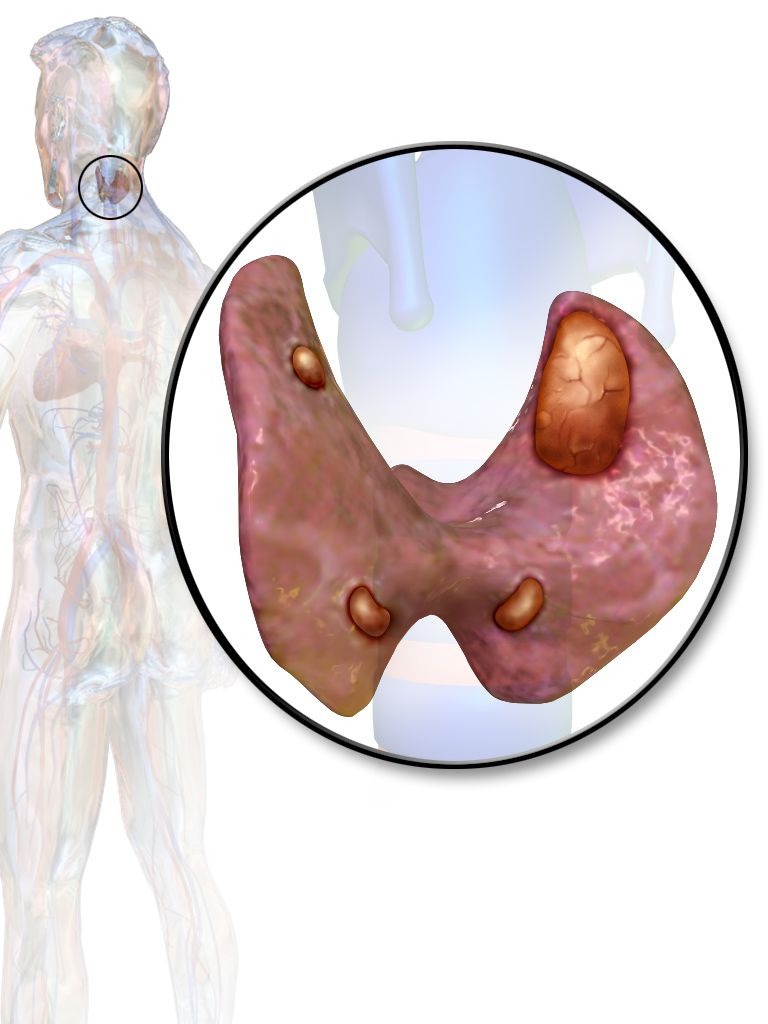
Parathyroid hormone in one paragraph
Parathyroid hormone (PTH) exists to defend the serum ionised calcium. When calcium falls, PTH rises and does three things: it pulls calcium out of bone by activating osteoclasts, it makes the kidney reabsorb calcium while dumping phosphate in the urine, and it activates vitamin D to 1,25-dihydroxycholecalciferol so the gut absorbs more calcium. Remember the reciprocal relationship: PTH drives calcium up and phosphate down. That is why primary hyperparathyroidism shows a high calcium with a low phosphate, and hypoparathyroidism shows a low calcium with a high phosphate.
Reference values you must know cold: total serum calcium roughly 8.5 to 10.5 mg/dL, ionised calcium roughly 4.5 to 5.6 mg/dL (1.12 to 1.4 mmol/L), phosphate 2.5 to 4.5 mg/dL, magnesium 1.8 to 2.4 mg/dL, and intact PTH about 10 to 65 pg/mL. Because roughly 40 percent of serum calcium is bound to albumin and only the ionised fraction is physiologically active, a hypoalbuminaemic patient can look hypocalcaemic while the physiologically active fraction is fine. Correct it: add 0.8 mg/dL to the measured calcium for every 1 g/dL that albumin sits below 4.0 g/dL. Better still, in the critically ill, ask for an ionised calcium.
Hyperparathyroidism and hypercalcaemia: stones, bones, groans and moans
Primary hyperparathyroidism is usually a single benign adenoma secreting PTH autonomously; a minority is hyperplasia, often part of multiple endocrine neoplasia. Secondary hyperparathyroidism is the appropriate response of healthy glands to chronic kidney disease, where phosphate retention and failed vitamin D activation drive calcium down; here calcium is low or low-normal while PTH is high. Tertiary disease is what happens when chronically stimulated glands become autonomous. The other big cause of hypercalcaemia on a med-surg floor is malignancy — PTH-related peptide from squamous tumours, or direct bone lysis in myeloma and breast cancer — and there the PTH is appropriately suppressed.
The classic mnemonic still earns its keep. Stones: nephrolithiasis, polyuria, nephrogenic diabetes insipidus from calcium’s effect on the collecting duct. Bones: bone pain, osteoporosis, pathological fracture. Abdominal groans: anorexia, nausea, constipation, peptic ulcer disease, pancreatitis. Psychic moans: fatigue, depression, confusion, and at high levels stupor and coma. Add muscle weakness with diminished deep tendon reflexes and, on the monitor, a shortened QT interval with bradyarrhythmias. Hypercalcaemia potentiates digoxin, so a patient on digoxin with a rising calcium is at real risk of toxicity.
A hypercalcaemic crisis — calcium above roughly 14 mg/dL with altered mentation — is managed in a fixed order. Volume first: these patients are profoundly dehydrated from calcium-induced polyuria and vomiting, so give isotonic saline at 200 to 300 mL per hour with careful attention to the older heart. Loop diuretics come after the patient is volume replete, and only if fluid overload threatens; giving furosemide to a dry patient worsens everything. Calcitonin 4 units/kg subcutaneously or intramuscularly every 12 hours works within hours but tachyphylaxes in 48. Intravenous bisphosphonates — zoledronic acid 4 mg over 15 minutes, or pamidronate — take two to four days but hold the calcium down; check renal function and dental status first, since osteonecrosis of the jaw is a real if uncommon risk. Denosumab is used when bisphosphonates cannot be. Cinacalcet, a calcimimetic, tricks the parathyroid’s calcium-sensing receptor into reading calcium as high and turns PTH off. Dialysis is the backstop.
Nursing care for chronic hypercalcaemia is mundane and effective: encourage 3 to 4 litres of fluid daily unless contraindicated, mobilise the patient because immobility resorbs bone, add fibre for constipation, screen every medication for thiazides, lithium and calcium-containing antacids, and institute fall precautions. Parathyroidectomy cures primary disease, and the postoperative watch is the mirror image — a sudden drop into hypocalcaemia, sometimes dramatic in “hungry bone syndrome” where demineralised bone soaks up calcium for days.
Hypoparathyroidism and tetany: the emergency you can see at the bedside
The commonest cause of hypoparathyroidism is surgical — the glands removed, devascularised or bruised during thyroidectomy, radical neck dissection or parathyroidectomy. Autoimmune destruction, congenital absence in DiGeorge syndrome, and severe hypomagnesaemia (which blocks PTH secretion and PTH action) are the others. Falling ionised calcium destabilises nerve membranes and lowers the threshold for depolarisation, so the presentation is neuromuscular irritability that escalates.
Early: perioral and fingertip numbness and tingling, muscle cramps, anxiety. Then hyperactive reflexes, carpopedal spasm, and the two named signs — Chvostek sign, a twitch of the ipsilateral facial muscles when you tap the facial nerve just anterior to the earlobe, and Trousseau sign, carpal spasm after inflating a blood-pressure cuff 20 mmHg above systolic for three minutes. Trousseau is the more specific of the two. Late and lethal: laryngospasm with stridor, bronchospasm, seizures, a prolonged QT interval that can degenerate into torsades de pointes, and hypotension with decreased contractility. On the postoperative thyroid patient, a complaint of tingling around the mouth at hour 20 is not anxiety — it is the first rung of a ladder that ends in laryngospasm.
Acute symptomatic hypocalcaemia is treated with IV calcium gluconate, typically 1 to 2 grams diluted in 50 to 100 mL of D5W infused over 10 to 20 minutes, followed by a continuous infusion if symptoms persist, with the patient on continuous cardiac monitoring throughout because rapid administration causes bradycardia and hypotension. Calcium gluconate is preferred peripherally; calcium chloride contains three times the elemental calcium but is a severe vesicant and should go through a central line. Check and replace magnesium — hypocalcaemia that will not correct is hypomagnesaemia until proven otherwise. Never mix calcium with sodium bicarbonate or phosphate in the same line; it precipitates. Long term, patients take oral calcium carbonate or citrate with calcitriol (activated vitamin D, because a patient without PTH cannot activate their own), and increasingly recombinant PTH. Teach seizure precautions, a high-calcium and low-phosphate diet, and the tingling-means-call rule.
| Hypercalcaemia (high PTH or malignancy) | Hypocalcaemia (low PTH) | |
|---|---|---|
| Calcium / phosphate | Calcium high, phosphate low | Calcium low, phosphate high |
| Neuromuscular | Weakness, hyporeflexia, lethargy, confusion | Tingling, cramps, tetany, hyperreflexia, seizures |
| Gastrointestinal | Constipation, nausea, ulcer, pancreatitis | Diarrhoea, abdominal cramping |
| ECG | Shortened QT, bradyarrhythmia, digoxin sensitivity | Prolonged QT, risk of torsades de pointes |
| Airway risk | Low | High — laryngospasm and stridor |
| Emergency drug | Isotonic saline, then calcitonin and a bisphosphonate | IV calcium gluconate; correct magnesium |
Falling calcium is an airway and rhythm emergency — perioral tingling after neck surgery buys the patient a cardiac monitor, an ionised calcium, a magnesium level and calcium gluconate at the ready, not reassurance.
Section 4The Adrenal Glands: Too Much Cortisol, Too Little Cortisol, and the Medulla
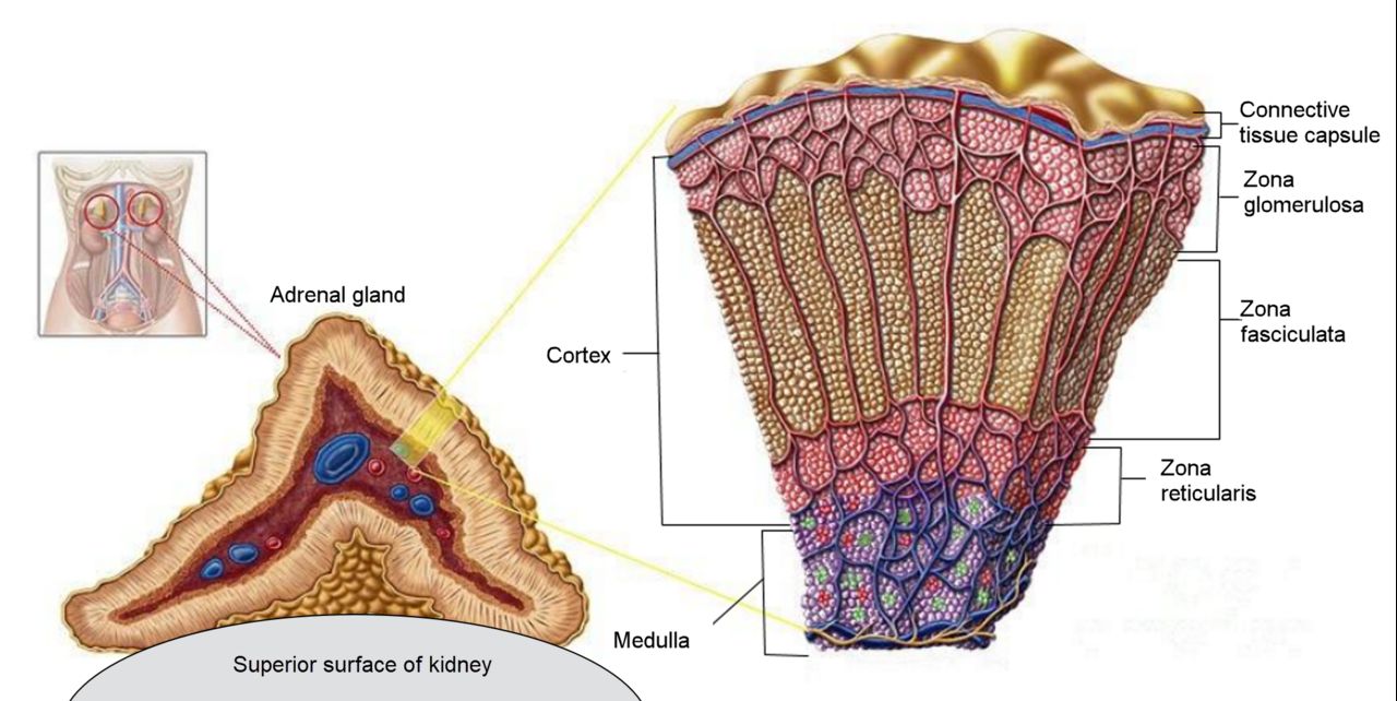
Cortisol excess: Cushing syndrome
Cortisol is a survival hormone: it raises glucose, breaks down protein and fat for fuel, suppresses inflammation and immunity, supports vascular tone, and retains sodium and water while wasting potassium. Give too much for too long and every one of those actions becomes a disease. Central obesity with a buffalo hump and moon face while the arms and legs waste; thin, fragile skin with purple striae wider than a centimetre; easy bruising and terrible wound healing; hyperglycaemia and frank diabetes; osteoporosis with vertebral compression fractures; hypertension; hypokalaemia and metabolic alkalosis; suppressed immunity with masked infection; mood lability, insomnia and psychosis; hirsutism and menstrual irregularity in women. The single most common cause is exogenous — the patient on prednisone for rheumatoid arthritis, transplant or inflammatory bowel disease. Endogenous causes split into ACTH-dependent (a pituitary corticotroph adenoma, which is properly called Cushing disease, and ectopic ACTH from small-cell lung cancer) and ACTH-independent (an adrenal adenoma or carcinoma).
Screening uses any two of three: 24-hour urinary free cortisol, late-night salivary cortisol (which exploits the fact that cortisol should be at its nadir at midnight), and the 1 mg overnight dexamethasone suppression test, where a morning cortisol above 1.8 mcg/dL fails to suppress. Once excess is confirmed, a plasma ACTH localises it: suppressed ACTH means the adrenal is the source; normal or high ACTH means pituitary or ectopic, and high-dose dexamethasone testing plus imaging or inferior petrosal sinus sampling sorts those. Treatment is surgical when there is a tumour — transsphenoidal resection for Cushing disease, adrenalectomy for an adrenal source — with ketoconazole, metyrapone, osilodrostat or mifepristone used medically. For exogenous Cushing, the answer is the lowest effective steroid dose and a slow taper, never an abrupt stop.
Nursing priorities read straight off the pathophysiology. Protect the skin: paper tape, gentle turning, no adhesive removal without an adhesive remover, meticulous pressure-injury prevention. Protect the bones: fall precautions, assistive devices, no independent ambulation until you have assessed proximal muscle strength, and remember a vertebral fracture can present as new back pain with no trauma. Protect against infection: hand hygiene, screening visitors, and a very low threshold for suspecting infection because the usual fever and leucocytosis may be blunted. Monitor glucose, potassium, weight and blood pressure daily. Teach a diet high in protein, calcium and vitamin D, low in sodium and refined carbohydrate. And after adrenalectomy or successful pituitary surgery, the patient swings the other way — they now need glucocorticoid replacement and are at risk of adrenal crisis for months.
Cortisol deficiency: Addison disease and adrenal crisis
Primary adrenal insufficiency, Addison disease, destroys the whole cortex, so the patient loses cortisol and aldosterone and adrenal androgens. Autoimmune adrenalitis is the leading cause in developed countries; tuberculosis, adrenal haemorrhage (including Waterhouse–Friderichsen syndrome in meningococcaemia), metastasis, HIV and drugs such as ketoconazole and etomidate account for the rest. Secondary insufficiency comes from a pituitary that cannot make ACTH — and by far the most common version is iatrogenic, from a patient who has taken more than about 5 mg of prednisone daily for more than three weeks and then stopped or got sick.
The distinction is clinically useful. In primary disease, ACTH is sky-high and its precursor peptide stimulates melanocytes, so the patient has hyperpigmentation of skin creases, scars, gums and the buccal mucosa; aldosterone is absent, so sodium is low, potassium is high, and the patient is salt-craving, orthostatic and volume depleted with a mild metabolic acidosis. In secondary disease there is no hyperpigmentation, and because the renin–angiotensin–aldosterone axis is intact, potassium is usually normal. Both share fatigue that is out of proportion, anorexia, weight loss, nausea, vague abdominal pain, myalgia, hypoglycaemia and hypotension.
Diagnosis: an 8 am serum cortisol below 3 mcg/dL is essentially diagnostic and above 15 to 18 mcg/dL essentially excludes it; anything between goes to a cosyntropin stimulation test, where a peak cortisol below roughly 18 mcg/dL confirms insufficiency. A simultaneous ACTH separates primary from secondary. Maintenance therapy is hydrocortisone 15 to 25 mg daily in divided doses that mimic the diurnal rhythm — the larger portion on waking — plus fludrocortisone 0.05 to 0.2 mg daily for primary disease only, since secondary patients still make aldosterone.
Adrenal crisis is the emergency. Triggers are infection, surgery, trauma, vomiting that prevents oral dosing, and abrupt steroid withdrawal. Presentation is profound hypotension and shock refractory to fluids and pressors, severe weakness, vomiting and abdominal pain that can mimic an acute abdomen, fever, confusion, hyponatraemia, hyperkalaemia and hypoglycaemia. Management: large-bore IV access, hydrocortisone 100 mg IV push immediately followed by 50 mg every 6 hours or a 200 mg per 24-hour infusion, and aggressive volume with isotonic saline — often 1 litre in the first hour — using D5 normal saline if hypoglycaemic. If the diagnosis is not yet made and you must preserve the cosyntropin test, dexamethasone 4 mg IV is given instead because it does not cross-react with the cortisol assay. Treat the hyperkalaemia, monitor the rhythm, find and treat the trigger. Do not delay steroids for a confirmatory test in a shocked patient.
Teaching is what keeps these patients out of your ICU. Never stop the steroid. Wear medical-alert identification. Carry an emergency injection kit of hydrocortisone 100 mg intramuscular and know how to use it — and teach a family member too. Follow sick-day rules: for fever or moderate illness, double or triple the usual oral hydrocortisone dose for two to three days; for vomiting or diarrhoea that prevents keeping tablets down, use the injection and call immediately; for major surgery, expect stress-dose steroids. Weigh daily, report weight loss, dizziness or salt craving, and increase salt in hot weather or with heavy exercise.
The medulla and the mineralocorticoid: pheochromocytoma and hyperaldosteronism
A pheochromocytoma is a catecholamine-secreting tumour of the adrenal medulla. It presents in paroxysms with the classic triad of pounding headache, palpitations and profuse diaphoresis, on a background of hypertension that may be sustained or episodic and is often severe and drug-resistant. Attacks can be provoked by exertion, anaesthesia, certain drugs, and by palpating the abdomen — which is why you do not deep-palpate the abdomen of a patient being worked up for one. Diagnosis is by plasma free metanephrines or 24-hour urinary fractionated metanephrines, then CT or MRI; teach the patient to avoid caffeine, acetaminophen, tricyclics and strenuous activity before the collection, and keep 24-hour urine on ice with the correct preservative.
The surgical rule here is the one students most often reverse: alpha blockade must come before beta blockade. Phenoxybenzamine or doxazosin is started 10 to 14 days preoperatively with liberal salt and fluid intake to re-expand the contracted vascular volume; only once alpha blockade is established is a beta blocker added for reflex tachycardia. Give a beta blocker first and you remove beta-2 mediated vasodilation while leaving alpha-1 vasoconstriction unopposed — a hypertensive crisis. Postoperatively, expect hypotension as the catecholamine surge disappears, and monitor glucose, which often falls.
Primary hyperaldosteronism (Conn syndrome) is an aldosterone-producing adenoma or bilateral hyperplasia and is a common cause of secondary hypertension. Look for hypertension that resists three drugs, hypokalaemia with muscle weakness, cramps and polyuria, and a metabolic alkalosis, with sodium high-normal and no oedema (aldosterone escape). Screen with a plasma aldosterone-to-renin ratio — a high aldosterone with a suppressed renin is the signature. Treatment is adrenalectomy for a unilateral adenoma, or a mineralocorticoid receptor antagonist — spironolactone or eplerenone — for bilateral disease. Monitor potassium closely once the antagonist starts, because it flips the patient toward hyperkalaemia, and warn men that spironolactone causes gynaecomastia.
Delegation across all adrenal disease: assistive personnel may obtain orthostatic vital signs when the RN has judged the patient stable enough, record weights and intake and output, and assist with hygiene. The RN retains the assessment of mentation, the interpretation of orthostatic changes, all steroid teaching including sick-day rules, and every decision about a hypotensive or hypoglycaemic patient.
Cortisol excess is a slow disease of skin, bone, sugar and infection risk; cortisol deficiency is a fast disease of shock, hyponatraemia, hyperkalaemia and hypoglycaemia — and in a shocked patient with suspected adrenal crisis, hydrocortisone and saline come before any confirmatory test.
Section 5The Pituitary: Water, Growth and the Transsphenoidal Patient
SIADH: too much antidiuretic hormone, too much water
Antidiuretic hormone (ADH, vasopressin) is made in the hypothalamus and released from the posterior pituitary; it inserts aquaporin channels in the renal collecting duct so water is reabsorbed. In the syndrome of inappropriate antidiuretic hormone, ADH keeps being released despite a low serum osmolality. Water is retained, the serum is diluted, and the patient becomes hyponatraemic while remaining euvolemic — there is no oedema, because the retained water distributes across all compartments and the mild volume expansion triggers a compensatory natriuresis — suppressed aldosterone plus released natriuretic peptides — that dumps the extra sodium, which is also why the urine sodium is high. Causes cluster: small-cell lung carcinoma (ectopic ADH), central nervous system insult — stroke, meningitis, subarachnoid haemorrhage, head injury, pituitary surgery — pulmonary disease such as pneumonia, and drugs including SSRIs, carbamazepine, oxcarbazepine, cyclophosphamide and thiazides — thiazides strictly produce a clinically identical hyponatraemia rather than true SIADH, and in older adults the combination of a thiazide and an antidepressant is one of the commonest causes you will meet.
The laboratory signature is worth memorising as a set: serum sodium below 135 mEq/L, serum osmolality below 275 mOsm/kg, urine osmolality inappropriately concentrated above 100 and usually above 300 mOsm/kg, urine sodium above 30 to 40 mEq/L, with normal thyroid, adrenal and renal function and no diuretic on board. Uric acid is characteristically low. Clinically, symptoms track the rate of fall more than the absolute number: headache, nausea, anorexia and muscle cramps early; then confusion, lethargy, weight gain without oedema, and hyporeflexia; then seizures, coma and respiratory arrest from cerebral oedema, usually below 120 mEq/L or with a rapid drop.
Management starts with fluid restriction, typically 800 to 1000 mL per 24 hours, counting everything including ice chips, the water with medications and the gravy on the tray. Add salt tablets or a high-sodium diet, sometimes a loop diuretic, and consider a vasopressin receptor antagonist (tolvaptan, conivaptan) or demeclocycline for chronic cases; tolvaptan requires inpatient initiation because it can correct sodium too quickly. For a seizing or comatose patient, hypertonic 3 percent saline is given — commonly 100 mL over 10 minutes, repeatable — through a monitored line. The nursing non-negotiable is the correction rate: raise the serum sodium by no more than about 8 mEq/L in 24 hours in a patient with chronic hyponatraemia. Correcting faster causes osmotic demyelination syndrome, in which the patient improves for a day or two and then develops dysarthria, dysphagia, spastic quadriparesis and locked-in syndrome that is often permanent. Check sodium every 2 to 4 hours during active correction, maintain strict intake and output, weigh daily on the same scale, institute seizure precautions, perform neurological checks every 2 to 4 hours, and provide frequent oral care because thirst under fluid restriction is genuinely miserable.
Diabetes insipidus: too little antidiuretic hormone, too much urine
Diabetes insipidus is the mirror image. In central DI the posterior pituitary does not release ADH — after head trauma, pituitary surgery, tumour, infection or infiltrative disease. In nephrogenic DI the kidney cannot respond to ADH — from lithium (the classic), demeclocycline, chronic hypokalaemia, hypercalcaemia or intrinsic renal disease. Either way, water pours out. Expect polyuria of more than 3 litres per day, often 5 to 20, with urine that is pale and dilute: specific gravity below 1.005 and urine osmolality below 300 mOsm/kg. Serum goes the other way — sodium above 145 mEq/L and serum osmolality above 295 mOsm/kg. The patient has extreme thirst and craves cold water, and as long as they can drink and the thirst mechanism works, they stay compensated. The danger is the patient who cannot drink: the postoperative patient, the intubated patient, the confused older adult. That patient becomes hypernatraemic and hypovolaemic fast, with tachycardia, hypotension, dry mucous membranes, weight loss, and neurological deterioration from cerebral cell shrinkage.
A water-deprivation test with desmopressin distinguishes the types: after deprivation, central DI concentrates the urine when desmopressin is given, nephrogenic DI does not. Copeptin assays are replacing the older test in many centres. Treatment of central DI is desmopressin — intranasal, oral or parenteral — titrated to urine output and serum sodium, with the caution that over-treatment produces iatrogenic SIADH. Nephrogenic DI is treated by removing the offending drug and using a low-sodium, low-protein diet with a thiazide diuretic (paradoxically antidiuretic here) or amiloride for lithium-induced disease.
Nursing care is measurement discipline. Hourly urine output in the acute phase, with a standing rule to report output greater than 200 to 250 mL per hour for two consecutive hours. Specific gravity every void or every shift. Daily weights at the same time on the same scale — a kilogram is a litre. Strict intake and output, serum sodium and osmolality on schedule, free access to water at the bedside for the patient who can drink, and safety measures for the patient with polyuria at night. Assistive personnel can measure and record output, weights and specific gravity; the RN interprets the trend and makes the call.
| SIADH | Diabetes insipidus | |
|---|---|---|
| ADH | Too much | Too little, or kidney cannot respond |
| Urine output | Low, concentrated | Massive, dilute |
| Urine specific gravity | Above 1.030 | Below 1.005 |
| Serum sodium | Low, below 135 mEq/L | High, above 145 mEq/L |
| Serum osmolality | Below 275 mOsm/kg | Above 295 mOsm/kg |
| Weight | Gains, without oedema | Loses |
| Main risk | Cerebral oedema and seizure | Hypovolaemic shock and hypernatraemia |
| Treatment | Fluid restriction; hypertonic saline if seizing; vaptans | Replace water; desmopressin for central; thiazide for nephrogenic |
Tumours, transsphenoidal surgery and hypopituitarism
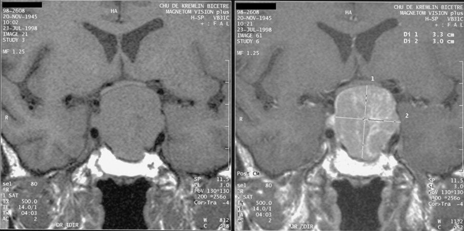
Anterior pituitary tumours are almost always benign adenomas that cause trouble two ways: by secreting a hormone, or by taking up space. Mass effect gives headache and, when the tumour presses upward on the optic chiasm, bitemporal hemianopsia — ask the patient about clipping doorframes or missing cars merging beside them, and perform confrontation visual-field testing on every one of these patients every shift. Prolactinoma is the most common secreting type, causing galactorrhoea, amenorrhoea and infertility in women and erectile dysfunction, low libido and gynaecomastia in men; it is one of the few tumours treated medically first, with the dopamine agonists cabergoline or bromocriptine, which shrink it. Teach patients to take dopamine agonists with food at bedtime and to rise slowly, because orthostatic hypotension and nausea are common.
Growth-hormone excess causes gigantism before the epiphyses fuse and acromegaly after: enlarging hands, feet, jaw and tongue, coarsening features, rings and shoes that no longer fit, deepened voice, arthralgia, carpal tunnel syndrome, sleep apnoea, insulin resistance, hypertension, cardiomyopathy and colon polyps. Diagnosis is an elevated IGF-1 with failure of GH to suppress during an oral glucose tolerance test. Transsphenoidal resection is first-line, with octreotide or lanreotide, pegvisomant or cabergoline as adjuncts. Anticipate a difficult airway from soft-tissue overgrowth and flag it to anaesthesia. Hypopituitarism — from a large tumour, surgery, radiation, Sheehan syndrome after postpartum haemorrhage or pituitary apoplexy — means replacing each deficient axis, and the order matters: always give glucocorticoid before levothyroxine, because thyroid hormone accelerates cortisol clearance and can precipitate adrenal crisis in someone whose ACTH is also gone.
Transsphenoidal hypophysectomy, done through the nose and sphenoid sinus, has a distinctive postoperative plan. Elevate the head of bed 30 degrees. Absolutely no nose blowing, no sneezing with the mouth closed, no straws, no coughing without support, and no straining — give stool softeners and antiemetics preemptively, because a Valsalva can blow open the sellar repair. Expect a nasal drip pad and change it, documenting the amount and character of drainage. Watch for cerebrospinal fluid leak: clear drainage that tests positive for glucose or shows a halo sign on gauze, persistent postnasal drip, a salty or sweet taste, or a severe positional headache. Report it immediately; meningitis is the consequence. Perform oral care with rinses only and no toothbrushing near the incision line for about ten days, and warn the patient that upper-lip and gum numbness may last weeks. Monitor for the triphasic response: diabetes insipidus in the first one to three days as injured cells stop releasing ADH, then SIADH around days five to ten as damaged cells dump their stores, then possibly permanent DI. That means hourly urine output, specific gravity and serial sodium levels are not optional. Finally, monitor vision and mental status, and know that these patients go home on glucocorticoid replacement with full sick-day teaching.
The posterior pituitary is a water switch: SIADH holds water and drowns the brain in dilute sodium, diabetes insipidus dumps water and dries the brain out — and urine specific gravity with a serum sodium tells you in two minutes which one you are looking at.
The chapter in one breath
Endocrine disease is a conversation between the pituitary and a target gland, and reading the pair of hormones tells you which speaker is broken. A high TSH with a low free T4 is a failing thyroid, treated with levothyroxine taken on an empty stomach, started low in older adults and anyone with coronary disease, and rechecked in six to eight weeks. Decompensation runs in two directions: myxedema coma, where hypothermia and hypoventilation kill and hydrocortisone must precede IV thyroid hormone, and thyroid storm, where fever and tachyarrhythmia kill and the sequence is beta blocker, thionamide, then iodine at least an hour later, then steroid, and never aspirin. After thyroidectomy you watch three things — the airway for haematoma, the voice for recurrent laryngeal nerve injury, and the fingertips for hypocalcaemia. Calcium runs opposite to phosphate and opposite to the QT interval; low calcium means tetany, laryngospasm and IV calcium gluconate with a magnesium level. Adrenal disease is cortisol excess that erodes skin, bone and immunity, or cortisol deficiency that presents as refractory shock with low sodium, high potassium and low glucose and is treated with hydrocortisone and saline before any test. In the pituitary, SIADH retains water and dilutes sodium, diabetes insipidus dumps it — and specific gravity settles the question fast.
Words worth knowing
| Term | What it means |
|---|---|
| Negative feedback | The target hormone circulating back to shut off the hypothalamic and pituitary signals that produced it; the basis of every paired endocrine lab. |
| Primary vs secondary disorder | Primary means the target gland is broken (trophic hormone moves opposite to target hormone); secondary means the pituitary or hypothalamus is broken (both move in the same direction). |
| Trophic hormone | A pituitary hormone whose job is to stimulate another gland — TSH, ACTH, FSH, LH. |
| Myxedema | Non-pitting, boggy oedema from glycosaminoglycan deposition in hypothyroidism, classically periorbital and facial with generalised swelling of the hands and feet. The separate entity pretibial myxedema is Graves dermopathy over the shins and occurs in hyperthyroidism. |
| Myxedema coma | Decompensated hypothyroidism with hypothermia, hypoventilation, hyponatraemia, hypoglycaemia and depressed consciousness; a respiratory emergency. |
| Thyroid storm | Life-threatening thyrotoxicosis with fever above 38.5 degrees Celsius, heart rate above 140, delirium and high-output cardiac failure. |
| Thionamide | Methimazole or propylthiouracil; blocks thyroid peroxidase and therefore hormone synthesis. Agranulocytosis is the warning toxicity. |
| Wolff–Chaikoff effect | The transient block of iodide organification and thyroid hormone synthesis caused by a large iodine load; combined with an acute block of hormone release, it is why SSKI works within hours in storm and why the effect escapes after 10 to 14 days. |
| Exophthalmos | Forward protrusion of the globes from retro-orbital tissue expansion in Graves ophthalmopathy; risks corneal drying and ulceration. |
| Recurrent laryngeal nerve | The nerve running beside the thyroid whose injury causes hoarseness unilaterally and airway obstruction bilaterally; assessed by asking the patient to speak. |
| Chvostek sign | Facial twitch on tapping the facial nerve anterior to the earlobe; a sign of hypocalcaemic neuromuscular irritability. |
| Trousseau sign | Carpal spasm after a blood-pressure cuff is held above systolic for three minutes; more specific than Chvostek for hypocalcaemia. |
| Corrected calcium | Measured calcium plus 0.8 mg/dL for every 1 g/dL that albumin falls below 4.0 g/dL; prevents mislabelling a low-albumin patient as hypocalcaemic. |
| Calcimimetic | A drug such as cinacalcet that makes the parathyroid calcium-sensing receptor read calcium as high, turning PTH secretion off. |
| Cosyntropin stimulation test | Synthetic ACTH given IV or IM with cortisol drawn at 30 and 60 minutes; a peak below roughly 18 mcg/dL confirms adrenal insufficiency. |
| Dexamethasone suppression test | 1 mg at 11 pm with an 8 am cortisol; failure to suppress below 1.8 mcg/dL screens positive for Cushing syndrome. |
| Adrenal crisis | Acute cortisol deficiency with refractory hypotension, hyponatraemia, hyperkalaemia and hypoglycaemia; treated with IV hydrocortisone and isotonic saline immediately. |
| Sick-day rules | The patient teaching that doubles or triples oral glucocorticoid during illness and switches to the intramuscular kit if vomiting. |
| Osmotic demyelination syndrome | Irreversible pontine injury from correcting chronic hyponatraemia faster than about 8 mEq/L in 24 hours. |
| Triphasic response | The post-pituitary-surgery sequence of diabetes insipidus, then SIADH, then possible permanent diabetes insipidus. |