Endocrine Disorders: Diabetes Mellitus
Diabetes is the disease you will meet on every unit you ever work, attached to every other diagnosis you have already studied — the heart failure patient, the post-op abdomen, the pneumonia, the pressure injury that will not close. It is also the disease in which nursing judgement is measured in minutes: a capillary glucose of 48 mg/dL, a patient breathing deeply and smelling faintly of acetone, an insulin drip running while the potassium is 3.1 mEq/L. This chapter builds the reasoning from the inside out. First the mechanism — why absent insulin produces ketones and why resistant insulin produces dehydration. Then the assessment and the numbers that define the disease. Then the drugs, organised by what they actually do, because that is what tells you when they cause hypoglycaemia and when they do not. Then the two hyperglycaemic emergencies side by side, with the treatment sequence you will be expected to defend: fluids first, potassium before insulin, insulin last. Then the slow complications and the teaching that prevents them.
By the end of this chapter you can
- Explain how absolute versus relative insulin deficiency produces the differing presentations of type 1 and type 2 diabetes mellitus, including the role of insulin resistance, lipolysis and ketogenesis.
- Apply the four diagnostic criteria for diabetes (A1C, fasting plasma glucose, 2-hour OGTT, random glucose with symptoms) and identify prediabetes from the same panel.
- Perform a focused assessment for hyperglycaemia, hypoglycaemia and the complication surveillance schedule, including monofilament testing and urine albumin-to-creatinine ratio.
- Compare insulins by onset, peak and duration and predict the hypoglycaemia window for each; demonstrate safe mixing, storage and high-alert administration practice.
- Classify non-insulin agents by mechanism — biguanide, sulfonylurea, DPP-4 inhibitor, GLP-1 receptor agonist, SGLT2 inhibitor, thiazolidinedione — and state the monitoring that matters for each.
- Differentiate diabetic ketoacidosis from hyperosmolar hyperglycaemic state by laboratory profile and sequence the treatment: fluids, then potassium, then insulin.
- Recognise and treat hypoglycaemia using the rule of 15, IV dextrose or glucagon, and identify hypoglycaemia unawareness.
- Teach sick-day rules, foot care, injection-site rotation and when to call, and construct an SBAR escalation for a deteriorating patient.
- Modify assessment and glycaemic targets for the older adult, and delegate appropriately within the RN scope.
Section 1Two Ways the Pancreas Fails
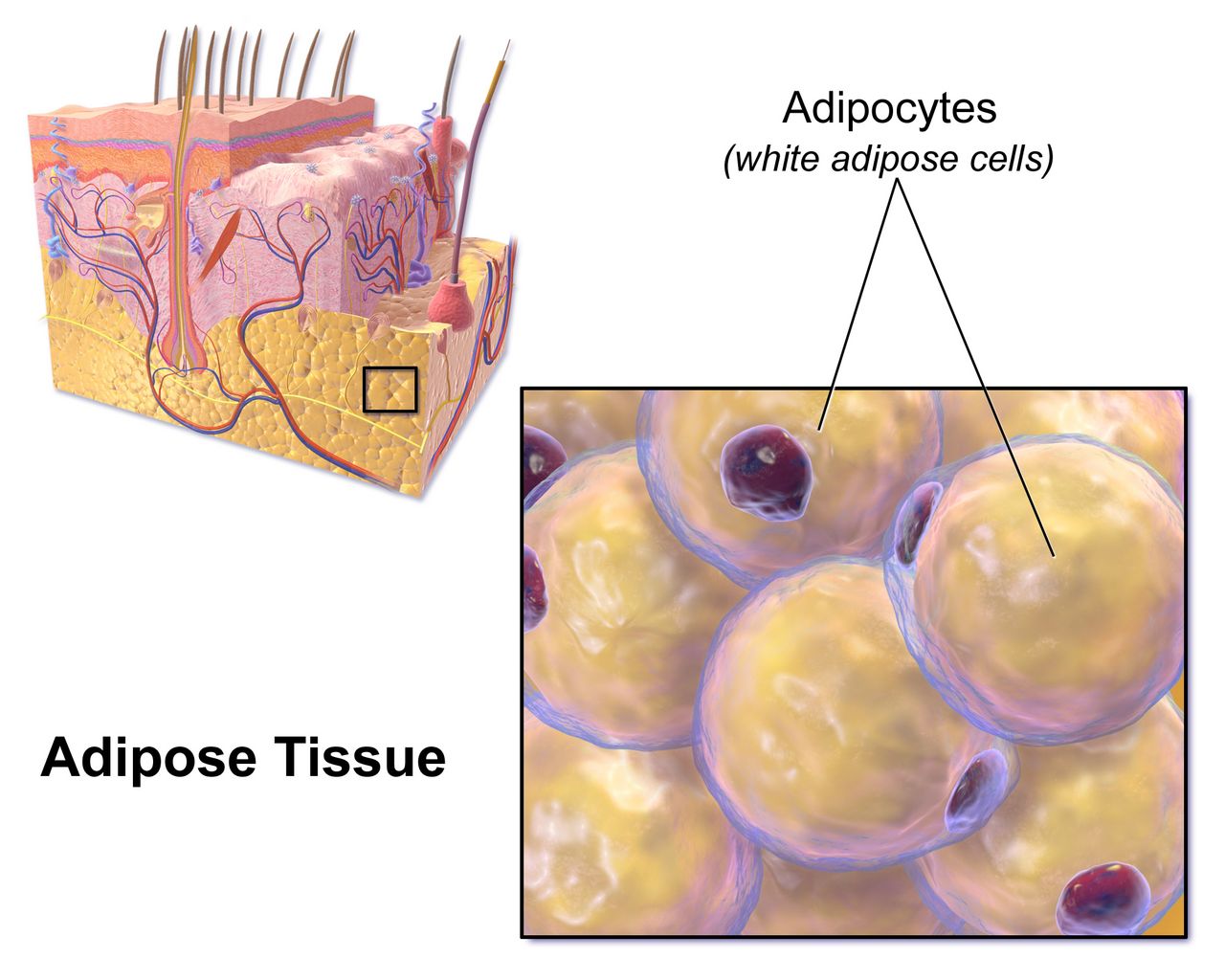
Insulin is the storage signal, and everything follows from that
Beta cells in the islets of Langerhans release insulin when blood glucose rises after a meal. Insulin does three things worth memorising because every sign of diabetes traces back to one of them. It opens GLUT4 transporters in muscle and adipose tissue so glucose leaves the blood and enters cells. It tells the liver to stop making new glucose — to suppress glycogenolysis and gluconeogenesis. And it suppresses lipolysis, keeping fatty acids locked inside adipocytes. Alpha cells in the same islet release glucagon, which does the opposite, and the fed-fasted balance between the two normally keeps plasma glucose in a remarkably narrow band of roughly 70 to 140 mg/dL across a whole day of eating and sleeping.
Remove insulin and all three brakes release at once. Glucose cannot enter muscle, so it accumulates in plasma. The liver, seeing no insulin signal, interprets the situation as starvation and pours out still more glucose. Adipose tissue releases free fatty acids, which the liver converts to ketone bodies — acetoacetate, beta-hydroxybutyrate and acetone — as an alternative fuel. That is the whole of diabetic ketoacidosis in one sentence: the cells are starving in a sea of sugar, and the liver keeps setting the table.
Once plasma glucose exceeds the renal threshold of roughly 180 mg/dL, the proximal tubule cannot reabsorb it all and glucose spills into urine. Glucose is osmotically active, so it drags water and electrolytes with it. This osmotic diuresis is why the classic presentation is polyuria and polydipsia, why the patient is volume depleted before anyone thinks to check a set of orthostatics, and why sodium, potassium, magnesium and phosphate all leave the body through the bladder while the serum values may still look deceptively normal.
Type 1: the beta cells are gone
Type 1 diabetes is autoimmune destruction of beta cells — a T-cell mediated insulitis that unfolds over months to years in a genetically susceptible person, often after a viral trigger. By the time hyperglycaemia appears, roughly 80 to 90 percent of beta-cell mass is already lost. Autoantibodies to glutamic acid decarboxylase 65 (GAD65), islet antigen 2, insulin and zinc transporter 8 are typically detectable, and C-peptide — the fragment cleaved from proinsulin when the body makes its own insulin — is low or undetectable. That C-peptide value is how a laboratory distinguishes a patient making no insulin from one making plenty but using it badly.
Presentation is fast and dramatic: days to a few weeks of polyuria, polydipsia, polyphagia and weight loss, often in a child, adolescent or young adult, and roughly a third of new cases arrive already in ketoacidosis. Because there is no endogenous insulin at all, these patients are ketosis prone and require exogenous insulin from diagnosis, permanently. There is no such thing as skipping the basal dose. A brief “honeymoon phase” of partial remission after starting insulin can fool families into thinking the disease resolved — it has not, and teaching should say so plainly.
Do not assume type 1 means young. Latent autoimmune diabetes in adults (LADA) is a slower autoimmune process presenting in the thirties, forties or later, frequently misclassified as type 2 until oral agents fail unusually quickly. A lean adult whose glucose control deteriorates within a year or two of diagnosis, or who arrives with ketones, deserves antibody and C-peptide testing.
Type 2: the insulin is there and nobody is listening
Type 2 diabetes begins with insulin resistance in muscle, liver and fat, usually driven by visceral adiposity, inactivity and genetics. The pancreas compensates by making more insulin, and for years it succeeds — the patient is hyperinsulinaemic and euglycaemic and has no idea. Over time the beta cells cannot sustain the output, secretion declines, and glucose rises first after meals, then fasting. Because there is still some circulating insulin, lipolysis stays largely suppressed and ketones usually do not accumulate. That single fact is why type 2 patients decompensate into a hyperosmolar, non-ketotic state rather than into ketoacidosis.
Onset is insidious, and the diagnosis is frequently made by a complication rather than a symptom. A patient presents with a non-healing foot ulcer, recurrent vaginal candidiasis, blurred vision, an unexpected myocardial infarction or a routine A1C drawn before surgery. Look for acanthosis nigricans — velvety hyperpigmentation in the axillae and posterior neck — and for the rest of the metabolic syndrome: central obesity, hypertension, elevated triglycerides, low HDL.
Two other categories matter on a medical-surgical floor. Secondary diabetes follows pancreatitis, pancreatectomy, Cushing syndrome, acromegaly, or drugs — glucocorticoids above all, plus thiazides, atypical antipsychotics, tacrolimus and some antiretrovirals. Steroid-induced hyperglycaemia is a daily reality: prednisone given each morning produces a predictable afternoon and evening glucose surge with a relatively normal fasting value, so the insulin adjustment belongs in the middle of the day, not at bedtime. Gestational diabetes is hyperglycaemia first recognised in pregnancy; it usually resolves at delivery but confers a lifetime type 2 risk that warrants screening every one to three years afterwards.
Making the diagnosis
Four tests establish diabetes, and any one of them will do — but in an asymptomatic patient the abnormal result must be confirmed, either by repeating the same test on a new sample or by a second different test from the same sample. A patient with unequivocal hyperglycaemic symptoms and a random glucose of 200 mg/dL or more needs no confirmation. Note that the A1C reflects average glycaemia over roughly the preceding three months, the lifespan of a red cell, and is therefore unreliable in haemoglobinopathies, recent transfusion, haemolysis, iron-deficiency anaemia, pregnancy and advanced chronic kidney disease.
| Test | Normal | Prediabetes | Diabetes |
|---|---|---|---|
| A1C | Below 5.7% | 5.7–6.4% | 6.5% or higher |
| Fasting plasma glucose (no caloric intake 8 h) | Below 100 mg/dL | 100–125 mg/dL (impaired fasting glucose) | 126 mg/dL or higher |
| 2-hour plasma glucose during 75-g OGTT | Below 140 mg/dL | 140–199 mg/dL (impaired glucose tolerance) | 200 mg/dL or higher |
| Random plasma glucose | — | — | 200 mg/dL or higher with classic symptoms or hyperglycaemic crisis |
Screen asymptomatic adults beginning at age 35, and earlier at any age with a BMI of 25 kg/m² or more (23 in Asian Americans) plus one risk factor — first-degree relative with diabetes, high-risk race or ethnicity, history of cardiovascular disease, hypertension, HDL below 35 mg/dL or triglycerides above 250 mg/dL, polycystic ovary syndrome, physical inactivity, acanthosis nigricans, or prior gestational diabetes. Repeat at least every three years if normal, annually in prediabetes. Prediabetes is not a benign label: intensive lifestyle intervention aiming at seven percent weight loss and 150 minutes of weekly activity reduces progression more effectively than metformin does, and it is the single highest-yield teaching conversation in primary care.
Absent insulin means the liver makes ketones and the patient becomes acidotic; deficient-but-present insulin means the patient becomes profoundly dehydrated and hyperosmolar instead — the same hyperglycaemia takes two different exits.
Section 2Assessment, Diagnostics and Surveillance
The history and physical that actually changes management
Start with the glycaemic history, not the diagnosis list. What is the regimen, in units and in times of day — and what does the patient actually take at home, which is frequently a different answer? When was the last dose? Has anyone been checking glucose, how often, and what have the numbers looked like? Any hypoglycaemic episodes, and did the patient feel them coming? Any hospitalisations for high or low sugars? Who fills the prescriptions, and can they afford them? Insulin rationing is a real and lethal cause of ketoacidosis, and patients rarely volunteer it unless asked without judgement.
Symptoms of hyperglycaemia are the three P’s plus fatigue: polyuria, polydipsia, polyphagia, blurred vision from osmotic lens swelling, weight loss, slow wound healing, recurrent yeast or urinary infections. Symptoms of hypoglycaemia divide into two groups you should keep separate in your head because they appear at different thresholds. Autonomic or adrenergic symptoms come first, usually below 70 mg/dL — tremor, palpitations, anxiety, diaphoresis, hunger, pallor. Neuroglycopenic symptoms come as the brain runs out of substrate, usually below 54 mg/dL — confusion, slurred speech, blurred or double vision, incoordination, bizarre behaviour, seizure, coma.
The physical examination is largely a search for complications. Take a supine and standing blood pressure. Inspect the mouth for candidiasis and periodontal disease. Auscultate for carotid bruits. Palpate dorsalis pedis and posterior tibial pulses and note capillary refill and hair distribution on the lower legs. Take both shoes and both socks off — every time, on every diabetic patient, no exceptions — and inspect the plantar surfaces, the heels and between every toe. Examine injection sites for lipohypertrophy, the rubbery subcutaneous thickening that develops where a patient injects repeatedly into the same spot, and which makes insulin absorption erratic and unpredictable.
Glycaemic monitoring: A1C, fingersticks and continuous sensors
The A1C measures the fraction of haemoglobin that has been non-enzymatically glycated, and it correlates to an estimated average glucose: an A1C of 6% corresponds to roughly 126 mg/dL, 7% to about 154, 8% to about 183, 9% to about 212, and 10% to about 240. Check it every three months when therapy has changed or the target is not met, and every six months when the patient is stable at goal. The general adult target is below 7%, but a target is a clinical judgement, not a reflex — below 6.5% for a young patient with long life expectancy and no hypoglycaemia risk, and a looser ceiling of below 8% for a frail elder with limited life expectancy, cognitive impairment, advanced complications or hypoglycaemia unawareness, where the harm of a low sugar plainly outweighs the benefit of a tight one. Read that looser figure as an upper limit to stay under, never as a number to climb to; and in very poor health or at end of life, stop managing to an A1C at all and manage instead to glucose values, avoiding both hypoglycaemia and symptomatic hyperglycaemia.
Capillary glucose targets for most non-pregnant adults are 80 to 130 mg/dL before meals and below 180 mg/dL at the peak, one to two hours after the start of a meal. Continuous glucose monitoring adds a metric worth knowing by name: time in range, the percentage of readings between 70 and 180 mg/dL, with a goal above 70 percent, less than 4 percent of time below 70 mg/dL and less than 1 percent below 54 mg/dL. Sensors report interstitial glucose, which lags plasma by five to fifteen minutes, so when a sensor value and the patient’s symptoms disagree — or when the glucose is falling rapidly — confirm with a fingerstick before you act.
Two patterns of morning hyperglycaemia are commonly confused and are distinguished by a single 0200–0300 glucose check. The dawn phenomenon is a physiological pre-waking surge of growth hormone and cortisol; the 3 a.m. glucose is normal or high and rises further by morning, and the fix is more or later-timed basal insulin. The Somogyi effect is rebound hyperglycaemia after unrecognised nocturnal hypoglycaemia; the 3 a.m. glucose is low, and the fix is less evening insulin or a bedtime snack. Adding insulin to a Somogyi pattern makes the patient worse, which is exactly why the 3 a.m. check is worth the interrupted sleep.
Complication surveillance is a schedule, not an impulse
The chronic complications of diabetes are silent until they are not, so the surveillance calendar is itself a nursing intervention. Ask about it at every encounter and document the gaps — discharge teaching that includes “you are due for your eye exam” is worth more than another lecture on carbohydrates.
| Surveillance | Frequency | Target or action threshold |
|---|---|---|
| A1C | Every 6 months at goal; every 3 months if not | Individualised; below 7% for most adults |
| Dilated retinal examination | Type 2 at diagnosis; type 1 within 5 years of onset; then annually | Refer any retinopathy to ophthalmology |
| Urine albumin-to-creatinine ratio and eGFR | Annually (type 2 from diagnosis; type 1 after 5 years) | Albuminuria at 30 mg/g or higher with hypertension → ACE inhibitor or ARB (monitor potassium and creatinine) |
| Comprehensive foot examination with 10-g monofilament | Annually; visual inspection every visit | Loss of protective sensation → podiatry, protective footwear |
| Blood pressure | Every visit | Below 130/80 mmHg for most |
| Lipid panel | Annually | Moderate-intensity statin ages 40–75; high-intensity with added risk |
| Immunisations | Influenza annually; pneumococcal, hepatitis B, COVID-19 per schedule | Diabetes is a standing indication for all of these |
The 10-gram Semmes-Weinstein monofilament test deserves a specific description because students are often asked to perform it. With the patient’s eyes closed, touch the filament perpendicular to the plantar skin at standard sites until it buckles into a C shape, hold about one second, and ask the patient to say when and where it is felt. Avoid callus, ulcer and scar. Inability to feel the buckling filament at any tested site means loss of protective sensation — that patient can stand on a tack, or wear a shoe with a pebble in it all day, and never know. Everything about their footwear teaching changes from that moment.
Assessment in diabetes is two jobs at once: find today’s glucose problem, and audit the surveillance schedule that prevents the complication you will otherwise meet in five years.
Section 3Insulin and the Non-Insulin Agents
Insulin by onset, peak and duration
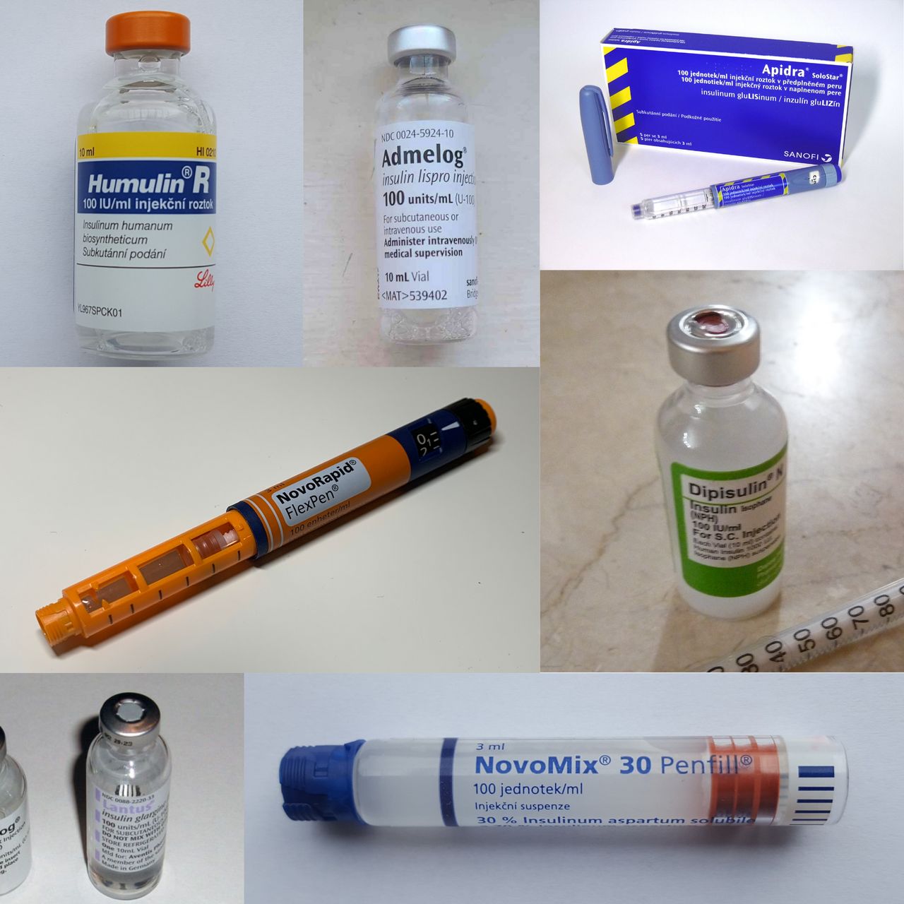
Every insulin question you will ever be asked reduces to a timeline. If you know when the drug peaks, you know when the patient is most likely to go low, when the tray must be in front of them, and when to schedule the fingerstick. Memorise the table below as a shape, not as trivia.
| Class and examples | Onset | Peak | Duration | Nursing timing |
|---|---|---|---|---|
| Rapid-acting: lispro, aspart, glulisine | 10–30 min | 30 min–3 h | 3–5 h | Give 0–15 min before eating; tray must be present. Never give if the meal may not arrive. |
| Ultra-rapid: faster aspart, inhaled human insulin | Within 15 min (inhaled about 12 min) | About 30–60 min | About 2.5–3 h | Inhaled form is contraindicated in asthma and COPD (bronchospasm). |
| Short-acting: regular (U-100) | 30 min–1 h | 2–4 h | 5–8 h | Give 30 min before the meal. The only insulin given IV. |
| Intermediate: NPH (isophane) | 1–2 h | 4–12 h | 12–18 h | Cloudy; roll gently to resuspend. The 4–12 h peak is the classic nocturnal hypoglycaemia window. |
| Long-acting: glargine U-100, detemir | 2–4 h (detemir 1–3 h) | No pronounced peak | Up to 24 h (detemir often twice daily) | Same time daily. Do not mix with any other insulin in a syringe. |
| Ultra-long: degludec, glargine U-300 | About 1–6 h | No peak | Beyond 24 h (degludec beyond 42 h) | Flexible dosing time; steady state takes 3–4 days, so do not chase numbers daily. |
Mixing rules are narrow and absolute. Only NPH and regular are mixed in the same syringe: inject air into the NPH (cloudy) vial, inject air into the regular (clear) vial, then draw up the regular first — clear before cloudy, so that no cloudy NPH contaminates the clear vial. Administer within five minutes of mixing. Glargine, detemir and degludec are never mixed with anything and never diluted. Premixed products such as 70/30 NPH-regular or 75/25 lispro protamine-lispro carry the peak profile of their intermediate component and demand a fixed, reliable meal schedule.
Insulin is a high-alert drug and should be handled like one
Insulin appears near the top of every list of medications most frequently involved in fatal error. Use an insulin syringe only — never a tuberculin or standard syringe — and match the syringe to the concentration. Never write or accept the abbreviation “U” for units; “10U” has been read as 100. Perform an independent double check of the drug, concentration, dose and pump settings with a second nurse per facility policy, particularly for any insulin infusion, any paediatric dose and any concentrated product (U-200 lispro, U-300 glargine, U-500 regular).
Insulin pens are single-patient devices, period. Even with the needle changed, biological material can be aspirated back into the cartridge, and pens have transmitted bloodborne infection between patients. Label the pen with the patient name and keep it in that patient’s drawer. Prime with two units before each injection to clear air, inject at a 90-degree angle, and count to ten before withdrawing the needle so the full dose is delivered rather than tracking back out.
Absorption varies by site — abdomen is fastest, then upper arm, then thigh, then buttock — so teach the patient to use one anatomic region consistently for a given injection and to rotate systematically within it, about an inch apart, avoiding a two-inch radius around the umbilicus. Exercise of a limb, heat, massage and hot showers all accelerate absorption from that site. Unopened vials and pens are refrigerated until the printed expiry; once in use, most products are kept at room temperature and discarded after 28 days (some, including detemir and degludec, allow longer — check the package). Never freeze insulin, never use it if it is discoloured, clumped or frosted-looking, and do not inject cold insulin, which stings.
Non-insulin agents, grouped by what they do
Whether a drug causes hypoglycaemia on its own is entirely a function of mechanism. Agents that force insulin release regardless of glucose — sulfonylureas and meglitinides — do. Agents that improve sensitivity, slow absorption, promote glucose loss in urine or act only in the presence of a rising glucose generally do not, unless combined with insulin or a secretagogue.
| Class (examples) | Mechanism | Key adverse effects | Nursing implications |
|---|---|---|---|
| Biguanide: metformin | Reduces hepatic glucose output; improves insulin sensitivity | GI upset, metallic taste, vitamin B12 deficiency, rare lactic acidosis | First-line in type 2. Take with food; extended release at night. Contraindicated if eGFR below 30 mL/min/1.73 m². Withhold at the time of iodinated contrast when eGFR is 30–60 or with acute kidney injury and restart after 48 h with stable renal function. No hypoglycaemia alone. |
| Sulfonylureas: glipizide, glimepiride, glyburide | Close beta-cell potassium channels, forcing insulin release | Hypoglycaemia, weight gain, disulfiram-like reaction with alcohol | Give about 30 min before a meal; never on an empty, skipped-meal day. Long duration means prolonged hypoglycaemia requiring admission. Avoid glyburide in older adults (Beers criteria). |
| Meglitinides: repaglinide, nateglinide | Same channel, rapid on and off | Hypoglycaemia (shorter) | Take with each meal; skip the dose if the meal is skipped. |
| Thiazolidinedione: pioglitazone | PPAR-gamma agonist; increases peripheral insulin sensitivity | Fluid retention, weight gain, heart failure exacerbation, fractures, macular oedema | Contraindicated in NYHA class III–IV heart failure. Daily weights; report a gain above 3 lb in a day or oedema and dyspnoea. Full effect takes 4–12 weeks. |
| DPP-4 inhibitors: sitagliptin, linagliptin, saxagliptin | Block degradation of endogenous incretins | Arthralgia, pancreatitis, urticaria; heart failure hospitalisation (saxagliptin, alogliptin) | Weight neutral, oral, modest A1C effect. Renal dose adjustment for most except linagliptin. |
| GLP-1 receptor agonists: semaglutide, liraglutide, dulaglutide, exenatide (tirzepatide is dual GIP/GLP-1) | Glucose-dependent insulin release, suppressed glucagon, delayed gastric emptying, satiety | Nausea, vomiting, pancreatitis, gallstones; thyroid C-cell tumours in rodents | Weight loss and cardiovascular benefit. Contraindicated with personal or family history of medullary thyroid carcinoma or MEN 2. Titrate slowly; small low-fat meals. Delayed emptying raises aspiration risk — anaesthesia must know before any sedation or surgery. |
| SGLT2 inhibitors: empagliflozin, dapagliflozin, canagliflozin | Block renal glucose reabsorption; glucose exits in urine | Genital mycotic and urinary infection, volume depletion and orthostasis, euglycaemic DKA, rare Fournier gangrene | Cardiovascular and kidney protection independent of glucose. Teach perineal hygiene and fluid intake. Hold 3–4 days before surgery or during acute illness. Check ketones for DKA symptoms even when glucose is near normal. |
| Alpha-glucosidase inhibitors: acarbose, miglitol | Delay intestinal carbohydrate breakdown | Flatulence, bloating, diarrhoea | Take with the first bite of each meal. If hypoglycaemia occurs on combined therapy, treat with glucose or dextrose, not sucrose — table sugar cannot be split. |
| Amylin analogue: pramlintide | Slows gastric emptying, suppresses glucagon, increases satiety | Severe hypoglycaemia with insulin, nausea | Separate syringe from insulin, separate site. Mealtime insulin is reduced by 50% when started. |
Glycaemic management in the hospitalised patient
Inpatient targets are deliberately looser than outpatient ones because the hospitalised patient eats unpredictably, receives steroids, goes NPO for procedures and has less physiological reserve for a low. Aim for 140 to 180 mg/dL for most critically ill and non-critically ill inpatients; a tighter 110 to 140 band is reasonable only for selected patients when it can be achieved without hypoglycaemia. Below 100 mg/dL, reassess the regimen; below 70, treat and change something.
A basal-bolus-correction regimen — long-acting insulin for background needs, rapid-acting insulin with meals, and a correction scale for the gap — is superior to sliding-scale insulin used alone. Sliding scale alone is reactive: it treats a high number after it has already happened, produces rollercoaster control, and is specifically inappropriate as sole therapy in type 1 diabetes. Time your fingersticks to the regimen: before meals and at bedtime for the eating patient, every four to six hours for the NPO patient, and hourly for anyone on an insulin infusion.
Watch the situations that predictably destabilise glucose: enteral feeding started or interrupted (an abrupt tube-feed hold with prandial coverage already given is a classic hypoglycaemic event), parenteral nutrition, glucocorticoids, octreotide, vasopressors, dialysis and any acute illness. And know that hypoglycaemia in a hospitalised patient is a reportable safety event in most facilities, not a private nursing footnote.
Know when each insulin peaks and you know when your patient will crash; know each oral agent’s mechanism and you know whether it can cause a low at all.
Section 4The Acute Crises: Hypoglycaemia, DKA and HHS
Hypoglycaemia is the emergency that kills first
Between a glucose of 40 and a glucose of 400, treat the 40. Hyperglycaemia damages the patient over hours to years; hypoglycaemia damages the brain in minutes. Level 1 hypoglycaemia is a glucose below 70 mg/dL, level 2 is below 54 mg/dL (clinically significant neuroglycopenia), and level 3 is severe hypoglycaemia of any value causing cognitive impairment requiring another person’s assistance.
For the conscious patient who can swallow safely, use the rule of 15: give 15 grams of fast-acting carbohydrate, wait 15 minutes, recheck. Fifteen grams is four glucose tablets, 4 ounces (half a cup) of juice or regular soda, one tablespoon of honey or sugar, or 8 ounces of milk. Repeat the cycle until the glucose is above 70 mg/dL, then give a snack containing protein and complex carbohydrate if the next meal is more than an hour away. Avoid chocolate, ice cream and peanut butter as the initial treatment — fat slows gastric emptying and delays the rise. Never put food or fluid in the mouth of a patient who is not fully awake and able to protect the airway.
For the unresponsive patient, the patient having a seizure, or anyone NPO or unable to swallow: if IV access exists, give 25 to 50 mL of 50% dextrose (12.5 to 25 g) by slow IV push per protocol, then start or resume a dextrose-containing infusion, because a D50 push wears off quickly. If there is no IV access, give glucagon 1 mg intramuscularly or subcutaneously, or 3 mg intranasally, and turn the patient onto their side — glucagon frequently causes vomiting. Glucagon mobilises hepatic glycogen, so it works poorly in the malnourished, the alcohol-intoxicated and anyone with depleted stores; those patients need dextrose. Recheck in 15 minutes, feed as soon as the patient is alert and safe, then investigate the cause. A sulfonylurea-induced or long-acting-insulin-induced low will relapse: that patient stays, gets a dextrose infusion and serial checks, and does not go home from the emergency department.
Hypoglycaemia unawareness is the loss of adrenergic warning symptoms, seen with autonomic neuropathy, long-duration diabetes, repeated lows and beta-blocker therapy. Beta blockers blunt tremor and tachycardia but not diaphoresis — so in a beta-blocked patient, sudden unexplained sweating is your warning sign. Treatment for unawareness is deliberately relaxing targets and scrupulously avoiding lows for several weeks, which restores symptom perception.
DKA and HHS side by side
Both are hyperglycaemic emergencies, both are usually precipitated by infection, non-adherence or a new stressor such as myocardial infarction, stroke, pancreatitis, trauma, surgery or a new drug. The difference is whether enough insulin remains to suppress lipolysis.
| Feature | Diabetic ketoacidosis | Hyperosmolar hyperglycaemic state |
|---|---|---|
| Typical patient | Type 1 (also type 2 under severe stress); often younger | Type 2, often older, often with limited access to water |
| Onset | Hours to 1–2 days | Days to weeks |
| Plasma glucose | Usually above 250 mg/dL (may be near normal in euglycaemic DKA) | Usually above 600 mg/dL, often above 1000 |
| Arterial pH | Below 7.30 (severe below 7.00) | Above 7.30 |
| Serum bicarbonate | Below 18 mEq/L (severe below 10) | Above 18 mEq/L |
| Ketones (serum beta-hydroxybutyrate) | Positive; 3.0 mmol/L or higher | Absent or small |
| Anion gap | Elevated, above 10–12 mEq/L | Normal or mildly elevated |
| Effective serum osmolality | Variable, often below 320 mOsm/kg | Above 320 mOsm/kg |
| Fluid deficit | About 6 L (roughly 100 mL/kg) | About 9–12 L (roughly 100–200 mL/kg) |
| Distinctive findings | Kussmaul respirations, fruity acetone breath, abdominal pain, nausea and vomiting | Profound dehydration, altered mental status, focal neurologic deficits, seizures; no Kussmaul, no acetone breath |
| Mortality | Roughly 1–5% | Roughly 5–20% |
Two calculations belong in your pocket. Anion gap equals sodium minus the sum of chloride and bicarbonate; it is the metric of ketoacid accumulation and its closure — not the glucose — tells you the DKA is resolving. Effective serum osmolality equals two times the sodium plus glucose divided by 18; above 320 mOsm/kg it correlates with the obtundation of HHS, and a patient who is not fully alert with an osmolality below 320 needs a hunt for another cause of altered mentation. Remember also that hyperglycaemia dilutes serum sodium: add roughly 1.6 mEq/L to the measured sodium for every 100 mg/dL of glucose above 100 to get the corrected value that guides fluid choice.
The treatment sequence — fluids, potassium, insulin, and dextrose
Fluids first. Start isotonic 0.9% sodium chloride at roughly 15 to 20 mL/kg in the first hour (commonly 1 to 1.5 L in an adult), guided by haemodynamics and cardiac and renal status. After the initial resuscitation, the fluid is chosen by the corrected sodium: if it is normal or high, switch to 0.45% sodium chloride at 250 to 500 mL/h; if it is low, continue 0.9%. Volume alone lowers glucose substantially by dilution and restored renal perfusion, and it is the intervention that reverses the shock physiology. Monitor intake and output hourly, and in the elderly or anyone with heart failure or chronic kidney disease, watch for the crackles and jugular distension of overshoot.
Potassium before insulin. This is the sentence to carry out of this chapter. Total body potassium is severely depleted in both crises — on the order of 3 to 5 mEq/kg — even though the serum value on arrival is often normal or high, because acidosis and insulin deficiency have shifted potassium out of cells. Insulin drives potassium straight back in, and if you start the drip on a hypokalaemic patient you can precipitate lethal arrhythmia and respiratory muscle weakness. The rule: if serum potassium is below 3.3 mEq/L, hold the insulin and replace potassium (about 20 to 30 mEq/h) until it exceeds 3.3. If potassium is 3.3 to 5.2, run insulin and add 20 to 30 mEq of potassium to each litre of fluid to keep it in the 4 to 5 range. If it is above 5.2, withhold potassium, start insulin, and recheck in two hours. Confirm urine output before giving potassium, and keep the patient on continuous cardiac monitoring throughout.
Then insulin. Regular insulin is the only insulin given intravenously: a continuous infusion at 0.1 unit/kg/h, with or without an initial 0.1 unit/kg bolus (0.14 unit/kg/h if no bolus is used). Prime the tubing with the insulin solution before connecting it, because insulin adsorbs to plastic and the first millilitres are effectively wasted. Aim for a glucose fall of 50 to 75 mg/dL per hour; a faster drop risks cerebral oedema, particularly in children and adolescents, and a plateau usually means the fluid or insulin rate is inadequate. In HHS, insulin doses are lower and fluid resuscitation is proportionally more important.
Then dextrose. When the glucose reaches about 200 mg/dL in DKA, or about 250 to 300 mg/dL in HHS, change the fluid to one containing 5% dextrose and reduce the insulin infusion to roughly 0.02 to 0.05 unit/kg/h. This looks paradoxical to students — giving sugar to a hyperglycaemic patient — but the insulin must keep running to close the anion gap and stop ketogenesis, and dextrose is what makes that safe. DKA is resolved when the glucose is below 200 mg/dL plus two of: bicarbonate 15 mEq/L or higher, venous pH above 7.30, anion gap 12 mEq/L or less. Bicarbonate therapy is reserved for a pH below 6.9. Phosphate is replaced only for a level below 1.0 mg/dL with cardiac dysfunction, respiratory depression or haemolytic anaemia.
What you monitor and what makes you call
During active treatment expect hourly capillary glucose, basic metabolic panel every two to four hours, continuous cardiac monitoring, strict hourly intake and output, and neurologic checks. Falling mental status during treatment — especially in a young patient whose numbers are improving — is cerebral oedema until proven otherwise: headache, vomiting, bradycardia with rising blood pressure, then deterioration. Stop and escalate immediately.
Do not forget euglycaemic ketoacidosis. Patients taking an SGLT2 inhibitor, pregnant patients, and those who have been vomiting or eating nothing can be fully ketoacidotic with a glucose under 250 mg/dL. If the patient is nauseated, tachypnoeic and acidotic, check ketones and a gas regardless of how reassuring the glucose looks. And in every case, find the precipitant: culture, chest film, ECG, medication review. Treating the DKA without treating the pyelonephritis that caused it simply buys you a second admission.
Here is the escalation you should be able to give without notes. S: “This is Priya, RN, on 5-West with Mr. Alvarez in 512, a 24-year-old with type 1 diabetes admitted with vomiting.” B: “He was admitted this morning; he ran out of insulin four days ago.” A: “In the last hour his respirations have gone from 18 to 32 and deep, he is drowsy, his glucose is 486, his breath smells fruity and his blood pressure has dropped from 118/70 to 92/54 with a heart rate of 128.” R: “I think he is in DKA. I need you to see him now. I would like a stat basic metabolic panel, serum beta-hydroxybutyrate, venous blood gas, magnesium and phosphorus, a large-bore IV with isotonic fluid, and orders for an insulin infusion and cardiac monitoring.”
Fluids first, potassium before insulin, insulin next, dextrose when the glucose reaches 200 — and the anion gap, not the glucose, tells you when the ketoacidosis is actually over.
Section 5Chronic Complications, Teaching and Discharge
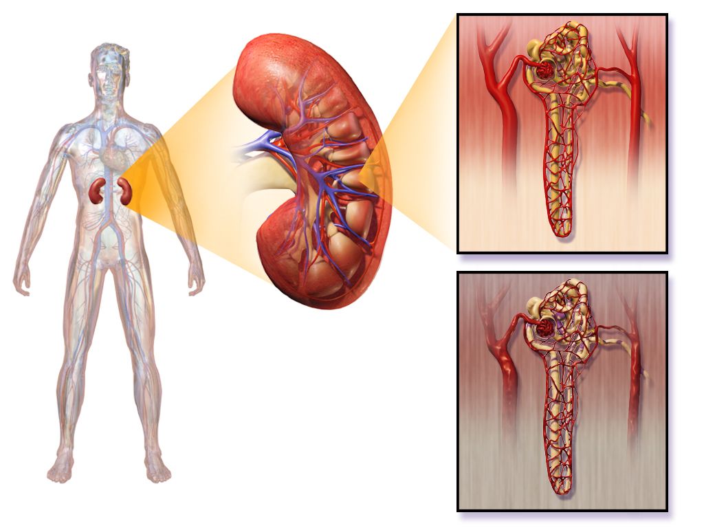
Microvascular disease: eyes, kidneys, nerves
Chronic hyperglycaemia glycates proteins and thickens basement membranes throughout the microcirculation. In the retina this produces diabetic retinopathy — non-proliferative changes of microaneurysms, dot-and-blot haemorrhages and hard exudates, progressing to proliferative retinopathy with fragile neovascular vessels that bleed into the vitreous or drag the retina off. It is asymptomatic until it is severe, which is the entire argument for the annual dilated examination. Sudden painless vision loss, a shower of new floaters or a “curtain” across the field is a same-day ophthalmology emergency. Diabetic macular oedema is the more common cause of gradual central vision loss and is treated with intravitreal anti-VEGF injections.
Diabetic kidney disease begins with hyperfiltration, then persistent albuminuria at a urine albumin-to-creatinine ratio of 30 mg/g or higher, then declining eGFR. An ACE inhibitor or ARB is indicated for albuminuria with hypertension and slows progression by reducing intraglomerular pressure; monitor potassium and creatinine after starting or increasing the dose, and expect a modest creatinine rise that is acceptable. SGLT2 inhibitors and, increasingly, non-steroidal mineralocorticoid antagonists add further renal protection. Blood pressure control below 130/80 mmHg matters at least as much as glucose control here. As eGFR falls, insulin clearance falls with it and insulin requirements paradoxically decrease — a patient with worsening kidney function who suddenly starts running low is telling you something about their kidneys, not their diet.
Diabetic neuropathy is most often a distal symmetric polyneuropathy in a stocking-glove distribution: numbness, tingling, burning and lancinating pain that is worse at night, with loss of vibratory sense and ankle reflexes early. Pharmacological options include pregabalin, gabapentin, duloxetine and amitriptyline; opioids are not first-line. Autonomic neuropathy is the quieter and more dangerous form — orthostatic hypotension, resting tachycardia, silent myocardial ischaemia, gastroparesis, neurogenic bladder with retention and overflow, erectile dysfunction, and hypoglycaemia unawareness. Gastroparesis produces early satiety, bloating and wildly erratic post-meal glucose; manage with small, frequent, low-fat, low-fibre meals, and note that metoclopramide carries a boxed warning for tardive dyskinesia and is limited to under 12 weeks.
Macrovascular disease and the diabetic foot
Atherosclerotic cardiovascular disease is the leading cause of death in diabetes, and diabetes is a coronary risk equivalent. Because of autonomic neuropathy, myocardial infarction may present without chest pain — as dyspnoea, fatigue, nausea, confusion or simply unexplained hyperglycaemia. Take “I just feel off” in a diabetic patient as seriously as crushing substernal pain in anyone else and obtain an ECG. Peripheral arterial disease produces claudication, diminished pulses, hair loss, dependent rubor and cool, shiny, thin skin; cerebrovascular disease produces stroke, which Chapter 3 takes up in detail.
The diabetic foot ulcer is where neuropathy, ischaemia and immune dysfunction meet, and it precedes the majority of non-traumatic lower-limb amputations. Neuropathy removes the pain that would otherwise stop the patient from walking on a wound; motor neuropathy deforms the arch and toes so pressure concentrates under the metatarsal heads; ischaemia starves the healing; hyperglycaemia impairs neutrophil function. Assessment of any ulcer records site, size in centimetres, depth, undermining, wound bed appearance, exudate, odour, surrounding callus or cellulitis, and whether bone is palpable on gentle probing — a positive probe-to-bone strongly suggests osteomyelitis. Management combines pressure offloading, debridement, moist wound care, targeted antibiotics for infection and vascular assessment. Note that a warm, red, swollen, relatively painless foot in a neuropathic patient may be Charcot neuroarthropathy rather than infection, and it requires immediate immobilisation to prevent midfoot collapse.
Foot teaching is concrete and non-negotiable. Inspect both feet daily, using a mirror or a family member for the soles. Wash daily in lukewarm water tested with a thermometer or the elbow — never the foot — and dry carefully between the toes. Moisturise the tops and bottoms but not between the toes. Cut nails straight across, or have podiatry do it. Never use over-the-counter corn removers, razor blades or heating pads. Never go barefoot, indoors or out. Shake out every shoe before putting it on. Break in new shoes 1 to 2 hours a day. Wear seamless, non-constricting socks. Report any blister, cut, redness, callus, ingrown nail or colour change the day it appears — not at the next scheduled visit.
Self-management teaching and sick-day rules
Discharge teaching is prioritised by what will kill the patient soonest if missed: hypoglycaemia recognition and treatment first, insulin administration technique second, sick-day rules third, then foot care, monitoring, nutrition and follow-up. Use teach-back for every psychomotor skill. Watch the patient draw up and inject their own dose before they leave, and watch the caregiver do it too. Ask directly about cost, refrigeration, vision, dexterity, literacy and transport, because the most elegant regimen fails against a broken refrigerator or a $300 copay.
Nutrition teaching centres on consistent carbohydrate intake, portion awareness (the plate method: half non-starchy vegetables, one quarter lean protein, one quarter starch) and carbohydrate counting for those on prandial insulin, where a typical starting ratio is one unit per 15 grams. Emphasise fibre, whole grains and limited added sugar and saturated fat. Alcohol inhibits hepatic gluconeogenesis and can cause delayed hypoglycaemia many hours later — teach patients to drink only with food, in moderation, and to check glucose before bed. Physical activity of 150 minutes per week improves insulin sensitivity for up to 48 hours; teach patients on insulin or secretagogues to check before, sometimes during and after exercise, to carry fast carbohydrate, and to avoid injecting into a limb about to be exercised. Do not exercise if the glucose is above 250 mg/dL with ketones present — exercise will worsen ketosis.
Sick-day rules prevent most avoidable hyperglycaemic crises, and they are counterintuitive enough that they must be taught explicitly and rehearsed. Illness raises counter-regulatory hormones, so glucose and insulin needs go up even when the patient is eating less.
| Sick day | What the patient does |
|---|---|
| Insulin | Never stop taking insulin, even when not eating. Basal continues. Doses often need to increase. |
| Glucose checks | Every 3–4 hours, day and night. |
| Ketone checks | Every 3–4 hours in type 1, and any time glucose exceeds 240 mg/dL or vomiting occurs. |
| Fluids | About 8 oz of fluid every hour while awake — sugar-free if glucose is high; carbohydrate-containing (broth, juice, regular soda, about 15 g per hour) if unable to eat solid food. |
| Food | Try to maintain usual carbohydrate intake in small, soft, frequent amounts. |
| Call the provider | Vomiting or diarrhoea persisting beyond 4 hours or unable to keep fluids down; glucose above 240 mg/dL on two consecutive checks with moderate or large ketones; fever above 101.5 °F not settling; rapid breathing, chest pain, weight loss, drowsiness or confusion; illness lasting beyond 1–2 days. |
The older adult, delegation and the red flags
Older adults present differently and are more fragile in both directions. Hypoglycaemia in an elder rarely looks like tremor and diaphoresis — it looks like a fall, a new confusion, slurred speech mistaken for stroke, or a family member saying “she is just not herself.” Check a glucose on every older adult with altered mental status, every time. Hyperglycaemia likewise presents atypically as new incontinence, delirium, weakness or dehydration, and because the thirst mechanism blunts with age and mobility may prevent reaching a glass of water, HHS develops quietly over days. Renal clearance declines, so sulfonylureas and renally cleared insulins accumulate; glyburide is on the Beers list and should be avoided. Glycaemic targets loosen deliberately — an A1C below 7.5% for a healthy older adult and below 8.0% with multiple comorbidities or cognitive impairment — upper limits, not goals to drift up to — while in very poor health or at end of life there is no A1C target at all, only glucose values managed to avoid hypoglycaemia and symptomatic hyperglycaemia. Polypharmacy, vision loss, arthritic hands and cost all bear directly on whether a regimen can be executed at home.
Delegation follows one principle: the nursing process cannot be delegated. Assessment, teaching, evaluation, clinical judgement and any unstable patient remain with the RN. Assistive personnel may, within facility policy and training, obtain point-of-care capillary glucose readings, record intake and output, assist with meals and positioning, obtain vital signs on stable patients, and report observations such as a reddened heel or an untouched tray — and they must be told explicitly which values to report immediately and to whom. Insulin administration, insulin infusion titration, evaluating whether a hypoglycaemic episode has resolved, initial teaching of injection technique, and the assessment of a new foot ulcer are RN responsibilities. A licensed practical or vocational nurse may administer subcutaneous insulin and reinforce previously taught content within state scope and policy, but the initial assessment, the initial teaching and the unstable patient stay with the registered nurse.
| Red flag | What it may mean | First action |
|---|---|---|
| Glucose below 54 mg/dL, or any low with confusion | Severe hypoglycaemia | Treat immediately — oral carbohydrate if alert and swallowing, otherwise IV dextrose or glucagon; recheck in 15 min; stay with the patient |
| Deep rapid respirations with fruity breath, vomiting, abdominal pain | DKA | Glucose, ketones, gas and metabolic panel; isotonic fluid; cardiac monitoring; escalate now |
| Glucose above 600 mg/dL with drowsiness or focal deficits in an older adult | HHS | Airway and neuro check, large-bore access, isotonic fluid, escalate; look for the infection that caused it |
| Falling level of consciousness during DKA treatment, with headache and vomiting | Cerebral oedema | Stop, escalate immediately; slow the correction; neurologic checks |
| Nausea, dyspnoea, acidosis with a near-normal glucose on an SGLT2 inhibitor | Euglycaemic DKA | Check ketones and a gas anyway; hold the agent; escalate |
| Fever, erythema, purulent or malodorous foot wound, probe-to-bone | Deep infection or osteomyelitis | Cultures, vascular and wound assessment, offloading, urgent provider notification |
| Sudden painless vision loss or a shower of floaters | Vitreous haemorrhage or retinal detachment | Same-day ophthalmology referral |
| Unexplained hyperglycaemia, fatigue or dyspnoea in a long-standing diabetic | Silent myocardial infarction | ECG and troponin; do not wait for chest pain |
The complications of diabetes are prevented at the bedside by three unglamorous habits: taking the socks off, checking a glucose on every confused patient, and making sure the patient can actually perform at home the regimen you documented as taught.
The chapter in one breath
Diabetes is one hyperglycaemia with two mechanisms. Type 1 is autoimmune destruction of beta cells — absolute deficiency, low C-peptide, ketosis prone, insulin required forever. Type 2 is insulin resistance followed by beta-cell exhaustion — insidious onset, enough residual insulin to suppress ketogenesis, so decompensation runs toward hyperosmolarity instead. Diagnosis rests on an A1C of 6.5% or higher, a fasting glucose of 126 mg/dL or higher, a 2-hour OGTT value of 200 mg/dL or higher, or a random 200 with symptoms; prediabetes occupies the band just below each. Management is glucose targets individualised to the patient, insulin chosen by its onset-peak-duration profile, and non-insulin agents understood by mechanism so you know which ones can drop a sugar. Acutely, hypoglycaemia outranks hyperglycaemia every time: treat the 40 before the 400. DKA brings acidosis, ketones, an anion gap and Kussmaul breathing; HHS brings glucose above 600, osmolality above 320 and profound obtundation without ketones. Both are treated in the same order — isotonic fluid first, potassium replaced before insulin is started if it is below 3.3 mEq/L, then a regular insulin infusion, then dextrose added when the glucose reaches about 200 so the drip can keep closing the gap. Chronically, the work is surveillance, foot care, sick-day rules and teaching that survives discharge.
Words worth knowing
| Term | What it means |
|---|---|
| Absolute insulin deficiency | No endogenous insulin production, as in type 1 diabetes after beta-cell destruction; obligates exogenous insulin and predisposes to ketoacidosis. |
| Insulin resistance | Reduced tissue response to circulating insulin, the initiating defect in type 2 diabetes; compensated by hyperinsulinaemia until beta cells fail. |
| C-peptide | Fragment cleaved from proinsulin during endogenous insulin production; low or absent in type 1, normal or high in early type 2. |
| A1C (glycated haemoglobin) | Percentage of haemoglobin glycated over the red cell lifespan; reflects roughly 3 months of average glycaemia. Diabetes at 6.5% or higher. |
| Osmotic diuresis | Water and electrolyte loss driven by glucose exceeding the renal reabsorption threshold near 180 mg/dL; cause of polyuria and volume depletion. |
| Ketogenesis | Hepatic conversion of free fatty acids to acetoacetate, beta-hydroxybutyrate and acetone when insulin is absent; the source of DKA acidosis. |
| Anion gap | Sodium minus (chloride plus bicarbonate); elevated above 10–12 mEq/L in DKA and the marker whose closure signals resolution. |
| Effective serum osmolality | Two times sodium plus glucose divided by 18; above 320 mOsm/kg correlates with the obtundation of HHS. |
| Kussmaul respirations | Deep, rapid, laboured breathing that blows off carbon dioxide to compensate for metabolic acidosis; characteristic of DKA, absent in HHS. |
| Euglycaemic DKA | Ketoacidosis with glucose below about 250 mg/dL, seen with SGLT2 inhibitors, pregnancy, starvation and vomiting; missed if you screen on glucose alone. |
| Rule of 15 | Treatment of conscious hypoglycaemia: 15 g of fast carbohydrate, recheck in 15 minutes, repeat until glucose exceeds 70 mg/dL. |
| Hypoglycaemia unawareness | Loss of adrenergic warning symptoms from autonomic neuropathy, repeated lows or beta blockade; the first sign may be neuroglycopenia. |
| Dawn phenomenon | Pre-waking hyperglycaemia from growth hormone and cortisol surge; 3 a.m. glucose is normal or high. |
| Somogyi effect | Rebound morning hyperglycaemia following unrecognised nocturnal hypoglycaemia; 3 a.m. glucose is low and evening insulin must be reduced. |
| Lipohypertrophy | Rubbery subcutaneous thickening at repeatedly used injection sites causing erratic insulin absorption; prevented by systematic site rotation. |
| Basal-bolus regimen | Long-acting insulin for background needs plus rapid-acting insulin at meals with a correction component; preferred over sliding scale alone. |
| Time in range | Percentage of continuous glucose monitor readings between 70 and 180 mg/dL; goal above 70% with less than 4% below 70 mg/dL. |
| Loss of protective sensation | Inability to perceive the 10-g monofilament; the patient cannot feel injury and requires intensified foot protection. |
| Charcot neuroarthropathy | Progressive destruction of bones and joints in a neuropathic foot, presenting warm, swollen and red with little pain; needs immobilisation and offloading. |
| Sick-day rules | Illness protocol: never stop insulin, check glucose and ketones every 3–4 hours, maintain fluids and carbohydrate, and call for persistent vomiting or ketones. |