High-Risk Pregnancy and Antepartum Complications
Most pregnancies are ordinary until, quite suddenly, one is not. A high-risk pregnancy is not a different kind of pregnancy so much as an ordinary one carrying a named threat — a placenta in the wrong place, an endothelium that has stopped behaving, a pancreas that cannot keep pace, a cervix that opens without pain. The nursing work is to know which threats announce themselves loudly and which arrive silently, to recognise the pattern before the numbers collapse, and to act in the correct order. Sequence matters enormously here. In obstetrics the intuitive first action is frequently the wrong one: you do not examine the bleeding cervix, you do not push a prolapsed cord back, you do not reach for the antidote before you stop the infusion. This chapter builds the mechanism first, then the assessment, then the management, then the complication that follows if the management fails — and it insists throughout on family-centred, non-blaming language, because a frightened person hears every word you choose.
By the end of this chapter you can
- Classify a pregnancy by risk using history, current findings and gestational age, and explain why risk is dynamic rather than assigned once at booking.
- Interpret the results of a non-stress test, biophysical profile, contraction stress test, amniotic fluid assessment and umbilical artery Doppler, and state the nursing action each result demands.
- Map early, variable and late fetal heart rate decelerations to head compression, cord compression and uteroplacental insufficiency, and carry out the intrauterine resuscitation sequence in order.
- Differentiate placenta praevia, abruptio placentae, vasa praevia and uterine rupture by pain, bleeding character, uterine tone and fetal heart rate, and name the first action for each.
- Apply the diagnostic criteria for gestational hypertension, pre-eclampsia with severe features, eclampsia and HELLP syndrome, including the severe-range blood-pressure treatment window.
- Monitor a person receiving magnesium sulfate, recognise the ordered sequence of toxicity, and carry out the antidote protocol with calcium gluconate.
- Manage gestational diabetes, pre-existing diabetes and hyperemesis gravidarum using target glucose values, insulin as first-line pharmacotherapy and thiamine-before-dextrose safety.
- Explain Rh alloimmunisation prophylaxis, PPROM latency care, group B Streptococcus prophylaxis and antenatal corticosteroid timing.
- Calculate weight-based doses in pregnancy, showing the arithmetic and checking every calculated dose against the maximum adult dose.
- Escalate a deteriorating antepartum patient using SBAR and delegate appropriately, retaining assessment, teaching, evaluation and the unstable patient for the registered nurse.
Section 1Naming the Risk and Watching the Fetus We Cannot See
Risk is a moving target, not a label
A high-risk pregnancy is one in which the pregnant person, the fetus or the newborn carries a greater-than-background probability of illness or death. That definition is deliberately broad because risk arrives from four directions: pre-existing medical conditions carried into the pregnancy (chronic hypertension, type 1 or type 2 diabetes, epilepsy, lupus, cardiac disease, thrombophilia, chronic kidney disease), obstetric history (prior preterm birth, prior stillbirth, prior pre-eclampsia, recurrent pregnancy loss, prior caesarean birth, cervical insufficiency), conditions arising during this pregnancy (pre-eclampsia, gestational diabetes, placenta praevia, multiple gestation, fetal growth restriction, infection), and the social and structural determinants that shape whether a person can reach care at all — distance, cost, insurance status, immigration status, intimate partner violence, food insecurity, racism inside the health system.
That last category is not a footnote. Outcome disparities in maternity care are large, persistent, and are not explained by individual behaviour. Black birthing people in the United States die of pregnancy-related causes at roughly three times the rate of white birthing people, and a substantial share of those deaths are judged preventable, most often because a symptom was reported and not believed. The nursing implication is concrete and repeatable: when a pregnant person tells you something is wrong, that report is data. Document it in their words, escalate it, and do not let it be resolved by reassurance alone.
Risk is also dynamic. A pregnancy classed as low risk at booking becomes high risk the afternoon the blood pressure reads 158/104, and a pregnancy flagged high risk for chronic hypertension may sail through with no superimposed disease at all. Reassess risk at every contact rather than treating the booking classification as permanent. Practically, that means every antenatal encounter includes blood pressure, weight, urine testing when indicated, fundal height after 24 weeks, fetal heart tones, and an open question about fetal movement, headache, visual change, epigastric pain, bleeding, fluid leakage and contractions.
Antepartum fetal surveillance and what each test actually measures
We cannot see the fetus, so we infer its oxygenation from its behaviour. A fetus with adequate oxygen and an intact autonomic nervous system moves, breathes, holds flexor tone, accelerates its heart rate and produces urine (and therefore amniotic fluid). Hypoxia switches these off in roughly the reverse order of their development: heart rate reactivity and fetal breathing are lost early, gross movement next, tone last, and reduced amniotic fluid signals chronic rather than acute compromise because it takes days of shunted renal blood flow to drop the volume. Knowing that hierarchy tells you what a given abnormal test means.
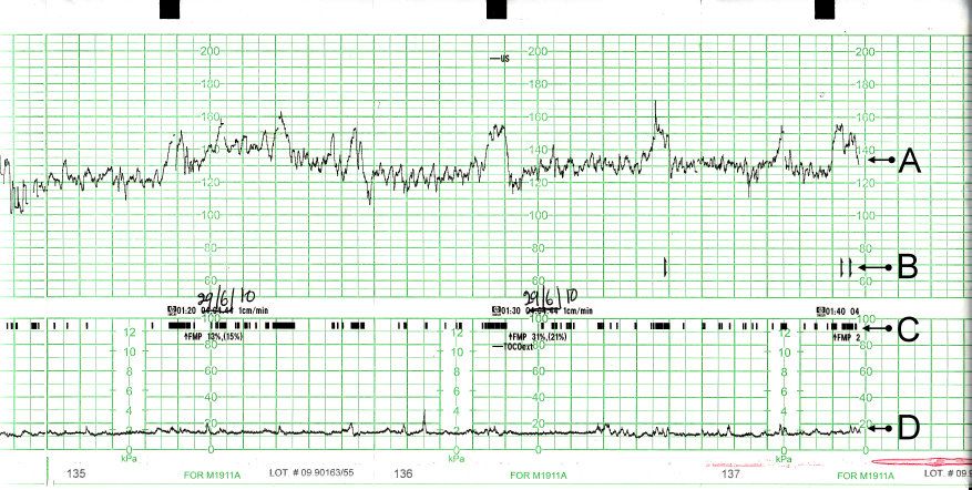
The non-stress test (NST) asks only whether the fetal heart rate accelerates with movement. At 32 weeks or more, reactive means at least two accelerations of at least 15 beats per minute above baseline lasting at least 15 seconds within a 20-minute window. Before 32 weeks the criterion drops to 10 beats per minute for 10 seconds because the autonomic nervous system is immature. A non-reactive tracing is far more often a sleeping fetus than a dying one — fetal sleep cycles run 20 to 40 minutes — so extend the test to 40 minutes and consider vibroacoustic stimulation before escalating. The NST has a high negative predictive value and a poor positive predictive value: a reactive test is genuinely reassuring, a non-reactive test mostly means do another test.
The biophysical profile (BPP) adds ultrasound to the NST and scores five parameters two points each. The contraction stress test (CST) asks a different question — whether the placenta has enough reserve to survive the transient perfusion drop of a contraction — and its scoring is counter-intuitive: negative (no late decelerations) is the good result. It is contraindicated when labour is dangerous, so avoid it in placenta praevia, PPROM, prior classical uterine incision or myomectomy entering the cavity, and cervical insufficiency. Umbilical artery Doppler is used in fetal growth restriction; absent or reversed end-diastolic flow signals critically raised placental resistance and prompts urgent obstetric review.
Three deceleration patterns carry almost the whole of intrapartum decision-making, and the mapping has to be automatic because the wrong label produces the wrong action. An early deceleration is gradual, mirrors the contraction with its nadir at the contraction peak, and reflects head compression — it is benign and requires no intervention. A variable deceleration is abrupt and V-shaped with no fixed relationship to the contraction, and reflects cord compression — reposition the patient and consider amnioinfusion; a sudden deep variable or a prolonged deceleration immediately after membranes rupture is umbilical cord prolapse until proven otherwise. A late deceleration is gradual, begins after the contraction has started and returns to baseline after it has finished, and reflects uteroplacental insufficiency — the placenta cannot meet oxygen demand, and this is the most ominous of the three. Recurrent late decelerations, recurrent variables, or a prolonged deceleration all trigger the same intrauterine resuscitation sequence, in order: reposition to the left or right lateral side, stop the oxytocin, give an intravenous bolus of isotonic crystalloid, apply oxygen at 10 litres per minute by non-rebreather mask, and notify the provider — then reassess, and prepare for expedited birth if the pattern does not recover.
| Test | What is measured | Normal / reassuring | Abnormal result and nursing action |
|---|---|---|---|
| Non-stress test | FHR accelerations with movement | ≥32 wk: 2 accels ≥15 bpm × ≥15 s in 20 min. <32 wk: ≥10 bpm × ≥10 s | Non-reactive → extend to 40 min, reposition left lateral, vibroacoustic stimulation, then BPP or CST |
| Biophysical profile | NST + breathing + movement + tone + fluid (2 points each) | 8–10 with normal fluid | 6 = equivocal, repeat in 24 h or deliver if term; ≤4 = deliver |
| Modified BPP | NST + amniotic fluid volume | Reactive NST + AFI >5 cm | Either abnormal → full BPP |
| Contraction stress test | Placental reserve under contractions (3 in 10 min, ≥40 s) | Negative = no late decelerations (good) | Positive = late decels with ≥50% of contractions → obstetric review, likely delivery |
| Amniotic fluid index | Sum of 4 quadrant pockets | 5–24 cm; single deepest pocket 2–8 cm | ≤5 cm oligohydramnios (renal, PPROM, placental insufficiency); ≥24 cm polyhydramnios (diabetes, anomaly) |
| Umbilical artery Doppler | Placental vascular resistance in FGR | Forward flow throughout diastole | Absent or reversed end-diastolic flow → urgent review, admission, steroids, timed delivery |
| Fetal movement counting | Maternal perception of movement | 10 distinct movements within 2 h, same time daily | Fewer than 10 in 2 h → come in the same day for NST; never advise waiting until morning |
Escalation, documentation and what you may not hand off
High-risk antepartum care generates a stream of borderline findings, and the failure mode is not usually ignorance — it is delay. Structured escalation shortens the delay. SBAR gives the receiving clinician the picture in the order they need it. A worked example for a patient at 33 weeks with new severe-range blood pressure sounds like this. Situation: “This is the RN on antepartum. Room 4, Ms Okoye, 33 weeks and 2 days, gravida 2 para 1, her blood pressure is 168 over 112 repeated at four minutes and she now has a headache that did not respond to paracetamol.” Background: “Admitted yesterday for gestational hypertension, no proteinuria on admission, platelets were 148.” Assessment: “I think this is pre-eclampsia with severe features. Reflexes are 3+, no clonus, urine output 25 millilitres over the last hour, fetal heart rate baseline 150 with moderate variability.” Recommendation: “I need you at the bedside now, I would like orders for IV labetalol and magnesium sulfate, and I have started repeat pre-eclampsia labs.” Notice that the recommendation is specific and time-bound. “Can you come see her when you get a chance” is not escalation.
Delegation on a high-risk antepartum unit follows one rule with no exceptions: the registered nurse retains assessment, teaching, evaluation and the unstable patient. Assistive personnel may take vital signs on stable patients, record intake and output, obtain daily weights, assist with hygiene and ambulation, and in many facilities perform capillary glucose checks — but the RN interprets every one of those numbers. A licensed practical or vocational nurse may reinforce teaching the RN has already delivered and give many routine medications, but titrated high-alert infusions such as magnesium sulfate and insulin, blood product administration, and the initial assessment of a patient who is bleeding, seizing or hypotensive remain with the RN. Delegating a task never delegates accountability for the outcome.
Documentation carries the same weight. Record the actual numbers, the time, who was notified, what was requested, what response was received, and what you did next. “Provider aware” is not a plan. If the response you get does not match the severity of the finding, use the chain of command — charge nurse, then the next clinician up — and document that you did. Every maternal mortality review that reads “the nurse recognised the deterioration” and still ends badly turns on the gap between recognition and effective escalation.
Antepartum surveillance tests have strong negative predictive value and weak positive predictive value — a reassuring result is trustworthy, an abnormal one is a prompt to escalate and test further, never a reason to wait and watch.
Section 2Bleeding in Pregnancy: Early Loss and Late Haemorrhage
Early pregnancy loss and ectopic pregnancy
Bleeding before 20 weeks occurs in roughly one in four pregnancies, and about half of those pregnancies continue normally. The clinical task is to sort threatened loss from established loss from ectopic implantation, because one of these three can kill within hours. Classification depends on the cervical os and on what has passed: threatened (bleeding, cramping, closed os, viable intrauterine pregnancy), inevitable (open os, bleeding, cramping), incomplete (some products passed, os open, bleeding often heavy), complete (all products passed, os closed, bleeding and cramping settling), missed (fetal demise retained, os closed, symptoms may have quietly disappeared), and septic (any of the above plus fever, foul discharge, uterine tenderness — a surgical and antibiotic emergency).
Ectopic pregnancy is implantation outside the uterine cavity, most often in the ampulla of the fallopian tube. Classic presentation is amenorrhoea of six to eight weeks, unilateral lower abdominal pain and light vaginal spotting; the spotting is decidual, so it is typically far less than the internal bleeding. Serum human chorionic gonadotropin rises more slowly than in a normal intrauterine pregnancy (a normal early rise is at least 35 to 50 percent over 48 hours), and transvaginal ultrasound should show an intrauterine gestational sac once hCG is around 3,500 milli-international units per millilitre. Rupture announces itself with sudden severe pain, signs of hypovolaemic shock, and referred shoulder pain from diaphragmatic irritation by blood in the peritoneum — the Kehr sign. Cervical motion tenderness is common.
Management depends entirely on stability. A stable, unruptured ectopic with a mass under about 3.5 to 4 centimetres, no fetal cardiac activity and hCG below roughly 5,000 may be treated medically with methotrexate 50 milligrams per square metre of body surface area intramuscularly, with hCG followed on days 4 and 7 expecting a fall of at least 15 percent. Teach the patient to avoid folic acid supplements (they antagonise the drug), avoid NSAIDs and alcohol, use sun protection, avoid gas-forming foods that confuse abdominal assessment, and to report severe abdominal pain immediately — some transient pain is expected, escalating pain with dizziness is rupture. A ruptured ectopic or an unstable patient goes to surgery; nursing priority is two large-bore intravenous lines, type and crossmatch, oxygen, and continuous monitoring while the theatre is readied.
Gestational trophoblastic disease — the hydatidiform mole — presents with bleeding that may contain grape-like vesicles, a uterus large for dates, markedly elevated hCG often above 100,000, severe nausea and vomiting, and pre-eclampsia appearing before 20 weeks, which is otherwise almost unheard of. Ultrasound shows the classic snowstorm pattern with no fetus in a complete mole. Treatment is suction curettage, and then the part that nursing follow-up must protect: serial hCG levels weekly until three consecutive negatives, then monthly for six months, with reliable contraception throughout, because a rising hCG signals choriocarcinoma and a new pregnancy would make the marker uninterpretable.
Late-pregnancy bleeding: praevia, abruption and vasa praevia
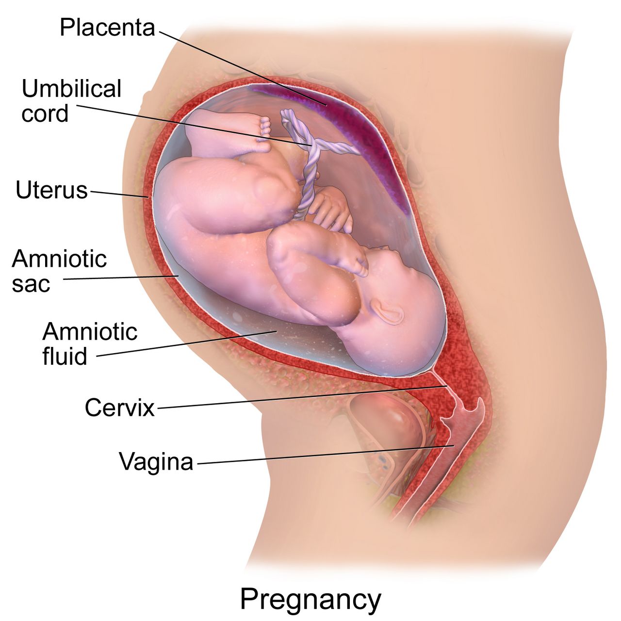
Bleeding after 20 weeks is treated as haemorrhage until proven otherwise. The three named causes separate cleanly on four features: pain, blood colour, uterine tone and fetal status. Placenta praevia is implantation of the placenta over the internal cervical os; a placental edge that lies within two centimetres of the os without covering it is termed low-lying, and both carry the same nursing prohibition. The classic presentation is painless, bright red bleeding, often in the late second or third trimester, with a soft, non-tender, relaxed uterus and a normal fetal heart rate, because the blood being lost is maternal and the placenta is still functioning. Malpresentation is common because the placenta blocks engagement. The single most important nursing rule is absolute: no digital vaginal examination and nothing in the vagina — no speculum from nursing, no cervical check, no intercourse, no tampons. A digital exam can perforate the placenta and cause exsanguinating haemorrhage in minutes. Transabdominal and transvaginal ultrasound are diagnostic and transvaginal scanning is safe because the probe is angled away from the os.
Abruptio placentae is premature separation of a normally implanted placenta. It presents with painful bleeding that is dark red, a uterus that is tender, rigid or board-like with elevated resting tone and frequent low-amplitude contractions, and a fetal heart rate that frequently deteriorates because the exchange surface has been lost. Roughly one in five abruptions is concealed: the blood collects behind the placenta and the external loss badly understates the true loss, so a patient can be in shock with very little visible bleeding. Any mismatch between visible blood and haemodynamic status should make you think concealed haemorrhage. Risk factors include hypertension in any form, blunt abdominal trauma including intimate partner violence, cocaine and other stimulant use, smoking, prior abruption, PPROM and sudden uterine decompression. Disseminated intravascular coagulation is the classic complication because thromboplastin from the decidua enters the circulation.
Vasa praevia is rarer and faster than either. Unprotected fetal vessels cross the internal os, and when the membranes rupture the vessels tear. The presentation is a small volume of painless bleeding coinciding with rupture of membranes, immediately followed by fetal bradycardia or a sinusoidal fetal heart rate pattern. The blood is fetal, and total fetal blood volume is only about 80 to 100 millilitres per kilogram — a term fetus has roughly 250 to 350 millilitres in total, so a loss that would barely register in an adult is exsanguinating. Vasa praevia is an immediate caesarean birth. Placenta accreta spectrum, in which the placenta invades the myometrium, is increasingly common because its dominant risk factor is the combination of a prior caesarean birth with a current praevia; it is planned for with delivery between 34 and 36 weeks in a centre with massive transfusion capability, and caesarean hysterectomy is often expected.
| Feature | Placenta praevia | Abruptio placentae | Vasa praevia | Uterine rupture |
|---|---|---|---|---|
| Pain | Painless | Painful, constant | Painless | Sudden sharp tearing pain, may then ease |
| Bleeding | Bright red, external | Dark red; concealed in ~20% | Small volume, fetal blood, at ROM | Variable, often concealed |
| Uterine tone | Soft, relaxed, non-tender | Rigid, board-like, tender, high resting tone | Normal | Contractions cease; loss of tone |
| Fetal heart rate | Usually normal initially | Late decelerations, bradycardia | Abrupt bradycardia or sinusoidal | Prolonged deceleration; presenting part recedes (station rises) |
| First nursing action | NOTHING in the vagina; left lateral, IV access, type and cross, fetal monitoring | Large-bore IV × 2, oxygen, left lateral, continuous EFM, coagulation studies, prepare for caesarean | Call for immediate caesarean; do not delay for confirmation | Prepare for immediate laparotomy; fluid and blood resuscitation |
Volume, coagulation and the haemorrhage response
Pregnancy hides blood loss. Plasma volume rises by 40 to 50 percent by the third trimester, so a healthy pregnant person can lose 1,000 to 1,500 millilitres — up to 25 percent of blood volume — before the blood pressure falls. Tachycardia, narrowing pulse pressure, rising respiratory rate, restlessness and cool clammy skin appear long before hypotension, and a falling urine output below 30 millilitres per hour is one of the earliest reliable signs of inadequate perfusion. Do not wait for a low blood pressure to declare haemorrhage; by then the compensation has already failed.
The standing antepartum haemorrhage response is: call for help and activate the obstetric haemorrhage protocol, place two large-bore intravenous catheters (16 or 18 gauge), give warmed isotonic crystalloid, send type and crossmatch with a full coagulation panel and fibrinogen, apply oxygen at 10 litres per minute by non-rebreather mask if there is evidence of compromise, position left lateral or with manual displacement of the uterus off the vena cava, insert an indwelling catheter to measure output hourly, monitor the fetus continuously, weigh pads and drapes for quantitative blood loss rather than estimating visually, and keep the patient and family informed in plain sentences. Fibrinogen deserves special attention: it rises physiologically in pregnancy to 400 to 600 milligrams per decilitre, so a “normal” adult value near 200 in a bleeding obstetric patient is a red flag for consumptive coagulopathy.
Although postpartum haemorrhage belongs to a later chapter, the sequence is worth fixing here because it is the single most tested and most misordered response in maternity nursing. When the uterus is boggy and bleeding after birth, the first action is to massage the fundus — not to start the oxytocin, not to call the provider, not to take a blood pressure. Massage stimulates the myometrium to contract and clamp the spiral arteries mechanically, which is faster than any drug can act. Massage first, then empty the bladder (a distended bladder displaces the uterus and prevents contraction), then give uterotonics as ordered while help is on the way.
Painless bright-red bleeding with a soft uterus is praevia and forbids any vaginal examination; painful dark bleeding with a rigid tender uterus is abruption and may conceal most of its blood loss behind the placenta.
Section 3Hypertensive Disorders and Magnesium Sulfate Safety
Mechanism and the four diagnostic categories
Pre-eclampsia begins in the placenta and ends in the maternal endothelium. In normal implantation, trophoblast remodels the spiral arteries into wide, low-resistance channels. In pre-eclampsia that remodelling is incomplete, the placenta is under-perfused, and it releases anti-angiogenic factors into the maternal circulation. Those factors injure endothelium everywhere at once, which explains why the disease is multi-system rather than merely hypertensive: vasospasm raises blood pressure, glomerular endothelial injury leaks protein, hepatic sinusoidal injury distends the liver capsule and produces right upper quadrant pain, cerebral vasospasm and oedema produce headache, visual scotomata and ultimately seizure, and platelet consumption drops the count. Fluid leaves the intravascular space, so these patients are simultaneously oedematous and intravascularly depleted — which is why aggressive fluid loading causes pulmonary oedema.
The four categories are distinguished by timing and by findings. Chronic hypertension is blood pressure at or above 140/90 before 20 weeks or persisting beyond 12 weeks postpartum. Gestational hypertension is new hypertension at or after 20 weeks without proteinuria or severe features, resolving postpartum. Pre-eclampsia is new hypertension at or after 20 weeks plus either proteinuria or, in the absence of proteinuria, any severe feature. Chronic hypertension with superimposed pre-eclampsia is a worsening picture on a pre-existing baseline and carries the highest risk. Proteinuria is defined as 300 milligrams or more in 24 hours, a protein-to-creatinine ratio of 0.3 or above, or a dipstick of 2+ when quantitative testing is unavailable. Critically, proteinuria is no longer required for the diagnosis — waiting for it delays treatment and has killed patients.
Prevention is real and under-used. Low-dose aspirin 81 milligrams daily, started between 12 and 28 weeks (ideally before 16 weeks) and continued until birth, meaningfully reduces pre-eclampsia in people with one high-risk factor (prior pre-eclampsia, chronic hypertension, pre-gestational diabetes, chronic kidney disease, autoimmune disease, multifetal gestation) or two moderate-risk factors (first pregnancy, age 35 or older, body mass index above 30, family history, sociodemographic risk, interpregnancy interval over 10 years). Antihypertensives compatible with pregnancy are labetalol, nifedipine, methyldopa and hydralazine; ACE inhibitors, angiotensin receptor blockers and direct renin inhibitors are contraindicated because of fetal renal injury and oligohydramnios.
| Severe feature | Threshold | What it reflects |
|---|---|---|
| Severe-range blood pressure | Systolic ≥160 or diastolic ≥110 on two occasions 4 h apart (or confirmed within minutes to permit treatment) | Risk of maternal stroke — the leading cause of death in pre-eclampsia |
| Thrombocytopenia | Platelets <100,000 per microlitre | Platelet consumption at injured endothelium |
| Impaired liver function | AST or ALT at least twice the upper limit of normal, or severe persistent right upper quadrant or epigastric pain | Hepatic ischaemia, capsular distension, risk of subcapsular haematoma |
| Renal insufficiency | Serum creatinine >1.1 mg/dL or doubling of baseline | Glomerular endothelial injury; normal pregnancy creatinine is LOW (0.4–0.8) |
| Pulmonary oedema | Clinical or radiographic | Capillary leak plus low oncotic pressure |
| New cerebral or visual symptoms | Headache unresponsive to analgesia, scotomata, blurred or double vision, altered mental state | Cerebral vasospasm and oedema — the immediate seizure warning |
Severe-range pressure, eclampsia, and what you do first
A confirmed severe-range blood pressure is a hypertensive emergency with a clock attached. Treatment should be given within 30 to 60 minutes of confirmation, and sooner is better. First-line agents are intravenous labetalol 20 milligrams over 2 minutes, repeated at escalating doses of 40 then 80 milligrams every 10 minutes to a cumulative maximum of 300 milligrams; intravenous hydralazine 5 to 10 milligrams over 2 minutes repeated every 20 minutes; or immediate-release oral nifedipine 10 milligrams repeated at 20 milligrams if intravenous access is not yet established. The goal is not normotension — it is 140 to 150 systolic over 90 to 100 diastolic, because dropping the pressure too far collapses placental perfusion. Avoid labetalol in asthma, decompensated heart failure and bradycardia.
Eclampsia is a generalised tonic-clonic seizure in a person with pre-eclampsia and no other cause. Nursing first actions, in order: stay with the patient and call for help, turn them onto their side, protect the head and clear the immediate environment, and do not restrain the limbs or place anything in the mouth. Do not attempt to assess the fetus during the seizure — the fetal heart rate will be abnormal, and the only useful intervention for the fetus is resuscitating the person carrying it. When the seizure ends, suction the airway, give oxygen at 10 litres per minute by non-rebreather, establish or confirm intravenous access, give the magnesium sulfate bolus as ordered, then assess the fetus and prepare for birth. Delivery is the only definitive treatment for pre-eclampsia, but the patient must be stabilised first; a caesarean during an active seizure endangers both patients.
Two timing facts change discharge teaching. First, roughly a third to a half of eclamptic seizures occur after birth, some days later. Second, postpartum pre-eclampsia can present up to six weeks after birth in someone whose pregnancy was entirely uncomplicated. Every postpartum patient therefore leaves with explicit warning signs: severe headache that does not respond to paracetamol, visual changes, right upper quadrant or epigastric pain, shortness of breath, sudden swelling of face or hands, or a seizure — and an instruction to present to an emergency department and say the words “I gave birth recently”.
Magnesium sulfate: therapeutic window, toxicity order and antidote
Magnesium sulfate is given to prevent and treat eclamptic seizures, not to lower blood pressure. It competes with calcium at the neuromuscular junction and stabilises the cerebral membrane. A typical regimen is a loading dose of 4 to 6 grams intravenously over 15 to 30 minutes followed by a maintenance infusion of 1 to 2 grams per hour, continued for 24 hours after birth. Given before 32 weeks it also confers fetal neuroprotection against cerebral palsy, usually as a 4-gram load followed by 1 gram per hour. It is a high-alert medication: it must run on a dedicated pump with a second-nurse independent double check of the concentration, rate and pump settings, and it must never be given by rapid intravenous push.
Toxicity is entirely predictable because it follows serum level in a fixed order, and that order is the assessment schedule. The therapeutic range is 4 to 7 milliequivalents per litre (about 4.8 to 8.4 milligrams per decilitre). Deep tendon reflexes disappear at roughly 9 to 12 milligrams per decilitre — before respiratory depression, which begins around 12 to 18, and long before cardiac arrest above 25 to 30. This is why hourly patellar reflex checks are not a ritual: the absent reflex is the early warning that buys you the time to act. Magnesium is cleared entirely by the kidney, so urine output is the other essential parameter; oliguria means the drug is accumulating even at an unchanged infusion rate.
Hold parameters before each hourly assessment are: respiratory rate at least 12 per minute, deep tendon reflexes present (at least 1+), urine output at least 30 millilitres per hour (or 100 millilitres over 4 hours), and level of consciousness intact. If any parameter fails, the sequence is fixed: stop the infusion first, then give the antidote — calcium gluconate 1 gram, which is 10 millilitres of a 10 percent solution, intravenously over approximately 3 minutes — then support ventilation with a bag-valve mask and oxygen, call for rapid response and the obstetric team, and send a stat magnesium level. Stopping first matters because giving calcium while magnesium continues to infuse is treating against a running tap. Keep calcium gluconate and a bag-valve mask physically at the bedside of every patient on magnesium.
HELLP syndrome
HELLP is haemolysis, elevated liver enzymes and low platelets — a severe variant of pre-eclampsia that can occur with only modest hypertension and, in a substantial minority, without proteinuria, which is exactly why it gets missed. The laboratory definition is haemolysis (lactate dehydrogenase at or above 600 international units per litre, schistocytes on smear, or bilirubin at or above 1.2 milligrams per decilitre), AST or ALT at least twice the upper limit of normal, and platelets below 100,000 per microlitre. The presenting complaint is often deceptively vague: malaise, nausea and vomiting that feels like influenza, and right upper quadrant or epigastric pain from hepatic capsular distension.
Any pregnant or recently pregnant person describing epigastric pain, especially with nausea, should have a blood pressure, platelet count and liver enzymes checked before the symptom is attributed to reflux or gastritis. Nursing care adds bleeding precautions to standard pre-eclampsia management: avoid intramuscular injections and non-essential venipuncture, apply prolonged pressure to puncture sites, use a soft toothbrush, avoid NSAIDs, and palpate the right upper quadrant gently or not at all because a subcapsular hepatic haematoma can rupture. Sudden shoulder pain, severe abdominal pain and shock in a HELLP patient suggests exactly that rupture and is a surgical emergency. Definitive treatment is birth, with magnesium sulfate for seizure prophylaxis, antihypertensives for severe-range pressures, corticosteroids for fetal lung maturity if gestational age allows, and platelet transfusion generally reserved for counts below 20,000 or for surgery.
On magnesium sulfate, deep tendon reflexes vanish before the respiratory rate falls — so if reflexes are absent, stop the infusion first, then give calcium gluconate 1 gram (10 mL of 10 percent) intravenously over about 3 minutes.
Section 4Metabolic, Endocrine and Nutritional Complications
Diabetes in pregnancy: the mechanism sets the risks
Human placental lactogen, progesterone, cortisol and growth hormone all antagonise insulin, and their concentrations climb through the second and third trimesters. This is a design feature: maternal insulin resistance keeps glucose in the circulation for the fetus. A pancreas with reserve simply secretes more insulin. A pancreas without reserve cannot, and maternal hyperglycaemia results. Glucose crosses the placenta freely; insulin does not. The fetus therefore receives the glucose load and responds by producing its own insulin, which is a powerful growth factor. That single sentence explains nearly every complication: macrosomia and shoulder dystocia, neonatal hypoglycaemia in the minutes after the cord is cut and the glucose supply stops while fetal hyperinsulinaemia persists, delayed surfactant production and respiratory distress, polycythaemia, hypocalcaemia and hyperbilirubinaemia.
Timing separates the two diseases. Pre-gestational diabetes exposes the embryo to hyperglycaemia during organogenesis, so it raises the risk of congenital anomalies — cardiac defects, neural tube defects and the rare but characteristic caudal regression syndrome — and of spontaneous loss. Preconception care aiming for a haemoglobin A1c below 6.5 percent is the single most effective intervention available, along with folic acid supplementation and switching off ACE inhibitors, ARBs and statins. Gestational diabetes appears in the second half of pregnancy, after organogenesis, so it does not raise anomaly risk but does drive macrosomia, hypertensive disorders, polyhydramnios, and a long-term maternal risk of type 2 diabetes of roughly 50 percent within 10 to 20 years — which is why the 4-to-12-week postpartum 75-gram oral glucose tolerance test is not optional follow-up.
Screening happens at 24 to 28 weeks, earlier if risk factors are present. The two-step approach gives a non-fasting 50-gram glucose challenge with a one-hour threshold of 130 to 140 milligrams per decilitre, and those who screen positive proceed to a fasting 100-gram three-hour test where two or more abnormal values diagnose gestational diabetes (fasting 95, one hour 180, two hours 155, three hours 140). The one-step approach uses a fasting 75-gram two-hour test where a single abnormal value diagnoses it (fasting 92, one hour 180, two hours 153). Glycaemic targets in pregnancy are tighter than outside it: fasting and pre-meal below 95, one hour after the start of a meal below 140, two hours after below 120. Nutrition therapy and physical activity are first-line; when pharmacotherapy is needed, insulin is the first-line agent because it does not cross the placenta, whereas metformin and glyburide both do.
The arithmetic habit: weight-based dosing and dose ceilings
Every dose that appears in this book from here forward is expressed per kilogram, because the patients on the other side of the birth are newborns and children, and children are not small adults. The habit starts in the antepartum unit. Enoxaparin for venous thromboembolism in pregnancy is prophylactic at 40 milligrams subcutaneously daily but therapeutic at 1 milligram per kilogram every 12 hours: for a 78-kilogram patient that is 78 milligrams every 12 hours, and a nurse who accepts a pre-printed “80 mg” without checking the documented weight has skipped the calculation. Vancomycin for group B streptococcal prophylaxis in severe penicillin allergy is 20 milligrams per kilogram every 8 hours with a maximum of 2 grams per dose: for a 90-kilogram patient, 20 × 90 = 1,800 milligrams, which is under the cap and therefore given; for a 120-kilogram patient, 20 × 120 = 2,400 milligrams, which exceeds the cap and is given as 2 grams.
That last step is the rule that prevents the most dangerous paediatric error: a weight-based dose must always be checked against the maximum adult dose, and the smaller number wins. Paracetamol for a child is 15 milligrams per kilogram per dose every 4 to 6 hours. For a 30-kilogram child: 15 × 30 = 450 milligrams, well within limits. For a 70-kilogram adolescent: 15 × 70 = 1,050 milligrams — which exceeds the 1,000-milligram adult single dose, so the dose given is 1,000 milligrams. A large adolescent is not entitled to a larger-than-adult dose simply because the multiplication says so.
| Calculation | Rule | Worked example | Safety check |
|---|---|---|---|
| Per-dose medication | mg/kg/dose × weight, then compare with maximum adult dose | Paracetamol 15 mg/kg for 70 kg = 1,050 mg → give 1,000 mg | The smaller of (calculated dose, adult maximum) is the dose |
| Capped antibiotic dose | mg/kg/dose with a stated ceiling per dose | Vancomycin 20 mg/kg for 120 kg = 2,400 mg → give 2 g | Confirm a current, documented weight in kilograms — never a reported weight in pounds |
| Therapeutic enoxaparin | 1 mg/kg subcutaneously every 12 h | 78 kg → 78 mg q12h | Hold 24 h before neuraxial anaesthesia (12 h for prophylactic dosing) |
Hyperemesis gravidarum, anaemia and cholestasis
Hyperemesis gravidarum is not severe morning sickness; it is intractable vomiting producing weight loss greater than 5 percent of pre-pregnancy weight, dehydration, ketonuria and electrolyte disturbance — classically hypokalaemia and a hypochloraemic metabolic alkalosis from loss of gastric acid. It typically begins before 9 weeks. Management escalates from non-pharmacological measures (small frequent dry carbohydrate snacks, eating before rising, separating fluids from solids, ginger, avoiding triggering smells) to pyridoxine (vitamin B6) 10 to 25 milligrams every 6 to 8 hours with or without doxylamine 12.5 milligrams, then to promethazine, metoclopramide or ondansetron, then to intravenous rehydration and, rarely, enteral or parenteral nutrition.
One safety rule governs the intravenous phase: give thiamine before any dextrose-containing fluid in a person who has been vomiting for more than about three weeks. Dextrose consumes the last of a depleted thiamine store and can precipitate Wernicke encephalopathy — confusion, ataxia and ophthalmoplegia — which is preventable and sometimes irreversible. Thiamine 100 milligrams intravenously is the usual dose. Equally important is the language: this condition is frequently dismissed as psychological or exaggerated, and patients arrive expecting not to be believed. Weigh them, check ketones and electrolytes, and treat the physiology.
Iron-deficiency anaemia is defined in pregnancy as haemoglobin below 11 grams per decilitre in the first and third trimesters and below 10.5 in the second, reflecting the physiological dilution of plasma volume expansion. Daily requirement rises to about 27 milligrams of elemental iron; treatment of established deficiency uses 100 to 200 milligrams of elemental iron daily, taken with a vitamin C source and away from milk, calcium supplements, tea and antacids. Warn about dark stools (expected) and constipation (manage with fluid, fibre and a stool softener). Intrahepatic cholestasis of pregnancy presents with intense itching of the palms and soles, worse at night, with no rash; the diagnostic test is serum total bile acids, abnormal at 10 micromoles per litre or above and severe at 100 or above, where stillbirth risk rises sharply. Treatment is ursodeoxycholic acid 10 to 15 milligrams per kilogram per day with antenatal surveillance and planned birth, usually at 36 to 37 weeks and earlier if bile acids are severe.
Insulin is first-line pharmacotherapy in pregnancy because it does not cross the placenta, and every weight-based dose — maternal or paediatric — is calculated per kilogram and then capped at the maximum adult dose.
Section 5Infection, Immune Incompatibility and Preterm Birth
Infection in pregnancy and prophylaxis that works
Pregnancy shifts immune function toward tolerance of the fetus, and that shift makes several infections more severe. Urinary tract infection deserves particular respect: progesterone relaxes ureteral smooth muscle and the growing uterus compresses the ureters, producing physiological hydronephrosis and urinary stasis. Asymptomatic bacteriuria, which would be ignored outside pregnancy, is treated in pregnancy because up to 30 percent of untreated cases ascend to pyelonephritis, and pyelonephritis in pregnancy causes preterm labour, sepsis and acute respiratory distress syndrome. Flank pain, fever and costovertebral angle tenderness in a pregnant patient is an admission, not an outpatient prescription.
Group B Streptococcus colonises the vagina and rectum of about a quarter of pregnant people and is harmless to them, but transmitted during birth it causes early-onset neonatal sepsis, pneumonia and meningitis. Universal screening by vaginal-rectal culture is performed at 36 0/7 to 37 6/7 weeks. Intrapartum prophylaxis — not antenatal treatment, which does not prevent recolonisation — is penicillin G 5 million units intravenously as a load then 2.5 to 3 million units every 4 hours until birth. Prophylaxis is considered adequate once at least 4 hours have elapsed before birth. Indications for prophylaxis without a culture include GBS bacteriuria at any point in this pregnancy, a previous infant with invasive GBS disease, or unknown status with birth before 37 weeks, membranes ruptured 18 hours or more, or intrapartum fever.
The TORCH group — toxoplasmosis, other (syphilis, varicella, parvovirus B19, Zika, hepatitis B), rubella, cytomegalovirus and herpes simplex — matters because these organisms cross the placenta and injure the developing fetus. Teaching is largely preventive: avoid changing cat litter and eating undercooked meat (toxoplasmosis); hand hygiene around toddlers’ saliva and urine, the main source of cytomegalovirus; rubella immunity is checked at booking and the vaccine is given postpartum because it is a live vaccine and contraindicated in pregnancy, with the advice to avoid conception for 28 days. Active genital herpes lesions or prodromal symptoms at the onset of labour indicate caesarean birth. Untreated syphilis causes stillbirth and congenital syphilis and is entirely preventable with penicillin.
Rh alloimmunisation
An Rh-negative pregnant person carrying an Rh-positive fetus can be exposed to fetal red cells at any point where the placental barrier is breached — birth, miscarriage, ectopic pregnancy, chorionic villus sampling, amniocentesis, external cephalic version, abdominal trauma, abruption. The maternal immune system produces anti-D antibodies. The first pregnancy is usually unaffected because sensitisation occurs at delivery, but those IgG antibodies cross the placenta in every subsequent pregnancy and haemolyse fetal red cells, producing fetal anaemia, high-output heart failure, hydrops fetalis and death.
Prevention is one of the great successes of obstetric care. Rh(D) immune globulin 300 micrograms intramuscularly is given at 28 weeks and again within 72 hours of birth if the newborn is Rh-positive, and additionally after any sensitising event; a 50-microgram microdose is used for events before 12 weeks. One 300-microgram dose neutralises approximately 30 millilitres of fetal whole blood (15 millilitres of fetal red cells), so after abruption or trauma a Kleihauer-Betke test quantifies the fetomaternal haemorrhage and determines how many doses are required. Two antibody tests are easily confused: the indirect Coombs (antibody screen) is performed on the pregnant person and detects circulating antibody, while the direct Coombs is performed on the newborn’s cord blood and detects antibody already attached to fetal cells. Rh(D) immune globulin is useless once sensitisation has occurred — a positive indirect Coombs means the window has closed, and management shifts to middle cerebral artery Doppler surveillance for fetal anaemia and, if needed, intrauterine transfusion.
Preterm labour and PPROM
Preterm labour is regular uterine activity with documented cervical change before 37 0/7 weeks. Its symptoms are notoriously vague — low dull backache, pelvic pressure, menstrual-like cramping, increased or changed vaginal discharge, and contractions that may be painless — and patients routinely delay presenting because the sensations do not match the dramatic picture they expect. Teach the specific symptoms, and teach the response: empty the bladder, lie down on the left side, drink two or three glasses of water, and if contractions continue at four or more per hour after an hour, come in. Two tests refine the decision: fetal fibronectin has an excellent negative predictive value, so a negative test makes birth within the next 7 to 14 days very unlikely, and transvaginal cervical length under 25 millimetres before 24 weeks identifies elevated risk. Vaginal progesterone is used for a short cervix; cerclage is used for cervical insufficiency, which classically presents as painless dilation in the second trimester.
Tocolytics are widely misunderstood. They do not meaningfully prolong pregnancy; they buy roughly 48 hours, and that window exists to complete antenatal corticosteroids and to transfer to a facility with an appropriate neonatal unit. Nifedipine and indomethacin are common first choices; indomethacin is restricted to before 32 weeks because it constricts the fetal ductus arteriosus and reduces amniotic fluid. Terbutaline is short-term only and causes maternal tachycardia, tremor and hyperglycaemia — a maternal heart rate above 120 is a hold parameter. Antenatal corticosteroids are the intervention that actually changes newborn survival: betamethasone 12 milligrams intramuscularly every 24 hours for two doses, or dexamethasone 6 milligrams intramuscularly every 12 hours for four doses, given between 24 0/7 and 33 6/7 weeks (and in selected late-preterm situations). Benefit begins about 24 hours after the first dose and is optimal from 48 hours to 7 days. Expect a transient rise in maternal blood glucose — in a patient with diabetes this can be substantial and requires close monitoring. Magnesium sulfate before 32 weeks is added for fetal neuroprotection.
Preterm premature rupture of membranes (PPROM) is rupture before 37 weeks and before labour begins. Confirm with a sterile speculum examination showing pooling, nitrazine paper turning blue (alkaline amniotic fluid) and ferning on a dried slide — and perform no digital examination, because each digital exam shortens latency and introduces organisms. Nursing priorities are temperature every 2 to 4 hours, fetal heart rate for tachycardia above 160 (an early sign of chorioamnionitis), uterine tenderness, the character and odour of fluid, and white cell trend. Latency antibiotics prolong the pregnancy and reduce infection: ampicillin 2 grams intravenously every 6 hours plus erythromycin for 48 hours, followed by 5 days of oral amoxicillin and erythromycin. Amoxicillin-clavulanate is avoided because of an association with neonatal necrotising enterocolitis. Expectant management generally continues to 34 0/7 weeks, and chorioamnionitis, abruption, cord prolapse or non-reassuring fetal status ends it immediately. The greatest immediate hazard after any rupture with an unengaged presenting part is umbilical cord prolapse, so check the fetal heart rate immediately after membranes rupture, every time.
| Emergency | Recognition | FIRST action | Never do this |
|---|---|---|---|
| Umbilical cord prolapse | Sudden variable or prolonged deceleration or bradycardia after ROM; cord palpated or seen | Keep a gloved hand in the vagina and lift the presenting part off the cord; position knee-chest or Trendelenburg; call for immediate caesarean; oxygen 10 L/min | Do not push the cord back in; do not repeatedly handle it; do not remove your hand until the surgeon takes over |
| Magnesium sulfate toxicity | Deep tendon reflexes absent, then RR <12, slurred speech, oliguria | Stop the infusion, then calcium gluconate 1 g (10 mL of 10%) IV over ~3 min; support ventilation | Do not give the antidote while the magnesium continues to run |
| Eclamptic seizure | Generalised tonic-clonic activity in pre-eclampsia | Call for help, turn on the side, protect the head, then oxygen and magnesium bolus | Do not restrain limbs, put anything in the mouth, or leave to fetch equipment |
| Postpartum haemorrhage | Boggy fundus, heavy or continuous bleeding, rising pulse | Massage the fundus, then empty the bladder, then uterotonics as ordered | Do not leave to page the provider before massaging; do not massage a firm fundus |
| Shoulder dystocia | Turtle sign; head delivers and retracts; shoulders will not deliver | Call for help, McRoberts (hyperflex thighs to abdomen) plus suprapubic pressure; note the time | Never apply fundal pressure — it drives the shoulder harder into the symphysis |
| Uterine rupture | Sharp tearing pain, contractions cease, loss of tone, presenting part recedes, prolonged deceleration, shock | Call the obstetric emergency team and prepare for immediate laparotomy; two large-bore IVs, blood, oxygen | Do not give oxytocin; do not delay surgery for imaging |
| Supine hypotension | Dizziness, pallor, nausea, falling BP when lying flat after 20 weeks | Turn to the left side or manually displace the uterus | Do not start with fluids or vasopressors before repositioning |
In every obstetric emergency the life-saving move is the first one — lift the presenting part off a prolapsed cord, stop the magnesium before giving calcium, massage the fundus before anything else — and the intuitive alternative is usually the harmful one.
The chapter in one breath
High-risk pregnancy is ordinary pregnancy plus a named, trackable threat, and nursing owns the surveillance that finds it early. Antepartum testing infers fetal oxygenation from fetal behaviour: the non-stress test, biophysical profile, contraction stress test, amniotic fluid volume and umbilical artery Doppler all reassure powerfully when normal and demand escalation when not. Bleeding sorts by pain and tone — painless bright red with a soft uterus is praevia and forbids vaginal examination, painful dark bleeding with a rigid tender uterus is abruption and may conceal most of its loss, painless bleeding at rupture with sudden fetal bradycardia is vasa praevia and is an immediate caesarean. Hypertensive disease is endothelial injury, diagnosable without proteinuria, treated for severe-range pressures within 30 to 60 minutes, and prevented from seizing with magnesium sulfate — whose toxicity announces itself by absent reflexes before respiratory depression, and whose antidote, calcium gluconate 1 gram, follows stopping the infusion. Diabetes is fetal hyperinsulinaemia driven by maternal glucose, managed with insulin because insulin does not cross the placenta. Every dose is per kilogram, capped at the adult maximum. Every emergency is won or lost on the first action.
Words worth knowing
| Term | What it means |
|---|---|
| Abruptio placentae | Premature separation of a normally implanted placenta; painful dark bleeding with a rigid, tender uterus and up to 20 percent concealed loss. |
| Amniotic fluid index | Sum of the deepest fluid pocket in each of four uterine quadrants; normal 5–24 cm. |
| Biophysical profile | Five-parameter fetal assessment (NST, breathing, movement, tone, fluid) scored 2 points each; 8–10 reassuring, 4 or less means deliver. |
| Calcium gluconate | Antidote to magnesium sulfate toxicity; 1 gram (10 mL of a 10 percent solution) intravenously over about 3 minutes, given after the infusion is stopped. |
| Chorioamnionitis | Intra-amniotic infection; maternal fever, uterine tenderness, foul fluid, maternal and fetal tachycardia — ends expectant management of PPROM. |
| Eclampsia | Generalised tonic-clonic seizure in a person with pre-eclampsia; may occur for the first time postpartum. |
| Ectopic pregnancy | Implantation outside the uterine cavity, usually tubal; unilateral pain, spotting, and shoulder pain (Kehr sign) once ruptured. |
| Fetal fibronectin | Cervicovaginal glycoprotein test whose strength is its negative predictive value for birth within 7 to 14 days. |
| HELLP syndrome | Haemolysis, elevated liver enzymes, low platelets; often presents as epigastric or right upper quadrant pain with only modest hypertension. |
| Hydatidiform mole | Gestational trophoblastic disease with a uterus large for dates, hCG above 100,000, snowstorm ultrasound and pre-eclampsia before 20 weeks. |
| Hyperemesis gravidarum | Intractable vomiting with weight loss over 5 percent, ketonuria and electrolyte disturbance; give thiamine before dextrose-containing fluids. |
| Intrahepatic cholestasis of pregnancy | Nocturnal palmar and plantar itching without rash; bile acids 10 micromoles per litre or more, severe at 100 or more. |
| Kleihauer-Betke test | Quantifies fetal red cells in the maternal circulation to determine how many doses of Rh(D) immune globulin are needed. |
| Macrosomia | Birth weight above 4,000 to 4,500 grams; increases shoulder dystocia, operative birth and neonatal hypoglycaemia risk. |
| Magnesium sulfate | High-alert anticonvulsant for pre-eclampsia and neuroprotectant before 32 weeks; therapeutic 4–7 mEq/L, monitored by reflexes, respiratory rate and urine output. |
| Non-stress test | Fetal heart rate accelerations with movement; reactive at 32 weeks or more is 2 accelerations of 15 bpm for 15 seconds in 20 minutes. |
| Placenta accreta spectrum | Abnormally invasive placenta; strongest risk factor is prior caesarean birth combined with current praevia. |
| Placenta praevia | Placenta covering the internal os (edge within 2 cm but not covering it is low-lying); painless bright red bleeding with a soft uterus and an absolute prohibition on vaginal examination. |
| PPROM | Preterm premature rupture of membranes; confirmed by sterile speculum, pooling, nitrazine and ferning — never by digital examination. |
| Vasa praevia | Unprotected fetal vessels crossing the internal os; painless bleeding at rupture with immediate fetal bradycardia, requiring emergency caesarean. |