Biopsychology
Every thought you have ever had is a physical event. So is every feeling, every memory, and every choice. Each one is a pattern of electricity and chemistry moving through living cells. Biopsychology is the part of psychology that takes this idea seriously. It asks how the nervous system, and the glands that tune it, give rise to mind and behavior. This chapter starts with one working unit, the neuron. Then it follows the signal outward. You will see how one cell fires and how it passes a message to the next cell across a synapse. You will see how those cells are wired into the main lines of the nervous system and how the brain groups them into regions with jobs. Last, you will meet a slower chemical message service, the endocrine system, which sets the body's background mood. Once you know these pieces, the rest of psychology stops feeling like magic. It starts to feel like biology.
By the end of this chapter you can
- Label the main parts of a neuron and say what each part does.
- Describe the resting potential and walk through the steps of the action potential.
- Explain how a signal crosses the synapse. Name the main neurotransmitters and their jobs.
- Tell drugs that act as agonists apart from drugs that act as antagonists.
- Map the parts of the nervous system: central vs peripheral, somatic vs autonomic, and sympathetic vs parasympathetic.
- Find the hindbrain, midbrain, and forebrain, and give the jobs of the four cortical lobes.
- Explain how the endocrine system sends signals with hormones, and how that differs from nerve signals.
- Link real cases and researchers to the brain and body systems they helped explain. These include Phineas Gage, Broca, Wernicke, Sperry, and Selye.
Section 1The neuron
The parts of a neuron
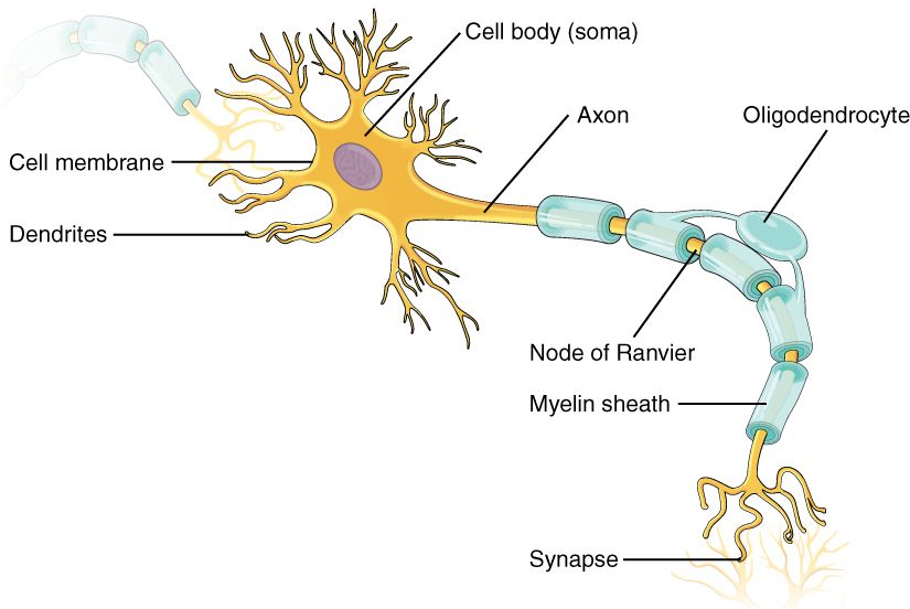
The neuron is the basic cell of the nervous system that handles information. All of psychology runs on neurons. That includes seeing, remembering, feeling, and moving. Your brain has about 86 billion of them, all wired together. A neuron does three jobs in order. It receives a signal. It decides whether to pass the signal on. Then it sends it.
The receiving end is a spray of branches called dendrites. They carry incoming signals toward the cell body, also called the soma. The cell body holds the nucleus. It also adds up everything that arrives. If the total is big enough, the signal leaves along one long fiber, the axon. The axon ends in small swollen tips called terminal buttons. There, the signal is handed off to the next cell.
Many axons are wrapped in a fatty white coating called the myelin sheath. It works like the plastic coating on a wire. Support cells lay it down. Myelin is not one smooth tube. It comes in segments with bare gaps between them. These gaps are the nodes of Ranvier, and they are the secret to how fast a myelinated axon can be.
Neurons do not work alone. They have a crew of helpers called glial cells, or glia. The brain has roughly 85 billion glia. That is about one glial cell for every neuron in the brain as a whole. In the cerebral cortex, though, glia outnumber neurons several times over. Glia build myelin, feed neurons, clean up waste, and hold everything in place. For a long time, people treated glia as simple packing material. An old textbook claim said there were ten glia for every neuron, and you may still hear it. Careful cell counts have retired both ideas. We now know glia help shape how signals travel.
| Part | Job |
|---|---|
| Dendrites | Take in signals from other neurons and carry them toward the cell body |
| Cell body (soma) | Holds the nucleus; integrates (adds up) all incoming signals to decide whether to fire |
| Axon | One long fiber that carries the outgoing signal away from the cell body |
| Myelin sheath | Fatty coating that speeds the signal; its gaps are the nodes of Ranvier |
| Terminal buttons | Axon tips that release neurotransmitter onto the next cell |
| Glial cells | Helper cells that make myelin, feed neurons, and protect them |
How a neuron fires: the action potential
At rest, a neuron is like a tiny charged battery. It keeps pumping charged particles, called ions, across its outer membrane. A pump called the sodium-potassium pump does much of this work. The result is that the inside of the cell sits at about −70 millivolts compared with the outside. This state is the resting potential. The cell is polarized, which means the inside and outside carry different charges. It is primed and waiting.
Signals from other neurons nudge that voltage up or down. Some push the cell toward firing. Others push it away. If enough push-toward signals add up, the voltage climbs to a tipping point near −55 mV. That tipping point is the threshold. Once the cell crosses it, the neuron fires.
Firing is the action potential. It is a quick electrical spike that races down the axon. It always runs in the same order. First, gates in the membrane fly open. Positively charged sodium (Na+) ions rush in. For a moment the inside turns positive. This step is depolarization. A split second later, the sodium gates shut and potassium (K+) ions flow out. That drags the voltage back down. This step is repolarization. The voltage often dips a little below rest before it settles.
During the spike and just after it, there is a short refractory period. In this window the cell cannot fire again, or it needs an extra strong push to do so. This keeps the signal moving one way down the axon. Then the cell settles back to rest, and the pump keeps the ion balance steady over time. The neuron is ready to go again.
One rule is key. The action potential is all-or-none. A neuron fires a full spike, or it does not fire at all. Think of a gun trigger. You cannot fire half a shot. So how does the brain know a strong touch from a light one? It does not use bigger spikes. Instead, a stronger stimulus makes neurons fire faster, and it makes more neurons fire.
In a myelinated axon, the spike does not crawl along the fiber. It leaps from one node of Ranvier to the next. This shortcut is called saltatory conduction. The name comes from the Latin word saltare, which means "to jump." This is why myelin matters so much. It can make a signal many times faster. It lets a signal travel from your spinal cord to your toe in a small part of a second.
- A neuron takes in signals at its dendrites, adds them up in the cell body, and sends the result down its axon to the terminals.
- The resting potential (about −70 mV) is a primed, polarized state. Crossing threshold (about −55 mV) makes the neuron fire.
- The action potential is an all-or-none spike. Na+ flows in (depolarize), then K+ flows out (repolarize).
- Myelin allows fast saltatory conduction. When myelin is damaged, signals and behavior suffer.
Section 2Neurotransmitters
Crossing the synapse
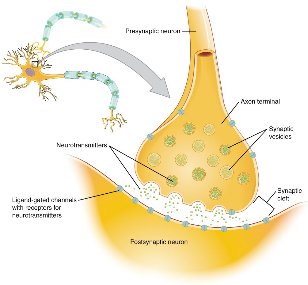
At most junctions, neurons do not touch. A very small gap sits between the end of one neuron and the dendrite of the next. This gap is the synaptic cleft. The whole junction is called the synapse. (A few synapses in the body are electrical and pass current straight through. Most, and the ones this chapter is about, are chemical.)
The electrical spike cannot jump the gap. So the message is turned into a chemical one. When the spike reaches the terminal buttons, tiny sacs called vesicles empty their contents into the cleft. What spills out are molecules of neurotransmitter. These chemical messengers drift across the gap. They lock onto receptors on the receiving neuron. Each receptor is shaped to fit a certain neurotransmitter, the way a lock fits one key.
The effect can be excitatory, which nudges the next neuron toward firing. Or it can be inhibitory, which nudges it away. Either way, the signal has to be shut off fast. If not, the receptor would keep going. The body turns off the signal in two ways. Enzymes in the cleft break the neurotransmitter apart. Or the sending neuron sucks the leftover molecules back up to use again. This second way is called reuptake. Reuptake will matter a lot in a moment, because many drugs for mental health conditions act on exactly this step.
The major neurotransmitters
Scientists have found dozens of neurotransmitters. A handful do most of the work that matters for psychology. Each is loosely tied to certain jobs. Each is also tied to certain disorders when its system runs too high or too low.
| Neurotransmitter | Linked with | When it is out of balance |
|---|---|---|
| Acetylcholine (ACh) | Muscle movement, learning, memory | Loss is linked to Alzheimer's disease |
| Dopamine | Reward, motivation, movement, pleasure | Loss of dopamine cells causes Parkinson's; too much activity in some pathways is linked to schizophrenia |
| Serotonin | Mood, sleep, appetite | Linked to depression, though not as a simple shortage |
| Norepinephrine | Alertness, arousal, the stress response | Tied to anxiety and mood |
| GABA | The brain's main inhibitory messenger; calming | Low activity linked to anxiety and seizures |
| Glutamate | The brain's main excitatory messenger; learning | Too much can overexcite and damage neurons |
| Endorphins | Pain relief and pleasure (the body's own opioids) | Copied by morphine and heroin |
A word of care about serotonin. You may hear that depression is "low serotonin." Research does not back that simple story. Large reviews have not found a clear serotonin shortage in most people with depression. Serotonin is part of the picture. So are stress, sleep, other brain chemicals, life events, and how brain networks change over time.
How drugs act: agonists and antagonists
Almost every drug that changes the mind does it by meddling with this chemical hand-off. The effects fall into two camps. An agonist is anything that increases a neurotransmitter's action. It might copy the neurotransmitter and bind its receptor directly. It might cause more of it to be released. Or it might block reuptake, so the neurotransmitter stays in the cleft longer. An antagonist does the opposite. It blocks or reduces a neurotransmitter's action. Often it plugs the receptor, so the real neurotransmitter cannot dock.
This split is the heart of psychopharmacology, the study of drugs that affect the mind. SSRIs are the most common antidepressants. They are agonists that block the reuptake of serotonin, which leaves more of it in the synapse. Many antipsychotic drugs are dopamine antagonists. They calm an overactive dopamine system. Caffeine, nicotine, opioids, and antihistamines all get their effects by pulling one of these levers.
- Signals cross the synapse as chemicals. Vesicles release neurotransmitter, which binds to receptors on the next neuron.
- The signal is cleared by enzymatic breakdown (enzymes break it apart) or by reuptake back into the sending neuron.
- Key neurotransmitters include dopamine, serotonin, acetylcholine, GABA, glutamate, and endorphins. Each is tied to certain jobs and disorders.
- Agonists increase a neurotransmitter's action. Antagonists block it. Most psychiatric drugs work one of these two ways.
Section 3The nervous system
Central and peripheral
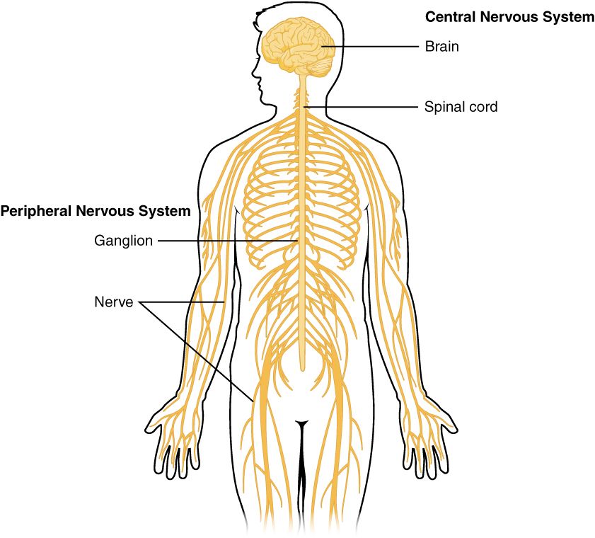
All those neurons are organized into two main parts. The central nervous system (CNS) is the brain and the spinal cord. It is the command center, where information is combined and decisions are made. Everything else is the peripheral nervous system (PNS). This is the huge web of nerves that runs out to the muscles, skin, and organs. Its job is to carry messages back and forth between the CNS and the rest of the body.
Sensory neurons carry messages in toward the CNS. Motor neurons carry messages out to muscles and glands. The spinal cord is not just a cable, either. It can run simple reflexes on its own. That is why your hand jerks off a hot stove before your brain even notices the pain.
Somatic and autonomic
The peripheral system splits again. This time the split is about whether you control the action. The somatic nervous system handles what you do on purpose. It carries sense information in. It also sends commands out to the skeletal muscles you control, as when you lift a cup, type, or walk. The autonomic nervous system runs the background jobs you never think about. These include heartbeat, digestion, breathing rate, pupil size, and sweating. The words "autonomic" and "automatic" sound alike for a reason. This system runs the body on autopilot.
Sympathetic and parasympathetic
The autonomic system has two branches that work against each other, like a gas pedal and a brake. The sympathetic nervous system is the gas pedal. It gets the body ready for action. This is the famous fight-or-flight response. When you face a threat, it speeds up your heart and widens your pupils. It sends sugar into your blood and shifts resources to your muscles, all in an instant.
The parasympathetic nervous system is the brake. It is sometimes called rest-and-digest. Once the danger passes, it slows the heart and restarts digestion. It brings the body back to a calm baseline. Both branches are almost always working at some level. Health depends on the balance between them. Trouble starts when the sympathetic gas pedal is held down too long, as happens in long-term stress.
| Organ | Sympathetic (“fight-or-flight”) | Parasympathetic (“rest-and-digest”) |
|---|---|---|
| Heart | Speeds up | Slows down |
| Pupils | Dilate (widen) | Constrict (narrow) |
| Digestion | Slowed | Stimulated |
| Lungs | Airways widen | Airways narrow |
| Adrenal glands | Release stress hormones | No direct role |
| Overall effect | Spend energy for action | Save energy, restore calm |
- The CNS (brain plus spinal cord) combines information. The PNS carries signals to and from the body.
- The somatic system controls the skeletal muscles you move on purpose. The autonomic system runs organs automatically.
- The sympathetic branch powers fight-or-flight. The parasympathetic branch brings back rest-and-digest.
- Health is the balance between the two. Keeping the sympathetic branch switched on for weeks is the biology of chronic stress.
Section 4The brain
Hindbrain, midbrain, forebrain
One useful way to read the brain is from the bottom up, with the oldest parts first. The hindbrain sits where the spinal cord enters the skull. It runs the machinery of staying alive. It includes three parts. The medulla controls heartbeat and breathing. The pons is a relay station that helps with sleep and arousal. The cerebellum, which means "little brain," coordinates balance, posture, and smooth, practiced movement.
The small midbrain sits just above the hindbrain. It helps you turn toward sudden sights and sounds. It also helps control movement and alertness. Together, the midbrain, pons, and medulla make up the brainstem, the brain's survival core. The cerebellum is part of the hindbrain but is not part of the brainstem. It attaches behind it.
The forebrain is the newest and largest region. It is where most of psychology lives. Deep inside it are several parts we will meet again and again. The thalamus is a relay station for the senses. It routes incoming signals to the right places. The hypothalamus controls hunger, thirst, body temperature, and the endocrine system. The limbic system includes the amygdala, which handles emotion, especially fear. It also includes the hippocampus, which helps form new memories. Draped over all of this is the wrinkled outer layer that makes us most human: the cerebral cortex.
The four cortical lobes
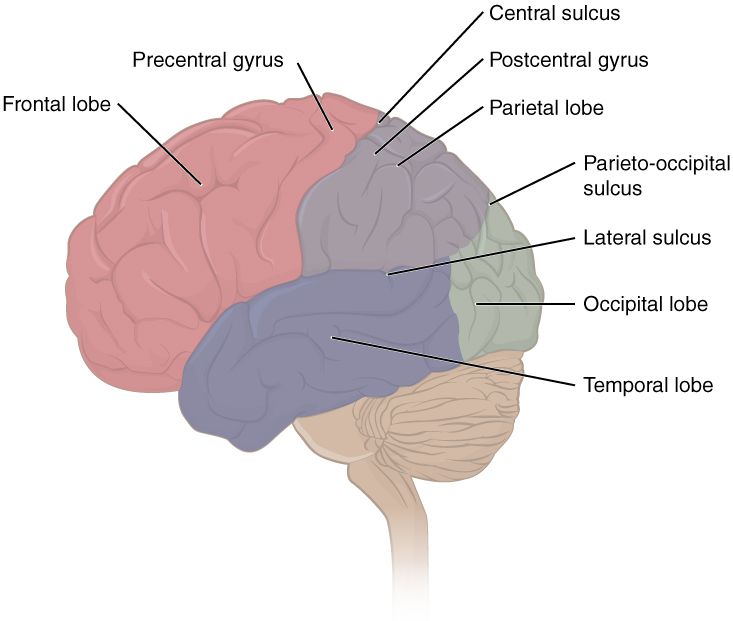
The cerebral cortex is the thin, folded sheet of neurons that covers the brain. It is split into two halves, called hemispheres. Each half is divided into four lobes, and each lobe has typical jobs.
- The frontal lobe sits at the front. It handles planning, judgment, personality, and voluntary movement. Its back strip is the motor cortex, which sends out movement commands. It also holds Broca's area, which is needed to produce speech.
- The parietal lobe sits just behind it. It handles touch, temperature, and body position. It does this along a strip called the somatosensory cortex.
- The temporal lobe sits at the side, near your ears. It handles hearing and understanding language. It is home to Wernicke's area, and it helps with memory.
- The occipital lobe sits at the very back. It is almost all about vision.
| Lobe | Location | Main jobs |
|---|---|---|
| Frontal | Front | Planning, judgment, personality, voluntary movement (motor cortex), speech production (Broca's area) |
| Parietal | Top, toward the back | Touch, temperature, pain, body position (somatosensory cortex) |
| Temporal | Sides | Hearing, understanding language (Wernicke's area), memory |
| Occipital | Back | Vision |
Two hemispheres, one brain
The cortex shows lateralization. That means the two hemispheres are not exact copies. For most people, language is centered in the left hemisphere. The right side leans toward spatial tasks, like reading a map or judging a face. Each hemisphere also controls the opposite side of the body. The left motor cortex moves your right hand.
A thick bundle of fibers called the corpus callosum links the two halves. In the 1960s, some people with severe seizures had this bundle cut to stop the seizures from spreading. Roger Sperry and Michael Gazzaniga studied these split-brain patients. If a picture was flashed only to the left hemisphere, the person could name it. If it was flashed only to the right hemisphere, the person often could not say what it was. But the left hand, which the right hemisphere controls, could still pick it out by touch. Sperry shared a Nobel Prize in 1981 for this work.
These findings are real. But a popular myth grew out of them. It says people are "left-brained" (logical) or "right-brained" (creative). Brain scans do not support this. A large study of over a thousand people found no sign that some people use one side more than the other overall. In a healthy brain, the two halves work as a team on almost every task.
None of this wiring is fully fixed, either. The brain shows great plasticity. That means it can rewire itself. After an injury, or with practice, healthy regions can take over lost jobs. This is strongest in young people, but it lasts across life.
- The hindbrain (medulla, pons, cerebellum) and the midbrain form the brain's survival core. The brainstem is that core minus the cerebellum.
- The forebrain holds the thalamus, hypothalamus, limbic system (amygdala, hippocampus), and cortex.
- The four lobes are frontal (planning, movement, speech), parietal (touch), temporal (hearing, language), and occipital (vision).
- Broca's area, Wernicke's area, split-brain studies, and Phineas Gage each tie a job to certain brain tissue. "Left-brained" and "right-brained" personalities are a myth, and the brain stays plastic.
Section 5The endocrine system
Glands and hormones
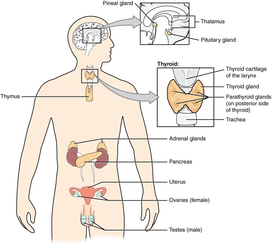
The nervous system is not the body's only message service. The endocrine system is a network of glands. These glands release chemical messengers called hormones straight into the bloodstream. The blood then carries them to organs far away. A neuron speaks privately to the one cell across its synapse. A gland broadcasts to the whole body at once. Any cell with the right receptor will answer.
The "master gland" is the pituitary. It is tucked under the brain, and it takes orders from the hypothalamus. This is the place where the nervous system and the endocrine system shake hands. The pituitary, in turn, directs the other glands.
| Gland | Key hormone(s) | Main effect |
|---|---|---|
| Pituitary | Growth hormone; hormones that signal other glands | The “master gland” directs other glands and controls growth |
| Thyroid | Thyroxine | Sets the body's energy use (metabolic rate) |
| Adrenal glands | Epinephrine (adrenaline), cortisol | Drive the stress response; arousal and energy |
| Pancreas | Insulin, glucagon | Keep blood sugar in balance |
| Gonads (ovaries and testes) | Estrogen, testosterone | Reproduction and sex characteristics |
| Pineal | Melatonin | Helps time sleep and the daily body clock |
Hormones versus nerve signals
Both systems use chemical messengers. Sometimes they even use close cousins. Epinephrine (adrenaline) travels in the blood as a hormone. Its close relative, norepinephrine, works as a neurotransmitter in the brain. But the two systems run on very different time scales, and that difference is the point.
Nerve signals are fast, precise, and brief. An action potential fires in about a thousandth of a second. It aims at one cell, and it is over almost as soon as it starts. Hormone signals are slow, spread out, and long-lasting. A hormone may take seconds to minutes to reach its target. It washes over the whole body, and its effects can last for hours. Think of how long it takes to calm down after a fright, long after the danger is gone. That lingering quality is why hormones shape mood and long-running states, rather than split-second reactions.
| Feature | Nerve signals | Hormone signals |
|---|---|---|
| Messenger | Neurotransmitter | Hormone |
| Route | Across a synapse to one cell | Through the bloodstream to the whole body |
| Speed | Milliseconds | Seconds to minutes |
| How long it lasts | Brief | Long-lasting |
| Precision | Targeted, private | Broadcast, spread out |
- The endocrine system is a network of glands that release hormones into the bloodstream.
- The hypothalamus and the pituitary gland link the nervous and endocrine systems and direct the other glands.
- Compared with nerve signals, hormones are slower, more spread out, and longer-lasting. They are the chemistry of mood and stress.
- Selye's work ties chronic stress and cortisol to real harm to health.
The chapter in one breath
- The neuron takes in signals at its dendrites, adds them up in its cell body, and sends them down its axon. Myelin speeds the signal.
- Crossing threshold sets off an all-or-none action potential. Na+ flows in to depolarize, and K+ flows out to repolarize.
- Signals cross the synapse as neurotransmitters. They bind to receptors and are then cleared by reuptake or by enzymes.
- Drugs act as agonists (boost a neurotransmitter) or antagonists (block it). SSRIs and antipsychotics work this way.
- The nervous system splits into central vs peripheral, somatic vs autonomic, and sympathetic (fight-or-flight) vs parasympathetic (rest-and-digest).
- The brain runs from the hindbrain and midbrain survival core up to the forebrain. Its cortex has four lobes: frontal, parietal, temporal, and occipital. The two hemispheres differ in some jobs but work as a team.
- The endocrine system signals with hormones through the blood. Hormones are slower, more spread out, and longer-lasting than nerve signals, and they drive the stress response.
ReferenceWords worth knowing
Here is a quick guide to the terms in this chapter. Skim it now, or come back any time a word trips you up.
- Action potential
- The quick, all-or-none electrical spike a neuron fires and sends down its axon.
- Agonist
- A drug or substance that increases a neurotransmitter's action. It may copy it, boost its release, or block its reuptake.
- Antagonist
- A drug or substance that blocks or reduces a neurotransmitter's action, often by sitting in its receptor.
- Autonomic nervous system
- The part of the peripheral system that runs automatic jobs like heartbeat and digestion. It has a sympathetic branch and a parasympathetic branch.
- Axon
- The one long fiber that carries a neuron's outgoing signal away from the cell body.
- Central nervous system (CNS)
- The brain and spinal cord, where information is combined and decisions are made.
- Cerebral cortex
- The wrinkled outer layer of the brain. It has two hemispheres and four lobes and handles higher mental jobs.
- Corpus callosum
- The thick bundle of fibers that connects the left and right hemispheres.
- Dendrite
- A branching fiber that takes in signals and carries them toward the cell body.
- Endocrine system
- The network of glands that release hormones into the bloodstream to signal organs far away.
- Glial cell
- A helper cell of the nervous system that makes myelin, feeds neurons, and protects them.
- Hormone
- A chemical messenger that a gland releases into the bloodstream.
- Lateralization
- The way some jobs lean more on one hemisphere than the other, such as language on the left for most people.
- Myelin sheath
- The fatty coating around many axons that speeds the signal. Its gaps are the nodes of Ranvier.
- Neuron
- The basic cell of the nervous system that handles information.
- Neurotransmitter
- A chemical released across a synapse to carry a signal to the next neuron.
- Parasympathetic nervous system
- The autonomic branch that calms the body and restores it: “rest-and-digest.”
- Peripheral nervous system (PNS)
- All the nerves outside the brain and spinal cord. They connect the CNS to the body.
- Plasticity
- The brain's ability to rewire itself after injury or with practice.
- Resting potential
- The neuron's charged, polarized resting state, about −70 mV inside compared with outside.
- Reuptake
- The sending neuron taking leftover neurotransmitter back in.
- Somatic nervous system
- The part of the peripheral system that carries sense information and controls the skeletal muscles you move on purpose.
- Sympathetic nervous system
- The autonomic branch that gets the body ready for action: “fight-or-flight.”
- Synapse
- The junction between two neurons, including the tiny gap (synaptic cleft) the signal crosses.
- Threshold
- The level of charge (about −55 mV) a neuron must reach to fire an action potential.