Bacterial Diversity & Classification
There are more kinds of bacteria than there are kinds of everything else alive, and only a small fraction ever make a person sick — but that fraction is the fraction you will meet at the bedside. The trick to keeping the clinically important organisms straight is not memorizing a list; it is learning the handful of features microbiologists use to sort bacteria into groups. Shape, staining, oxygen needs, and a few special quirks together place almost any pathogen into a recognizable box, and the box tells you how it spreads, what illness it causes, and which drugs will work. This chapter walks through those sorting features one at a time and hangs the most important pathogens on them, so that a culture result or a Gram-stain report starts to read like a sentence you already understand.
By the end of this chapter you can
- Name the three basic bacterial shapes and the common arrangements, and read the clues hidden in a genus name.
- Explain what the Gram stain reveals and why gram-positive and gram-negative cell walls respond differently.
- Identify the major gram-positive pathogens — Staphylococcus, Streptococcus, and Clostridium/Clostridioides — and the diseases they cause.
- Identify the major gram-negative pathogens — E. coli, Salmonella, and Pseudomonas — and explain why endotoxin makes them dangerous.
- Describe the special bacteria — mycobacteria, spirochetes, and mycoplasmas — that break the usual staining and cell-wall rules.
- Classify bacteria by their oxygen requirements and explain why this changes how specimens are collected and where infections form.
- List the other axes — temperature, pH, salt, intracellular lifestyle — used to describe and identify bacteria.
- Connect each classification feature to a concrete nursing action at the bedside.
Section 1Shapes & arrangements
Three basic shapes
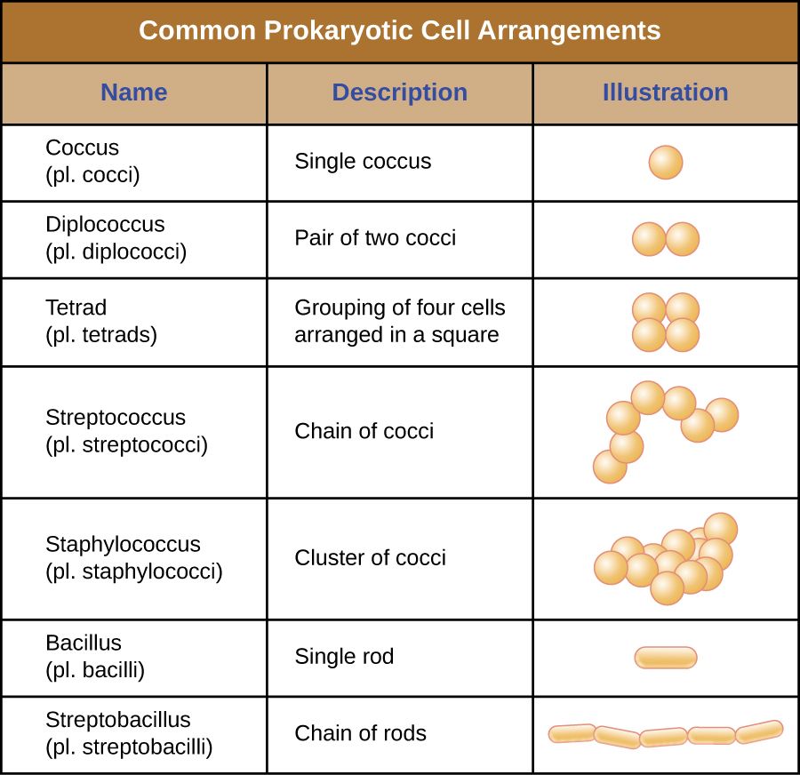
Under the microscope, the overwhelming majority of bacteria take one of three shapes, and the shape is the very first thing a lab reports because it narrows the field so quickly. A coccus (plural cocci) is round or spherical, like a tiny ball. A bacillus (plural bacilli) is a rod, shaped like a stick or a grain of rice. A spirillum (plural spirilla) is spiral or curved, coiled like a corkscrew or bent like a comma. Between these three there are in-between forms worth knowing: a coccobacillus is a short, plump rod that can be mistaken for a coccus, and a vibrio is a short comma-shaped curve — the shape that gives Vibrio cholerae its name.
Shape is not just description. It is built into the name of nearly every organism you will chart, so the moment you learn to hear it, the vocabulary of microbiology starts teaching itself. Staphylococcus and Streptococcus are spheres; Bacillus and Lactobacillus are rods; Spirochetes are coils. Reading the shape out of a genus name is a habit worth building on day one.
Arrangements: how the cells hang together
When a bacterium divides, the two daughter cells may drift apart or may stay stuck together, and the pattern they form is a second identifying clue — especially for cocci. The prefixes are logical once you learn them. Diplo- means the cells sit in pairs (diplococci, as in Neisseria). Strepto- means they form chains, like a string of beads (streptococci). Staphylo- means they clump in irregular grape-like clusters (staphylococci). Cocci can also form flat squares of four (tetrads) or cubical packets of eight (sarcinae). Rods more often line up end-to-end in short chains or side-by-side like a fence (a palisade), the arrangement typical of Corynebacterium.
| What you see | Name | Classic organism |
|---|---|---|
| Round, in grape-like clusters | Staphylococci | Staphylococcus aureus |
| Round, in chains | Streptococci | Streptococcus pyogenes |
| Round, in pairs | Diplococci | Neisseria, S. pneumoniae |
| Rod-shaped | Bacillus | E. coli, Clostridium |
| Comma / curved rod | Vibrio | Vibrio cholerae |
| Spiral / corkscrew | Spirochete | Treponema pallidum |
- The three basic shapes are coccus (sphere), bacillus (rod), and spirillum (spiral); vibrios and coccobacilli are in-between forms.
- Arrangement prefixes — diplo- (pairs), strepto- (chains), staphylo- (clusters) — add a second layer of identification, especially for cocci.
- Shape and arrangement are usually built into the genus name, so the name itself carries clues.
- The preliminary Gram-stain report (shape + arrangement + color) guides empiric therapy before the final identification arrives.
Section 2Gram-positive pathogens
What "gram-positive" means
The Gram stain, devised by Hans Christian Gram in 1884, sorts almost all bacteria into two great camps based on how their cell wall holds a purple dye. Gram-positive bacteria have a thick outer layer of peptidoglycan that traps the crystal-violet stain and turns the cells deep purple. Because that wall is exposed on the surface, gram-positive organisms are the classic targets of the penicillin family, which sabotages peptidoglycan as it is built. The three genera below account for a huge share of the gram-positive disease a nurse will ever see.
Staphylococcus — the cluster
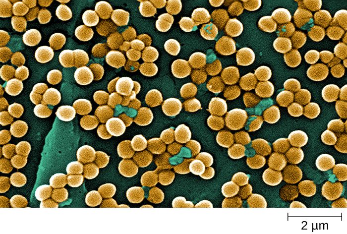
Staphylococcus are gram-positive cocci in grape-like clusters. The species that matters most, Staphylococcus aureus, is a spectacularly versatile pathogen: it lives harmlessly in the nose of roughly a third of healthy people, yet given a break in the skin it causes boils, abscesses, cellulitis, wound infections, bloodstream infections, and endocarditis, and its toxins cause a form of food poisoning and toxic shock syndrome. MRSA — methicillin-resistant S. aureus — is the resistant form that drives contact precautions on so many units. A related group, the coagulase-negative staphylococci such as S. epidermidis, are low-virulence skin residents that nonetheless cause trouble by colonizing catheters, prosthetic joints, and heart valves.
Streptococcus — the chain
Streptococcus are gram-positive cocci in chains, and they are sorted clinically by the way they break down blood on an agar plate and by surface antigens. Group A strep (S. pyogenes) causes strep throat, impetigo, cellulitis, scarlet fever, the flesh-destroying necrotizing fasciitis, and the delayed immune complications of rheumatic fever and post-streptococcal glomerulonephritis. Group B strep (S. agalactiae) colonizes the vagina in some pregnant patients and is a leading cause of newborn sepsis and meningitis — the reason for routine screening late in pregnancy. Streptococcus pneumoniae (a diplococcus) is a top cause of community-acquired pneumonia, otitis media, and meningitis, and is the target of the pneumococcal vaccines.
Clostridium — the spore-former
Clostridium and its relative Clostridioides are gram-positive rods with two dangerous properties: they are obligate anaerobes (Section 5), and they form endospores — dormant, armored survival capsules that shrug off heat, drying, and alcohol. The roll call is memorable: Clostridioides difficile (antibiotic-associated colitis), Clostridium tetani (tetanus), Clostridium botulinum (botulism), and Clostridium perfringens (gas gangrene and food poisoning). Because their spores resist alcohol, an alcohol hand rub does not remove C. difficile — hands must be washed with soap and water, and surfaces cleaned with a sporicidal (bleach) agent.
| Genus | Look | Key species & disease | Nursing flag |
|---|---|---|---|
| Staphylococcus | Cocci in clusters | S. aureus — skin/wound/blood; MRSA | Contact precautions for MRSA |
| Streptococcus | Cocci in chains | Group A (throat, necrotizing); Group B (newborn sepsis); pneumoniae (pneumonia) | Screen GBS in pregnancy; vaccinate for pneumococcus |
| Clostridium / Clostridioides | Rods, spore-forming, anaerobic | C. difficile, tetanus, botulism, gas gangrene | Soap & water, not alcohol, for C. diff |
- Gram-positive bacteria have thick peptidoglycan, stain purple, and are the classic targets of penicillins.
- Staphylococcus = cocci in clusters; S. aureus and MRSA cause skin, wound, and bloodstream infections.
- Streptococcus = cocci in chains; Group A (throat, necrotizing fasciitis, rheumatic fever), Group B (newborn sepsis), pneumoniae (pneumonia/meningitis).
- Clostridium/Clostridioides = anaerobic, spore-forming rods; C. diff spores resist alcohol, so wash with soap and water.
Section 3Gram-negative pathogens
What "gram-negative" means — and why endotoxin matters
Gram-negative bacteria have only a thin layer of peptidoglycan, so they lose the purple crystal violet during decolorizing and take up the pink counterstain instead. What makes them clinically distinct, though, is a second membrane wrapped outside the peptidoglycan — the outer membrane — whose outer face is studded with lipopolysaccharide (LPS). The lipid A portion of LPS is a potent endotoxin: when gram-negative bacteria die and break apart in the bloodstream, released endotoxin triggers a runaway inflammatory cascade — fever, falling blood pressure, and, at the extreme, septic shock. This is why a gram-negative bloodstream infection is a medical emergency, and why the sepsis alerts nurses respond to so often trace back to these organisms.
Escherichia coli — the everyday enteric
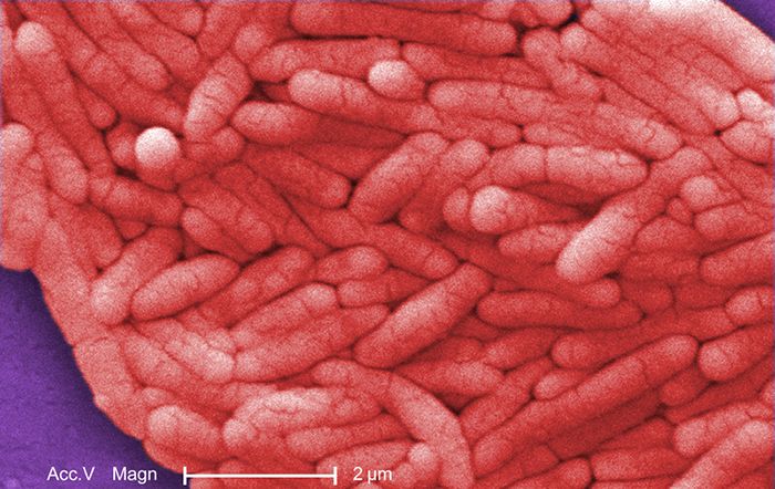
Escherichia coli is a gram-negative rod and a normal resident of the colon, where it is harmless. Out of place, it is the single most common cause of urinary tract infections, and it is a frequent cause of gram-negative sepsis and neonatal meningitis. Certain strains are outright intestinal pathogens: enterohemorrhagic E. coli (such as O157:H7) produces Shiga toxin and can cause bloody diarrhea and hemolytic uremic syndrome, a cause of acute kidney failure in children. Because E. coli is a facultative anaerobe, it thrives both in the oxygen-poor gut and in the bladder.
Salmonella — the food-borne invader
Salmonella are gram-negative rods spread by contaminated food and water and by contact with reptiles and poultry. Most infections are a self-limited gastroenteritis — the classic food poisoning from undercooked eggs or chicken — but Salmonella Typhi causes typhoid fever, a systemic illness with sustained high fever. Salmonella is a reminder that hydration and hand hygiene, not routine antibiotics, are the mainstay for uncomplicated infectious diarrhea in an otherwise healthy adult.
Pseudomonas aeruginosa — the opportunist of wet places
Pseudomonas aeruginosa is a gram-negative rod, an obligate aerobe, and one of the great opportunists of the hospital. It lives in moist reservoirs — sink drains, respiratory equipment, standing water, even flower vases — and it attacks patients whose defenses are down: it causes ventilator-associated pneumonia, infections of burns and chronic wounds, catheter-associated UTIs, and the chronic lung colonization seen in cystic fibrosis. It is famous for producing a blue-green pigment and a grape-like or "corn tortilla" odor, and for being intrinsically resistant to many antibiotics, which makes it hard to treat and a frequent driver of multidrug-resistant outbreaks.
| Organism | Oxygen | Signature disease | Where it lurks |
|---|---|---|---|
| Escherichia coli | Facultative anaerobe | UTI, gram-negative sepsis; O157:H7 → HUS | Colon (normal flora) |
| Salmonella | Facultative anaerobe | Gastroenteritis; typhoid fever | Eggs, poultry, reptiles |
| Pseudomonas aeruginosa | Obligate aerobe | VAP, burn/wound infection, CF lung | Water, moist equipment |
- Gram-negative bacteria stain pink and carry an outer membrane with LPS/endotoxin that can drive septic shock.
- E. coli is the top cause of UTIs and a common cause of gram-negative sepsis; some strains (O157:H7) cause bloody diarrhea and HUS.
- Salmonella is food-borne; most infections are self-limited gastroenteritis, but S. Typhi causes typhoid fever.
- Pseudomonas aeruginosa is a water-loving, drug-resistant opportunist behind ventilator pneumonia, burn/wound, and CF infections.
Section 4Special bacteria
Mycobacteria — the waxy, acid-fast rods
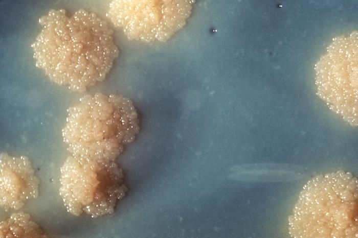
A few important bacteria refuse to fit the Gram-stain scheme, and the most important of these are the mycobacteria. Their cell wall is loaded with a waxy lipid called mycolic acid, which repels ordinary dyes; to see them you need a special acid-fast stain that forces dye into the wall and then resists decolorizing with acid-alcohol. Mycobacterium tuberculosis causes tuberculosis, and its features drive real nursing practice: the waxy wall makes it slow-growing (cultures take weeks) and hardy in the environment, it spreads through the air on tiny droplet nuclei, and it therefore requires airborne isolation — a negative-pressure room and a fitted N95 respirator, not a simple surgical mask. Its cousin Mycobacterium leprae causes leprosy (Hansen disease).
Spirochetes — the corkscrews
Spirochetes are long, thin, tightly coiled bacteria that move with a distinctive corkscrew motion driven by internal axial filaments (endoflagella). They are so slender that many cannot be seen on a routine Gram stain or grown on ordinary media, so they are usually diagnosed by blood tests rather than culture. The three to know are Treponema pallidum (syphilis), Borrelia burgdorferi (Lyme disease, spread by tick bite), and Leptospira (leptospirosis, from water contaminated with animal urine). Their stealthy, hard-to-detect nature is exactly why serologic testing and a careful history matter so much for these infections.
Mycoplasma — the wall-less minimalists
Mycoplasma are the smallest free-living bacteria and are unique in having no cell wall at all. Without a rigid wall they have no fixed shape (they are pleomorphic) and, crucially, no peptidoglycan for a penicillin to attack — so beta-lactam antibiotics are useless against them. Mycoplasma pneumoniae causes "walking pneumonia," a mild, drawn-out atypical pneumonia common in younger people, and is treated with a macrolide or tetracycline instead of a penicillin. The wall-less lesson is a clean illustration of selective toxicity from the other direction: no wall means no target.
| Group | What breaks the rules | Key organism / disease | Practical consequence |
|---|---|---|---|
| Mycobacteria | Waxy mycolic-acid wall; acid-fast | M. tuberculosis — TB | Airborne isolation; slow culture |
| Spirochetes | Thin coils; hard to stain/culture | T. pallidum (syphilis), B. burgdorferi (Lyme) | Diagnosed by serology, not culture |
| Mycoplasma | No cell wall at all | M. pneumoniae — walking pneumonia | Penicillins fail; use macrolide/tetracycline |
- Mycobacteria have a waxy mycolic-acid wall, are acid-fast, grow slowly, and (as M. tuberculosis) require airborne isolation.
- Spirochetes are thin corkscrews (Treponema, Borrelia, Leptospira) usually diagnosed by serology because they resist staining and culture.
- Mycoplasma have no cell wall, so beta-lactam antibiotics do not work; M. pneumoniae causes walking pneumonia.
- Each "special" group breaks a rule — a reminder to match staining, isolation, and drug choice to the organism's structure.
Section 5Oxygen & other groupings
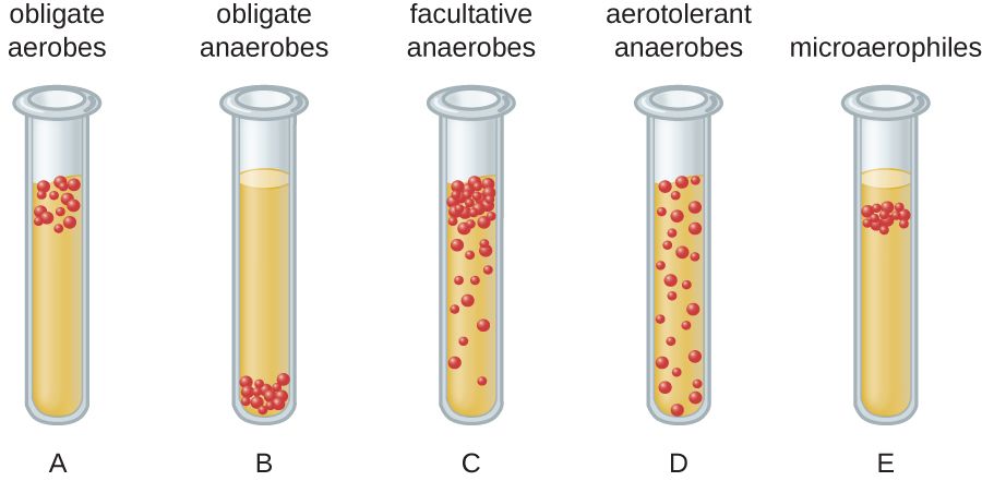
Sorting bacteria by their relationship to oxygen
Oxygen is life-giving to your cells, but to many bacteria it is either essential, irrelevant, or lethal — and how an organism relates to oxygen is a core classification axis with direct bedside consequences. An obligate aerobe must have oxygen to grow (Pseudomonas, Mycobacterium tuberculosis). An obligate anaerobe is poisoned and killed by oxygen and grows only where it is absent (Clostridium, Bacteroides). A facultative anaerobe is the flexible middle: it prefers oxygen but can switch to fermentation without it (E. coli, Staphylococcus, most of the organisms we culture easily). A microaerophile needs oxygen but only at low levels (Campylobacter, Helicobacter pylori), and an aerotolerant anaerobe does not use oxygen at all but survives being exposed to it.
Why oxygen grouping changes nursing care
This is not an academic distinction. Anaerobes flourish in exactly the places oxygen cannot reach: the depths of a puncture wound, the center of an abscess, dead and crushed tissue, the gut, and the pockets of the mouth — which is why deep, dirty, or bite wounds and gas gangrene are anaerobic problems. It also shapes specimen handling: because oxygen kills obligate anaerobes on contact, a specimen collected to look for them must be protected from air and transported to the lab quickly in special anaerobic conditions, or the organism dies before it can be grown and the culture falsely comes back negative.
| Group | Relationship to oxygen | Example |
|---|---|---|
| Obligate aerobe | Requires oxygen | Pseudomonas, M. tuberculosis |
| Obligate anaerobe | Killed by oxygen | Clostridium, Bacteroides |
| Facultative anaerobe | Uses oxygen if present, ferments if not | E. coli, Staphylococcus |
| Microaerophile | Needs low oxygen | Campylobacter, H. pylori |
| Aerotolerant anaerobe | Ignores oxygen but tolerates it | Some lactobacilli |
The other axes of classification
Oxygen is only one of several dials microbiologists turn. Bacteria are also grouped by temperature preference (most human pathogens are mesophiles that thrive near body temperature, 37°C), by pH and salt tolerance (a few extremophiles love acid or high salt), and by whether they can survive only inside host cells. Two clinically important groups are obligate intracellular bacteria — Chlamydia and Rickettsia — which cannot reproduce outside a host cell at all, so they too cannot be grown on standard plates and are diagnosed by other means. Put together, these axes — shape, Gram reaction, oxygen need, growth requirements, and lifestyle — are the coordinates that let a lab pin down almost any organism.
- Bacteria sort by oxygen into obligate aerobes, obligate anaerobes, facultative anaerobes, microaerophiles, and aerotolerant anaerobes.
- Anaerobes cause deep-wound, abscess, and dead-tissue infections, and their specimens must be protected from air during transport.
- Other axes include temperature, pH, salt, and an intracellular lifestyle (e.g., Chlamydia, Rickettsia).
- Combining all the features — shape, stain, oxygen, growth needs — is how the lab identifies an organism.
The chapter in one breath
- Bacteria are sorted first by shape (coccus, bacillus, spirillum) and arrangement (pairs, chains, clusters), clues usually built into the genus name.
- The Gram stain splits bacteria into gram-positive (thick peptidoglycan, purple) and gram-negative (thin wall + outer membrane, pink).
- Key gram-positive pathogens: Staphylococcus (clusters), Streptococcus (chains), and spore-forming anaerobic Clostridium/Clostridioides.
- Key gram-negative pathogens: E. coli, Salmonella, and Pseudomonas — their endotoxin (LPS) can drive septic shock.
- Special bacteria break the rules: mycobacteria are acid-fast, spirochetes are hard-to-stain coils, and mycoplasmas have no cell wall (so penicillins fail).
- Oxygen requirements (aerobe, anaerobe, facultative, microaerophile) shape where infections form and how specimens must be collected.
- Additional axes — temperature, pH, salt, and an intracellular lifestyle — complete the coordinate system labs use to identify any organism.
ReferenceWords worth knowing
A quick reference for the terms in this chapter — skim it now, or circle back whenever a word trips you up.
- Acid-fast
- A staining property of bacteria whose waxy, mycolic-acid wall resists decolorizing by acid-alcohol; the way mycobacteria are identified.
- Aerobe (obligate)
- A bacterium that requires oxygen to grow, such as Pseudomonas or M. tuberculosis.
- Anaerobe (obligate)
- A bacterium that is killed by oxygen and grows only where oxygen is absent, such as Clostridium.
- Arrangement
- The pattern bacterial cells form after dividing — pairs (diplo-), chains (strepto-), or clusters (staphylo-).
- Bacillus
- A rod-shaped bacterium (plural bacilli).
- Coccus
- A spherical or round bacterium (plural cocci).
- Endospore
- A dormant, highly resistant structure formed by Clostridium and Bacillus that survives heat, drying, and alcohol.
- Endotoxin
- The lipid A part of the lipopolysaccharide in a gram-negative outer membrane; released when cells die, it can trigger fever and septic shock.
- Facultative anaerobe
- A bacterium that uses oxygen when available but can grow without it, such as E. coli.
- Gram stain
- The staining procedure that divides bacteria into gram-positive (purple) and gram-negative (pink) by cell-wall structure.
- Gram-negative
- Bacteria with a thin peptidoglycan wall and an outer membrane bearing LPS; they stain pink.
- Gram-positive
- Bacteria with a thick peptidoglycan wall that traps crystal violet and stains purple.
- Lipopolysaccharide (LPS)
- The molecule on the gram-negative outer membrane whose lipid A component is endotoxin.
- Microaerophile
- A bacterium that needs oxygen but only at low levels, such as Campylobacter or H. pylori.
- MRSA
- Methicillin-resistant Staphylococcus aureus; a resistant strain that drives contact precautions.
- Mycolic acid
- The waxy lipid in the mycobacterial cell wall responsible for acid-fastness and environmental hardiness.
- Peptidoglycan
- The mesh-like polymer that forms the bacterial cell wall and is the target of penicillin-family drugs.
- Pleomorphic
- Having no fixed shape; describes wall-less bacteria such as Mycoplasma.
- Spirillum
- A spiral or corkscrew-shaped bacterium (plural spirilla).
- Spirochete
- A thin, tightly coiled bacterium that moves by internal axial filaments, such as Treponema pallidum.
- Vibrio
- A short, comma-shaped curved rod, as in Vibrio cholerae.