Prokaryotic vs Eukaryotic Cells
Almost every microbe that concerns a nurse is a single cell — and that cell is built one of two ways. It is either a prokaryote, a small cell with its DNA loose in the cytoplasm, or a eukaryote, a larger cell that locks its DNA inside a nucleus, exactly like your own cells do. Bacteria are prokaryotes; fungi and protozoa are eukaryotes; your patient is made of eukaryotic cells. That single architectural split is not just trivia — it is the reason an antibiotic can flood a patient's bloodstream and destroy the bacteria while leaving the patient unharmed. This chapter takes a bacterial cell apart piece by piece, from the wall that gives it shape to the spore that lets it survive a decade on a bedrail, and shows you why each structure matters at the bedside.
By the end of this chapter you can
- Contrast prokaryotic and eukaryotic cells by size, nucleus, organelles, and ribosomes.
- Explain how the prokaryote–eukaryote divide creates selective toxicity for antimicrobial drugs.
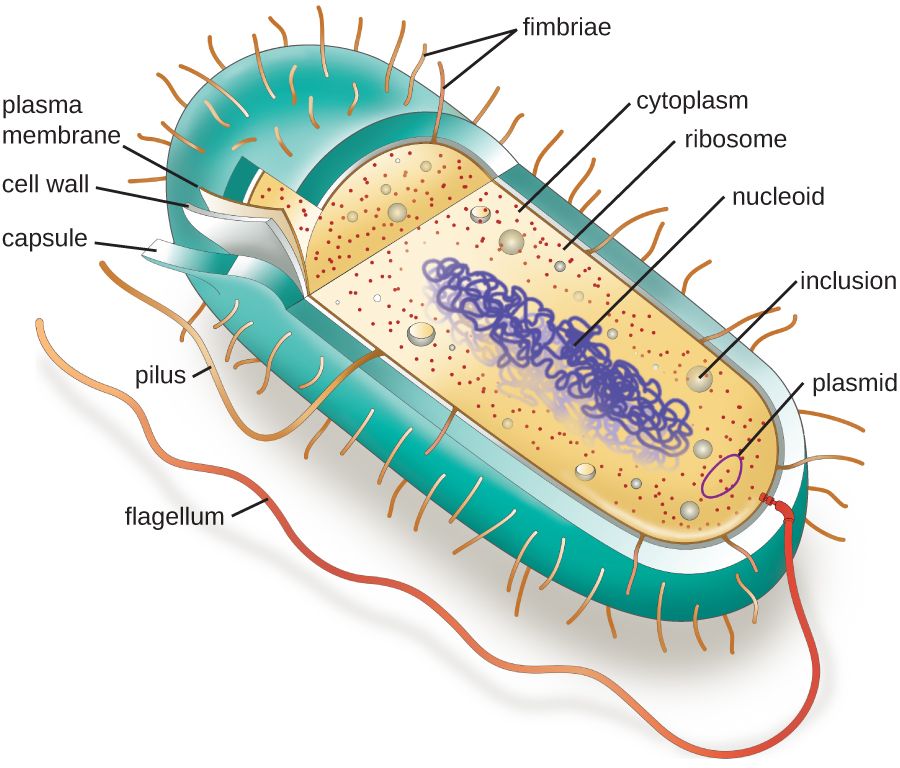
- Describe the interior of a bacterial cell — plasma membrane, cytoplasm with ribosomes, nucleoid, and plasmids — and what each part does.
- Explain the structure of peptidoglycan and contrast Gram-positive and Gram-negative cell walls.
- Connect the cell wall and endotoxin (LPS) to how antibiotics work and to Gram-negative sepsis.
- Describe the roles of the capsule, flagella, pili, and fimbriae in disease and gene transfer.
- Explain how endospores form, why they resist heat and disinfectants, and what that means for infection control.
Section 1Two kinds of cell
The single deepest divide in biology
Of all the ways you could sort living cells, one line runs deeper than any other: does the cell keep its DNA inside a membrane-bound nucleus, or not? Cells that do not are prokaryotes (from Greek roots meaning "before the nut," the "nut" being the nucleus). Cells that do are eukaryotes ("true nut"). This is not a small stylistic difference — it comes with a whole package of other features, and knowing which side of the line a microbe sits on tells you how it is built, how fast it grows, and which drugs can touch it.
Prokaryotes are the bacteria and the archaea. They are small — typically 0.2 to 2 µm across — and simple, with their single circular chromosome floating loose in the cytoplasm. Eukaryotes — the fungi, protozoa, plants, animals, and every cell in your patient's body — are usually ten to a hundred times larger across and far more elaborate inside. When you say a patient has a "bacterial" infection versus a "fungal" one, you are really naming which kind of cell you are fighting.
What prokaryotes lack — and what they have
The headline is what prokaryotes are missing. They have no nucleus, and no other membrane-bound organelles either — no mitochondria, no endoplasmic reticulum, no Golgi apparatus. All of that chemistry happens either in the open cytoplasm or against the inner face of the plasma membrane. But "simple" does not mean "defenseless." Prokaryotes make up for the lack of internal compartments with tough external gear: most wear a rigid cell wall, many add a slimy capsule, and many carry flagella to swim and pili to stick — the structures the rest of this chapter is about.
One difference is easy to overlook but medically enormous: the ribosome. Both cell types build proteins on ribosomes, but the machines are not the same size. Prokaryotes use a 70S ribosome; eukaryotes use a larger 80S ribosome. That size gap is a gift to medicine, because a drug can be designed to jam the 70S machine while ignoring the 80S one.
Why the difference is a drug target
Here is the payoff. Because bacterial cells have structures your cells simply do not have — a peptidoglycan wall, a 70S ribosome, unique enzymes — a drug can attack those targets and destroy the bacterium while leaving the patient's eukaryotic cells alone. This principle is called selective toxicity, and it is the entire reason antibacterial therapy is possible. Penicillin blocks cell-wall building, which human cells never do. Many antibiotics latch onto the 70S ribosome, which human cells do not have. The wider the gap between microbe and host, the safer the drug.
| Feature | Prokaryote (bacteria) | Eukaryote (fungi, protozoa, human) |
|---|---|---|
| Nucleus | Absent — DNA free in nucleoid | Present — DNA in membrane-bound nucleus |
| Typical size | 0.2–2 µm | 10–100 µm |
| Membrane-bound organelles | None | Mitochondria, ER, Golgi, etc. |
| DNA shape | Usually one circular chromosome | Multiple linear chromosomes |
| Ribosomes | 70S | 80S (cytoplasmic) |
| Cell wall | Usually peptidoglycan | Fungi: chitin; protozoa: none; human: none |
| Reproduction | Binary fission | Mitosis / meiosis |
- The deepest split among cells is prokaryote (no nucleus) vs eukaryote (nucleus + organelles).
- Bacteria and archaea are prokaryotes; fungi, protozoa, and human cells are eukaryotes.
- Prokaryotes are small, lack organelles, and use a 70S ribosome; eukaryotes use an 80S ribosome.
- Prokaryote-only structures give antibiotics their selective toxicity — the reason they cure without poisoning the patient.
Section 2Inside the bacterial cell
The plasma membrane: the real boundary
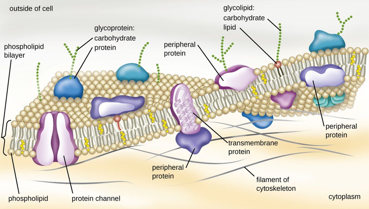
The layers that enclose a bacterium — the plasma membrane, the cell wall, and, in Gram-negatives, an outer membrane — are together called the cell envelope; everything the envelope encloses (cytoplasm, ribosomes, nucleoid, plasmids) is cell contents. Working from the outside in, the wall gives a bacterium its shape, but the plasma membrane is the layer that actually keeps the cell alive. It is a phospholipid bilayer — two sheets of fatty molecules with their water-loving heads facing out and their water-fearing tails tucked inside — peppered with proteins. It is selectively permeable, meaning it decides what crosses: nutrients in, wastes out, ions carefully balanced. Because prokaryotes have no mitochondria, the membrane also does a job your cells hand off to organelles: it runs the electron transport chain and generates the cell's energy right there in the membrane.
That makes the membrane a tempting drug target, but a risky one — bacterial and human membranes are chemically similar, so drugs that punch holes in membranes tend to be toxic. The polymyxins (colistin) do exactly this and are held in reserve as last-resort agents against highly resistant Gram-negative bacteria precisely because they can also injure the patient's kidneys.
Cytoplasm, ribosomes, and inclusions
Fill the membrane and you have the cytoplasm — a crowded, water-based gel where nearly all of the cell's chemistry happens. Suspended in it are tens of thousands of 70S ribosomes, the protein factories introduced in Section 1 and the target of whole families of antibiotics (aminoglycosides, tetracyclines, macrolides). Many bacteria also stockpile spare nutrients in inclusions — little granules of stored carbon, phosphate, or sulfur they can draw on when times are lean.
The nucleoid and plasmids
A prokaryote's DNA is not gone — it is just not gift-wrapped in a nucleus. The single, circular chromosome coils into a dense region called the nucleoid, which has no surrounding membrane, so the DNA sits in direct contact with the cytoplasm. This is one reason bacteria can grow so fast: transcription and translation happen in the same space, at the same time.
Many bacteria carry extra, much smaller loops of DNA called plasmids, separate from the chromosome. Plasmids are not usually needed for basic survival, but they often carry the genes that matter most to nurses: antibiotic-resistance genes, toxin genes, and the machinery to pass those genes to other bacteria. A plasmid can hop from one bacterium to another, which is how resistance spreads through a ward faster than any single organism could reproduce.
| Structure | What it is | Why it matters clinically |
|---|---|---|
| Plasma membrane | Phospholipid bilayer with proteins | Controls transport; makes energy; polymyxin target |
| Cytoplasm | Water-based gel filling the cell | Site of metabolism and growth |
| Ribosomes (70S) | Protein-building machines | Target of many antibiotics (selective toxicity) |
| Nucleoid | Region holding the circular chromosome | No membrane → fast, coupled gene expression |
| Plasmid | Small extra loop of DNA | Carries and spreads resistance & toxin genes |
- The plasma membrane is a selectively permeable phospholipid bilayer that also generates the cell's energy.
- The cytoplasm holds 70S ribosomes — the target of many antibiotics.
- The circular chromosome coils into a membrane-free nucleoid.
- Plasmids are small DNA loops that carry resistance and toxin genes and can move between bacteria.
Section 3The cell wall
Peptidoglycan: the mesh that holds shape
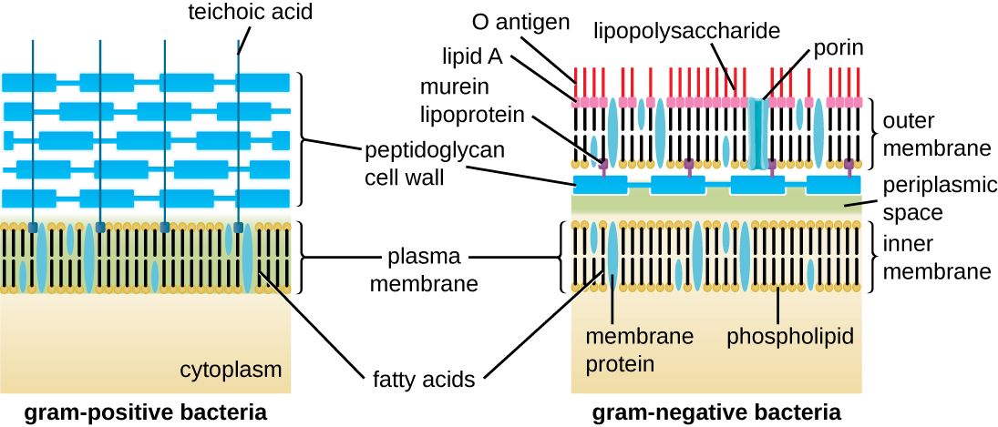
Outside the membrane, most bacteria wear a rigid cell wall made of peptidoglycan (also called murein) — a material found nowhere in the human body. Picture a chain-link fence wrapped completely around the cell: long sugar chains running in parallel, cross-linked to each other by short chains of amino acids. That cross-linking turns a floppy bag into a rigid mesh strong enough to resist the cell's internal osmotic pressure, which would otherwise make it swell and burst. The wall is what gives a bacterium its shape — rod, sphere, or spiral — and its very existence, absent from human cells, is why it is such a clean drug target.
This is exactly where the beta-lactam antibiotics work. Penicillins, cephalosporins, and carbapenems block the enzymes that stitch the peptidoglycan cross-links. The bacterium keeps trying to grow, the wall can no longer hold, and the cell ruptures. Because human cells have no wall to protect, these drugs are among the safest antibiotics we have.
Gram-positive vs Gram-negative walls
Not all walls are built alike, and the difference is the single most useful early clue in a microbiology lab. The Gram stain sorts bacteria into two great camps by how their walls hold a purple dye:
Gram-positive bacteria have a thick peptidoglycan layer on the outside. It traps the crystal-violet dye and the cell stays purple. Examples: Staphylococcus, Streptococcus, Enterococcus, Clostridioides.
Gram-negative bacteria have only a thin peptidoglycan layer, but they add a second, outer membrane on top of it. That outer membrane lets the purple dye wash out during decolorizing, so the cell picks up the pink counterstain and appears red/pink. Examples: Escherichia coli, Klebsiella, Pseudomonas, Neisseria.
Why the Gram stain still runs the show
The Gram result is not just a color — it predicts behavior. That extra outer membrane makes Gram-negative bacteria harder to kill, because it blocks many drugs from ever reaching the wall. And the outer membrane carries a molecule that matters enormously to nurses: lipopolysaccharide (LPS), better known as endotoxin. When Gram-negative bacteria die and break apart, LPS is released into the blood and triggers a violent immune response — fever, plummeting blood pressure, and, at the extreme, septic shock. This is why a Gram-negative bloodstream infection can spiral so fast.
| Feature | Gram-positive | Gram-negative |
|---|---|---|
| Peptidoglycan | Thick, on the surface | Thin, between two membranes |
| Outer membrane | No | Yes — carries LPS |
| Gram stain color | Purple (retains crystal violet) | Pink/red (takes counterstain) |
| Endotoxin (LPS) | No | Yes — drives septic shock |
| Examples | Staph, Strep, Enterococcus, C. diff | E. coli, Klebsiella, Pseudomonas |
- The wall is made of peptidoglycan, a human-free target — where beta-lactam antibiotics act.
- Gram-positive = thick peptidoglycan, stains purple; Gram-negative = thin wall + outer membrane, stains pink.
- The Gram-negative outer membrane blocks many drugs and carries LPS (endotoxin).
- Released endotoxin can drive fever, hypotension, and septic shock.
Section 4External structures
The capsule: a slippery shield
Some bacteria wrap themselves in a thick, sticky outer layer called a capsule (a looser version is called a slime layer). It is not needed to live, but where it matters, it matters a great deal. A capsule is a major virulence factor: it is slippery enough that the patient's immune cells struggle to grab and swallow the bacterium, a trick called resisting phagocytosis. This is why the most dangerous strains of Streptococcus pneumoniae, Haemophilus influenzae, and Neisseria meningitidis are the encapsulated ones. Reassuringly, the capsule is also what several key vaccines are aimed at — teach the immune system to recognize the capsule, and it can clear the organism before it causes meningitis or pneumonia.
Flagella: swimming and chemotaxis
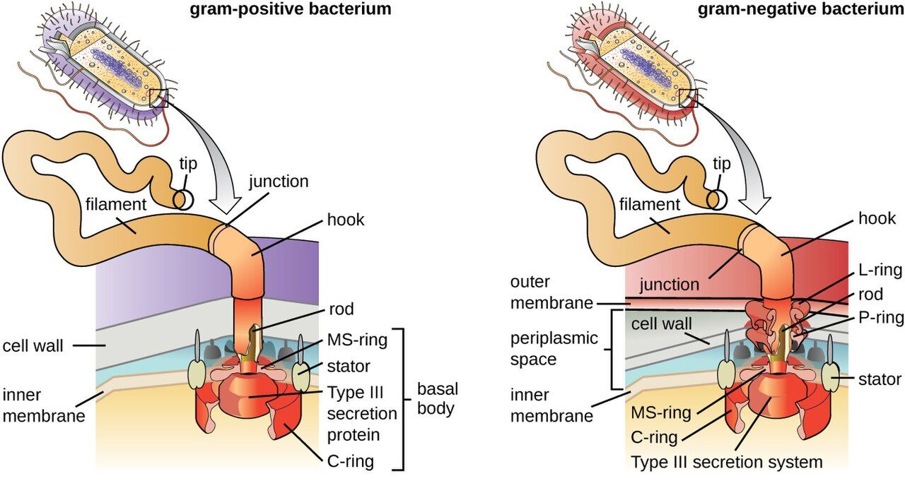
Flagella are long, whip-like tails that rotate like a propeller to push a bacterium through liquid. They are not random wanderers — bacteria steer toward nutrients and away from toxins in a directed movement called chemotaxis. Motility can be a virulence trait: it helps organisms like E. coli and Pseudomonas climb a urinary catheter or reach and colonize new tissue. The number and position of flagella (one end, both ends, or all over) is also a useful clue to a bacterium's identity.
Pili and fimbriae: sticking and swapping genes
Two shorter appendages round out the outside of the cell, and the naming trips students up, so hold them apart:
Fimbriae are numerous, short, hair-like fibers that a bacterium uses to attach to surfaces and to host cells. Attachment is the essential first step of nearly every infection — a microbe that cannot stick gets flushed away. Fimbriae are exactly how E. coli grips the lining of the bladder to start a urinary tract infection, and how bacteria anchor into the slimy communities called biofilms on catheters and implants.
Pili (specifically the sex pilus, or conjugation pilus) are usually longer and far fewer — often just one or two. Their job is different: a pilus builds a bridge to another bacterium and pulls it close so the two can swap DNA in a process called conjugation. This is one of the main ways antibiotic-resistance plasmids move from cell to cell.
| Structure | Main job | Clinical relevance |
|---|---|---|
| Capsule | Sticky outer coat | Resists phagocytosis; vaccine target |
| Flagella | Swimming (chemotaxis) | Motility helps reach and colonize tissue |
| Fimbriae | Attachment (many, short) | Adhesion to host cells; biofilms; UTIs |
| Pili (sex pilus) | DNA transfer (few, long) | Conjugation spreads resistance genes |
- The capsule resists phagocytosis (a virulence factor) and is a target of several vaccines.
- Flagella provide directed movement (chemotaxis) that can help bacteria colonize tissue.
- Fimbriae (many, short) mediate attachment — the first step of infection and of biofilms.
- Pili (few, long) transfer DNA by conjugation, spreading resistance genes.
Section 5Endospores
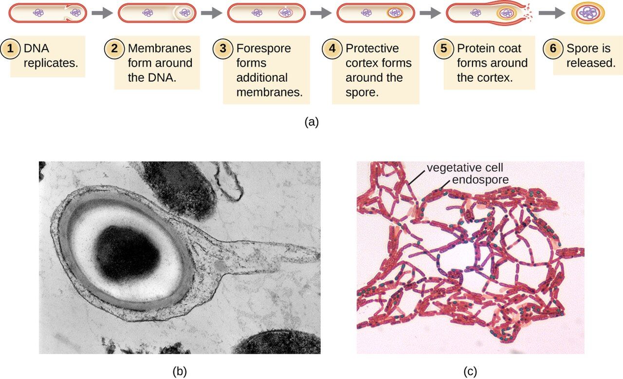
What an endospore is
A handful of Gram-positive bacteria — most importantly the genera Bacillus and Clostridioides/Clostridium — can do something no other microbes can: when conditions turn hostile, they seal a copy of their DNA inside an armored survival capsule called an endospore. The spore is not a way to reproduce — one cell makes one spore — it is a way to wait out disaster. Inside its tough coat, the spore is metabolically dormant: no growth, no chemistry, no vulnerability. It can persist for years, even decades, in soil, on surfaces, or on hospital equipment.
How a spore forms and wakes
The normal, actively growing bacterium is called the vegetative cell. When it senses starvation or other stress, it begins sporulation: it copies its chromosome, walls off a small portion of cytoplasm around that DNA, and wraps it in layer after protective layer — a dense core stripped of most of its water, a thick cortex, and a tough protein coat. The rest of the original cell then disintegrates, releasing the finished endospore.
The spore stays dormant until conditions improve. When nutrients return, it germinates — the coat cracks, water rushes back in, metabolism restarts, and it grows back into an ordinary, dividing vegetative cell. Crucially, the vegetative cell is the form that causes active disease and that ordinary antibiotics and disinfectants can kill; the spore is the form that survives to start the cycle again.
Why spores defeat routine disinfection
The endospore's low water content and layered armor make it extraordinarily resistant — to heat, drying, radiation, and most chemical disinfectants, including the alcohol-based hand rubs that work so well against almost everything else. This is not an academic point; it changes bedside practice. Two organisms make it real:
Clostridioides difficile spreads by spores that survive on bedrails, commodes, and hands. Alcohol gel does not kill them — so for a patient with C. diff, the rule is soap-and-water handwashing (which physically washes spores away) plus a sporicidal cleaner such as diluted bleach for surfaces. Clostridium tetani and Clostridium botulinum spores lurk in soil and improperly canned food. And because spores can shrug off boiling, true sterilization of instruments requires an autoclave — pressurized steam at 121°C — not just hot water.
| Organism | Disease | Why the spore matters |
|---|---|---|
| Clostridioides difficile | Antibiotic-associated colitis | Spores survive alcohol gel → soap & water + bleach |
| Clostridium tetani | Tetanus | Soil spores enter wounds |
| Clostridium botulinum | Botulism | Spores survive in improperly canned food |
| Bacillus anthracis | Anthrax | Durable spores; bioterror concern |
- An endospore is a dormant, armored survival form — not a method of reproduction.
- Made mainly by Gram-positive Bacillus and Clostridioides/Clostridium; the growing form is the vegetative cell.
- Sporulation packages DNA in protective layers under stress; germination revives it when conditions improve.
- Spores resist heat, drying, and disinfectants — hence soap-and-water for C. diff and autoclaving for sterilization.
The chapter in one breath
- Cells are prokaryotic (no nucleus — bacteria) or eukaryotic (nucleus + organelles — fungi, protozoa, human).
- Prokaryote-only structures — peptidoglycan wall, 70S ribosome — give antibiotics their selective toxicity.
- The cell envelope is the plasma membrane (transport + energy) plus the cell wall and, in Gram-negatives, an outer membrane; inside it sit the cytoplasm with 70S ribosomes and the DNA-holding nucleoid, while plasmids carry and spread resistance.
- The wall is peptidoglycan, the target of beta-lactams; Gram-positive = thick wall/purple, Gram-negative = thin wall + outer membrane/pink.
- The Gram-negative outer membrane carries LPS (endotoxin), a driver of septic shock.
- External structures — capsule (resists phagocytosis), flagella (motility), fimbriae (attachment), pili (gene transfer) — are key virulence and resistance-spreading tools.
- Endospores let Bacillus and Clostridioides survive heat and disinfectants — the reason C. diff needs soap-and-water and instruments need autoclaving.
ReferenceWords worth knowing
A quick reference for the terms in this chapter — skim it now, or circle back whenever a word trips you up.
- Capsule
- A thick, sticky outer layer around some bacteria that resists phagocytosis; a virulence factor and vaccine target.
- Cell envelope
- The layers enclosing a bacterium — the plasma membrane, the cell wall, and any outer membrane.
- Cell wall
- The rigid peptidoglycan layer outside the membrane that gives a bacterium its shape and resists osmotic bursting.
- Chemotaxis
- Directed movement by flagella toward nutrients or away from harmful chemicals.
- Conjugation
- Transfer of DNA (often a plasmid) between bacteria through a sex pilus.
- Endospore
- A dormant, highly resistant survival form made by some Gram-positive bacteria; not a means of reproduction.
- Endotoxin (LPS)
- Lipopolysaccharide in the Gram-negative outer membrane that triggers fever, hypotension, and septic shock when released.
- Eukaryote
- A cell whose DNA is enclosed in a membrane-bound nucleus and that contains organelles; fungi, protozoa, and human cells.
- Fimbriae
- Numerous short hair-like appendages used to attach to surfaces and host cells; enable biofilms and adhesion.
- Flagella
- Long whip-like appendages that rotate to propel a bacterium through liquid.
- Gram-negative
- Bacteria with a thin peptidoglycan wall plus an outer membrane; stain pink and carry endotoxin.
- Gram-positive
- Bacteria with a thick peptidoglycan wall and no outer membrane; retain crystal violet and stain purple.
- Nucleoid
- The membrane-free region of a prokaryote where the circular chromosome is coiled.
- Peptidoglycan
- The mesh of cross-linked sugars and amino acids that makes up the bacterial cell wall; the target of beta-lactam antibiotics.
- Pili (sex pilus)
- Long, few appendages that bridge two bacteria to transfer DNA by conjugation.
- Plasma membrane
- The selectively permeable phospholipid bilayer that controls transport and generates energy in prokaryotes.
- Plasmid
- A small circular loop of DNA separate from the chromosome that often carries resistance or toxin genes.
- Prokaryote
- A cell with no membrane-bound nucleus or organelles; bacteria and archaea.
- Ribosome (70S / 80S)
- The protein-building machine; prokaryotes use the smaller 70S, eukaryotes the larger 80S — a key antibiotic target.
- Selective toxicity
- Harming a microbe while sparing the host by targeting a structure the microbe has and the host lacks.
- Sporulation
- The process by which a stressed vegetative cell forms an endospore; reversed by germination.
- Vegetative cell
- The active, growing, dividing form of a bacterium — the form that causes disease and is killed by drugs and disinfectants.