The Musculoskeletal System
Two hundred and six bones, several hundred skeletal muscles, and every joint, tendon, ligament and bursa that connects them — the musculoskeletal system generates more clinical vocabulary than almost any other body system, and you will meet it constantly. It fills orthopedic notes, emergency department triage, rehabilitation goals, operative reports and long-term care charting. The good news is that this vocabulary is unusually orderly. A small set of roots names bone, cartilage, marrow, joint and muscle; a second set names individual bones; and the suffixes you already met in Chapters 2 and 3 attach to them with almost no irregularity. Once you can decode oste/o, arthr/o, my/o, chondr/o and -desis, a term such as osteochondrodysplasia stops being frightening and starts being readable. This chapter builds that decoding habit deliberately, then walks it through pathology, imaging, surgery and the abbreviations that appear on a real chart.
By the end of this chapter you can
- Decode terms built on the core skeletal roots oste/o, oss/e, chondr/o, myel/o and my/o, and explain why my/o and myel/o are not interchangeable.
- Name the combining form for each major bone of the axial and appendicular skeleton and use it to build an adjective or a diagnosis.
- Apply the Latin and Greek plural rules to musculoskeletal nouns such as vertebra, bursa, meniscus, phalanx and epiphysis.
- Distinguish arthr/o, articul/o, synovi/o, burs/o, ten/o, tendin/o, ligament/o and fasci/o, and use them in correctly formed terms.
- Interpret the suffixes of musculoskeletal disease — -algia, -malacia, -porosis, -penia, -sclerosis, -trophy, -asthenia, -plegia, -listhesis and -sarcoma.
- Describe the common fracture types and the difference between a sprain, a strain, a subluxation and a dislocation.
- Separate the surgical suffixes -ectomy, -otomy, -ostomy, -plasty, -desis, -clasis, -centesis and -rrhaphy in orthopedic context.
- Read the abbreviations that appear on orthopedic and rehabilitation charts, including Fx, ROM, ORIF, THA, DJD, HNP, DXA and the weight-bearing codes.
- Pronounce and spell high-frequency terms such as osteomyelitis, spondylolisthesis, arthrocentesis and rhabdomyolysis.
Section 1Bone, Cartilage and Marrow: the Core Roots
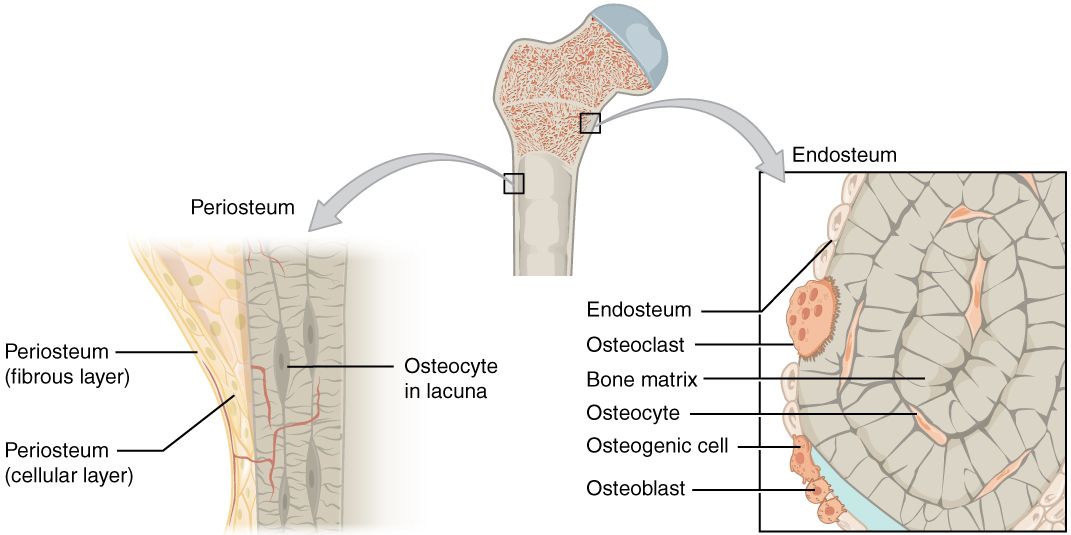
One root, one hundred terms: oste/o
Bone has two roots in medical language because medical language has two parents. The Greek root gives us oste/o, which does nearly all the work in diagnoses and procedures: osteoporosis, osteomyelitis, osteotomy, osteoarthritis. The Latin root gives us oss/e and oss/i, which survive mostly in anatomy and physiology: osseous (OSS-ee-us) tissue means bony tissue, and ossification (os-ih-fih-KAY-shun) is the process of forming bone. A third Latin form, ossicul/o, names the tiny ossicles of the middle ear. When you are unsure which root a clinician will reach for, bet on oste/o.
Take the term osteomyelitis and pull it apart deliberately: oste/o + myel + itis = bone + marrow + inflammation. It names an infection that involves both the bone and the marrow inside it, which is exactly why it is so hard to treat and why it often needs weeks of intravenous antibiotics rather than a short oral course. Now do the same with osteomalacia (os-tee-oh-mah-LAY-shah): oste/o + malacia = bone + softening, the adult form of vitamin D deficiency in which bone is present but poorly mineralized. Compare that to osteoporosis (os-tee-oh-por-OH-sis), oste/o + porosis = bone + porous condition, where the bone is normally mineralized but there is simply less of it. Two words, two different problems, and the suffix told you which was which.
The suffix -porosis has a milder cousin, -penia, meaning deficiency. Osteopenia (os-tee-oh-PEE-nee-ah) is bone density that is lower than normal but not yet low enough to be called osteoporosis. You will see the same suffix throughout the book: leukopenia, thrombocytopenia, and in this chapter sarcopenia (sar-koh-PEE-nee-ah), from sarc/o meaning flesh — the age-related loss of muscle mass that drives so much of the fall risk you will assess in older adults.
| Word part | Meaning | Example term | What the example means |
|---|---|---|---|
| oste/o | bone | osteotomy | surgical incision into a bone (often to realign it) |
| oss/e, oss/i | bone (Latin) | osseous | pertaining to or composed of bone |
| ossicul/o | small bone, ossicle | ossiculoplasty | surgical repair of the middle ear bones |
| myel/o | bone marrow; also spinal cord | myelogram | image of the spinal cord after contrast injection |
| chondr/o | cartilage | chondromalacia | softening of cartilage |
| cortic/o | outer layer, cortex | corticotomy | incision through the outer layer of bone |
| medull/o | inner portion, medulla | medullary cavity | the marrow-filled center of a long bone |
| calc/o, calci/o | calcium | hypocalcemia | low calcium level in the blood |
| orth/o | straight | orthopedics | the specialty that straightens musculoskeletal deformity |
| sarc/o | flesh, connective tissue | sarcopenia | deficiency (loss) of muscle flesh |
The anatomy of a long bone, decoded
Almost every structural word in a long bone is a prefix plus the suffix -physis, meaning growth. The diaphysis (dye-AF-ih-sis) is dia- + physis = through + growth, the long shaft. The epiphysis (eh-PIF-ih-sis) is epi- + physis = upon + growth, the widened end that sits upon the shaft. Between them is the metaphysis, meta- meaning beyond or change, the flared transition zone that contains the growth plate in children. The same suffix appears in symphysis (SIM-fih-sis), sym- + physis = together + growth, a joint where two bones are joined by fibrocartilage — the pubic symphysis being the one you will chart most often.
Wrapping the outside of the shaft is the periosteum (per-ee-OS-tee-um): peri + oste + um = around + bone + structure. Lining the medullary cavity is the endosteum, endo- meaning within. A student who can build periosteum from its parts can also build periostitis (inflammation of the periosteum) and recognize why a periosteal reaction shows up on x-ray when bone is irritated. This is the whole point of a word-part approach: one decoded term becomes a template for a dozen more.
Bone is a living, constantly rebuilt tissue, and three cell names carry that story. An osteoblast (OS-tee-oh-blast) uses -blast, an immature or building cell, and lays down new bone. An osteoclast (OS-tee-oh-klast) uses -clast, meaning to break, and resorbs bone. An osteocyte is the mature cell trapped in the matrix it built. Blast builds, clast crushes — a crude mnemonic, but it holds, and it will hold again in Chapter 8 when you meet the blast cells of blood.
| Word part | Meaning | Example term | What the example means |
|---|---|---|---|
| -physis | growth | epiphysis | the end of a long bone, upon the shaft |
| -blast | immature or forming cell | osteoblast | bone-forming cell |
| -clast | to break down | osteoclast | cell that resorbs bone |
| -cyte | cell | osteocyte | mature bone cell |
| peri- | around | periosteum | membrane around the bone |
| endo- | within | endosteum | membrane lining the inside of bone |
| -porosis | porous condition | osteoporosis | porous, low-density bone |
| -penia | deficiency | osteopenia | bone density below normal but above osteoporosis |
| -malacia | softening | osteomalacia | softening of bone from poor mineralization |
| -sclerosis | hardening | osteosclerosis | abnormal hardening or thickening of bone |
| -genesis | formation, production | osteogenesis | the formation of bone |
| -poiesis | formation (of cells) | hematopoiesis | blood cell formation, which occurs in red marrow |
Cartilage and marrow
Chondr/o names cartilage, the smooth, avascular tissue that caps joint surfaces and forms structures such as the costal cartilages and the intervertebral discs. Chondromalacia (kon-droh-mah-LAY-shah) patellae is softening of the cartilage behind the kneecap, a common cause of anterior knee pain in young athletes. Costochondritis is cost/o + chondr + itis = rib + cartilage + inflammation, a benign chest-wall pain that is frequently mistaken for cardiac pain in triage. A chondrosarcoma is a malignant tumor arising from cartilage; the suffix -sarcoma always signals a malignancy of connective tissue, in contrast to -oma alone, which usually indicates a benign growth (a chondroma is benign).
Myel/o is the root that catches students out, because it legitimately carries two meanings: bone marrow and spinal cord. Context decides. In osteomyelitis and myelosuppression, it means marrow. In myelogram and myelopathy, it means spinal cord. Nothing in the word itself resolves the ambiguity, so you resolve it from the neighbors: if the term also carries oste/o or sits in a hematology note, think marrow; if it carries a spinal or neurologic partner, think cord. Multiple myeloma, a malignancy of plasma cells in the marrow, uses the marrow sense and is a classic cause of pathologic fracture.
Bone vocabulary is built from a handful of roots — oste/o, chondr/o, myel/o — combined with position prefixes and condition suffixes, so decoding the parts is far more efficient than memorizing the whole.
Section 2Mapping the Skeleton, Bone by Bone
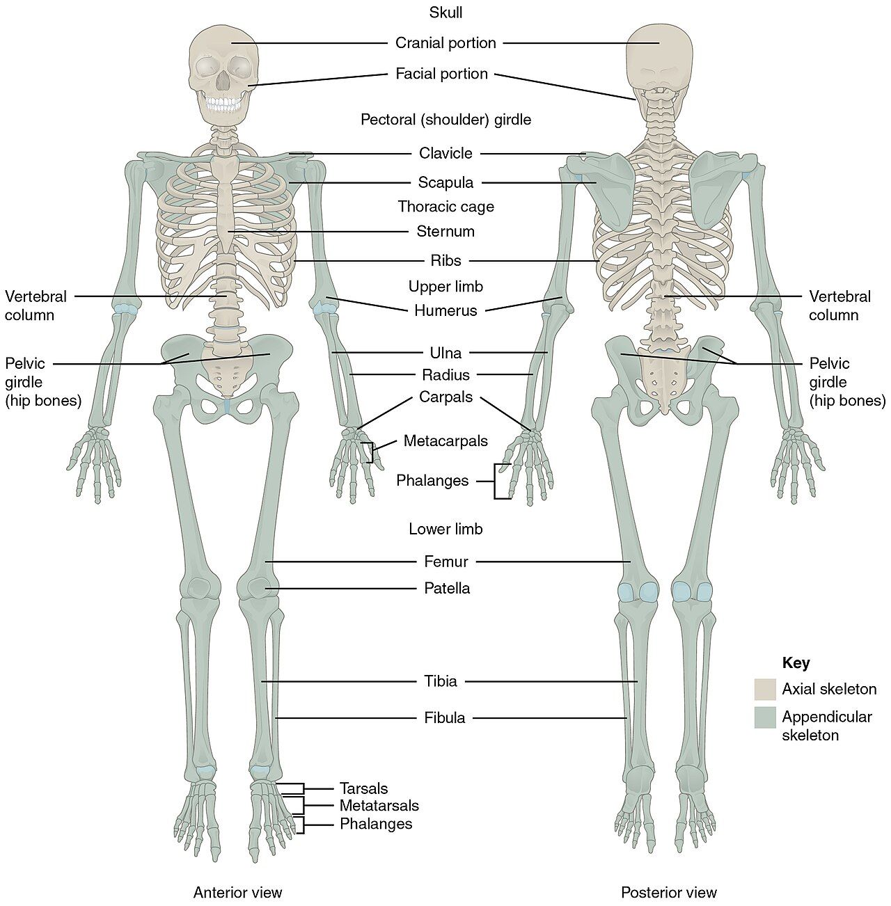
The axial skeleton: skull, spine, ribs and sternum
The axial skeleton is the central axis: cranium, facial bones, vertebral column, ribs and sternum. Its combining forms behave predictably. Crani/o gives craniotomy (incision into the skull, with the bone flap replaced) and craniectomy (a portion of skull removed and not replaced) — a single suffix separating two very different operations. Cost/o gives intercostal, between the ribs, the space where a chest tube goes and where you count retractions in a child with respiratory distress. Stern/o gives sternotomy, the midline incision used for open heart surgery, and the xiphoid (ZIF-oyd) process is the small cartilaginous tip you locate before chest compressions.
The spine has three roots, and knowing which one to reach for is a genuine skill. Vertebr/o is Latin and appears in anatomic adjectives: vertebral column, intervertebral disc. Spondyl/o is Greek and appears in diagnoses: spondylitis, spondylosis, spondylolisthesis. Rachi/o also means spinal column and survives in a few terms such as rachischisis (a congenital splitting of the vertebral column). Then there are the regional roots you already know from Chapter 4: cervic/o (neck), thorac/o (chest), lumb/o (lower back), sacr/o and coccyg/o. Charted numerically, the column reads C1 through C7, T1 through T12, L1 through L5, then the fused sacrum and coccyx. A note that says “L4-L5 HNP” is telling you the herniated disc sits between the fourth and fifth lumbar vertebrae.
The appendicular skeleton: girdles and limbs
The appendicular skeleton is everything that hangs off the axis: the shoulder girdle, the arms, the pelvic girdle and the legs. Nearly every one of these bones lends its Latin name directly to a combining form, so the terms are almost self-translating. Acromioclavicular breaks into acromi/o + clavicul + ar = acromion + clavicle + pertaining to, which is exactly the joint at the top of the shoulder that separates in a hard fall. Metacarpophalangeal is meta- + carp/o + phalange + al = beyond + wrist bones + finger bones + pertaining to, the knuckle joints that swell symmetrically in rheumatoid arthritis.
The pelvis is worth extra attention because it is three fused bones with three separate roots. Ili/o names the broad flaring ilium whose crest you palpate; ischi/o names the ischium (IS-kee-um) you sit on, the site of ischial pressure injuries in wheelchair users; pub/o names the pubis at the front. Where they meet, they form the acetabulum (as-eh-TAB-yoo-lum), the socket that receives the head of the femur and the structure replaced in a total hip arthroplasty.
| Combining form | Bone | Example term | What the example means |
|---|---|---|---|
| crani/o | skull | craniotomy | surgical incision into the skull |
| maxill/o | upper jaw | maxillofacial | pertaining to the upper jaw and face |
| mandibul/o | lower jaw | mandibular | pertaining to the lower jaw |
| vertebr/o | vertebra (anatomic use) | intervertebral | between the vertebrae |
| spondyl/o | vertebra (disease use) | spondylosis | degenerative condition of the vertebrae |
| rachi/o | spinal column | rachischisis | congenital splitting of the vertebral column |
| lamin/o | lamina (part of vertebral arch) | laminectomy | removal of the lamina to decompress the cord or nerve |
| cost/o | rib | intercostal | between the ribs |
| stern/o | sternum, breastbone | sternotomy | surgical incision through the breastbone |
| clavicul/o | clavicle, collarbone | subclavian | beneath the clavicle |
| scapul/o | scapula, shoulder blade | scapular | pertaining to the shoulder blade |
| acromi/o | acromion (tip of shoulder blade) | acromioplasty | surgical reshaping of the acromion |
| humer/o | humerus, upper arm bone | humeral | pertaining to the humerus |
| radi/o | radius (also x-ray, radiation) | radial pulse | pulse felt on the radius side of the wrist |
| uln/o | ulna | ulnar deviation | drift of the fingers toward the ulnar side |
| carp/o | wrist bones | carpal tunnel | the passage through the wrist bones |
| metacarp/o | hand bones | metacarpal | pertaining to the bones beyond the wrist |
| phalang/o | finger or toe bone | phalangeal | pertaining to a finger or toe bone |
| pelv/i, pelv/o | pelvis | pelvimetry | measurement of the pelvic dimensions |
| ili/o | ilium (flaring hip bone) | iliac crest | the upper ridge of the ilium |
| ischi/o | ischium (lower posterior hip bone) | ischial | pertaining to the sitting bone |
| pub/o | pubis (anterior hip bone) | suprapubic | above the pubic bone |
| acetabul/o | acetabulum, hip socket | acetabular | pertaining to the hip socket |
| femor/o | femur, thigh bone | femoral artery | artery running alongside the femur |
| patell/o | patella, kneecap | patellofemoral | pertaining to the kneecap and femur |
| tibi/o | tibia, shin bone | tibial | pertaining to the tibia |
| fibul/o, perone/o | fibula | peroneal nerve | nerve coursing near the fibula |
| tars/o | ankle bones (also edge of eyelid) | metatarsal | pertaining to the foot bones beyond the ankle |
| calcane/o | calcaneus, heel bone | calcaneal spur | bony outgrowth on the heel bone |
Making these words plural
Musculoskeletal nouns are overwhelmingly Latin and Greek, so English plural rules will fail you. “Two vertebras” and “both meniscuses” mark a note as careless. The rules are few and they repeat across every chapter of this book, so learning them here pays dividends in Chapter 10 and Chapter 12.
| Ending rule | Singular | Plural | Note |
|---|---|---|---|
| -a becomes -ae | vertebra, bursa, fascia, scapula, patella | vertebrae, bursae, fasciae, scapulae, patellae | The -ae is usually pronounced “ee”: ver-teh-BREE |
| -us becomes -i | humerus, radius, meniscus, calcaneus, malleolus | humeri, radii, menisci, calcanei, malleoli | Menisci is said men-ISS-kye |
| -um becomes -a | acetabulum, ischium, sternum, periosteum | acetabula, ischia, sterna, periostea | Do not confuse this -a plural with the -a singular above |
| -is becomes -es | epiphysis, diaphysis, symphysis, pubis | epiphyses, diaphyses, symphyses, pubes | Pronounced -eez: eh-PIF-ih-seez |
| -nx becomes -nges | phalanx | phalanges | fah-LAN-jeez |
| -ex, -ix becomes -ices | cortex, apex | cortices, apices | KOR-tih-seez |
| -oma becomes -omata (or -omas) | sarcoma, fibroma | sarcomata, fibromata | The English -s plural is now accepted in most charts |
| -os becomes -ossa | os (bone) | ossa | Formal anatomy: one os coxae, two ossa coxae |
Each named bone carries its own combining form, so an unfamiliar orthopedic term usually resolves the moment you identify which bone is hiding inside it — and its plural follows the Latin or Greek ending, not English.
Section 3Joints, Muscles and the Tissue Between
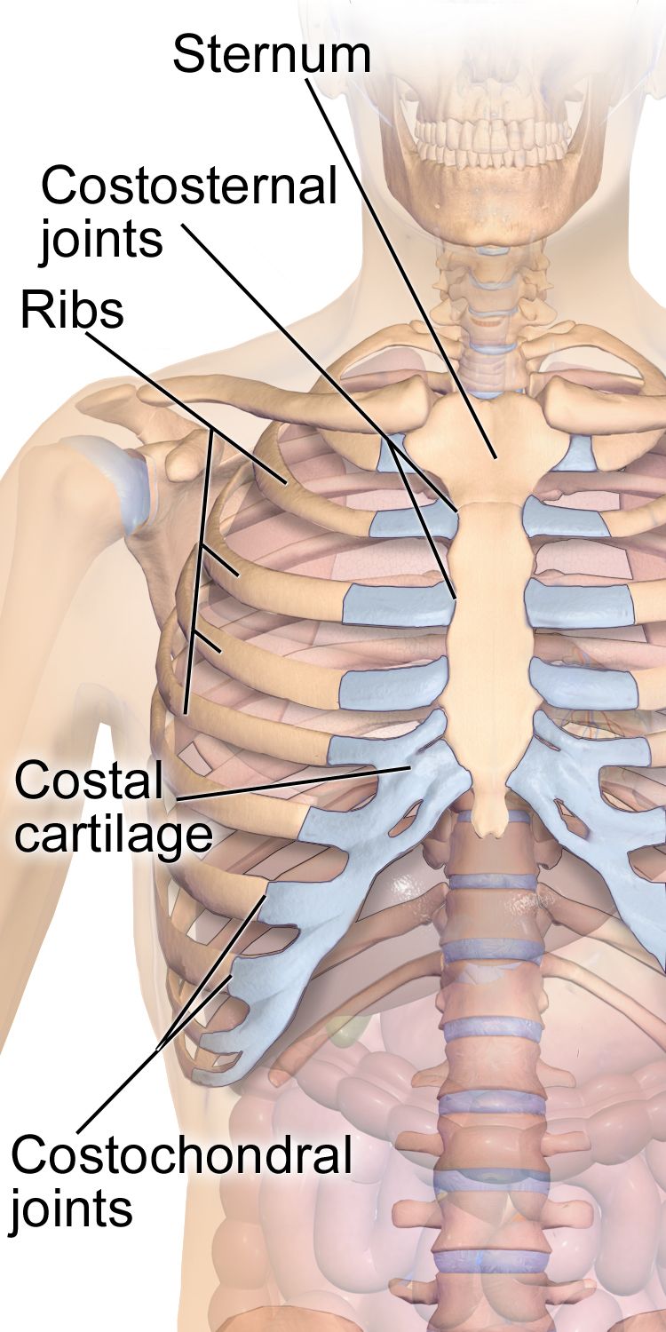
Joint roots and the structures inside a joint
A joint is where two bones meet, and again there is a Greek root and a Latin one. Arthr/o is Greek and dominates clinical terms: arthritis, arthroscopy, arthroplasty, arthrocentesis. Articul/o is Latin and supplies the descriptive words: articular cartilage, articulation. A synovial joint — the freely movable kind at the knee, hip and shoulder — is lined by a synovial (sih-NOH-vee-al) membrane that secretes synovial fluid, so synovi/o and synov/o both name that lining. Synovitis is inflammation of that membrane, and it is what makes an inflamed joint feel boggy rather than merely tender.
Near many joints sits a bursa (BER-sah), a small fluid-filled sac that cushions tendon against bone. Burs/o gives bursitis (ber-SYE-tis) and bursectomy. Ligaments connect bone to bone; tendons connect muscle to bone. Ligament/o is straightforward, but tendon has two forms — ten/o and tendin/o — and which one you use depends on the suffix. Tenorrhaphy (teh-NOR-ah-fee) is suturing a torn tendon; tenosynovitis (ten-oh-sin-oh-VYE-tis) is inflammation of a tendon and its sheath; but the inflammation of a tendon by itself is spelled tendinitis. You will see “tendonitis” in charts and even on medication labels, and it is widely understood, but tendinitis is the correctly built form: the Latin stem is tendin- (from tendo, tendinis), and the combining vowel o is dropped before the vowel-initial suffix -itis, exactly as it is in osteomyelitis.
| Word part | Meaning | Example term | What the example means |
|---|---|---|---|
| arthr/o | joint | arthralgia | joint pain |
| articul/o | joint (Latin) | articular surface | the joint-facing surface of a bone |
| synovi/o, synov/o | synovial membrane or fluid | synovitis | inflammation of the joint lining |
| burs/o | bursa, fluid-filled sac | bursitis | inflammation of a bursa |
| ten/o, tendin/o | tendon | tendinitis | inflammation of a tendon |
| ligament/o | ligament | ligamentous laxity | looseness of the bone-to-bone connectors |
| fasci/o | fascia, band of connective tissue | fasciotomy | incision through fascia to relieve pressure |
| menisc/o | meniscus, crescent cartilage | meniscectomy | surgical removal of a torn meniscus |
| ankyl/o | stiff, bent, fused | ankylosis | abnormal stiffening or fusion of a joint |
| -desis | surgical fusion, binding | arthrodesis | surgical fusion of a joint |
Muscle roots and the suffixes of strength
There are roughly 600 skeletal muscles, and three tissue types: skeletal (striated and voluntary), smooth (visceral and involuntary) and cardiac. My/o, myos/o and muscul/o all name muscle in general. Two specialized roots name the tissue subtype: rhabdomy/o is striated or skeletal muscle, and leiomy/o is smooth muscle. That distinction is not decorative — a leiomyoma is the benign uterine fibroid you will chart in Chapter 12, while a rhabdomyosarcoma is an aggressive malignancy of skeletal muscle seen mostly in children.
Rhabdomyolysis (rab-doh-my-OL-ih-sis) deserves a slow decode because it is a true emergency: rhabd/o + my/o + lysis = rod-shaped (striated) + muscle + breakdown. Skeletal muscle disintegrates, dumping myoglobin into the blood, which can obstruct the kidneys. It follows crush injury, prolonged immobility on a floor, extreme exertion or certain drugs, and the giveaway on the chart is a creatine kinase level in the thousands with tea-colored urine.
The suffixes that attach to muscle roots describe function rather than structure. -trophy means development or nourishment: atrophy (AT-roh-fee) is wasting from disuse or denervation, hypertrophy is enlargement from work, and dystrophy (DIS-troh-fee) is faulty development, as in muscular dystrophy. -asthenia means weakness, giving myasthenia. -plegia means paralysis and -paresis means partial weakness, so hemiplegia is one-sided paralysis and hemiparesis is one-sided weakness — a distinction that changes a stroke note materially. -tonia and ton/o refer to muscle tone: hypotonia is floppiness, dystonia is sustained abnormal contraction.
| Word part | Meaning | Example term | What the example means |
|---|---|---|---|
| my/o, myos/o | muscle | myositis | inflammation of muscle |
| muscul/o | muscle (Latin) | musculoskeletal | pertaining to muscles and bones |
| rhabdomy/o | striated, skeletal muscle | rhabdomyolysis | breakdown of skeletal muscle |
| leiomy/o | smooth muscle | leiomyoma | benign smooth muscle tumor (uterine fibroid) |
| fibr/o | fiber, fibrous tissue | fibromyalgia | pain in fibrous tissue and muscle |
| kinesi/o, -kinesia | movement | bradykinesia | abnormally slow movement |
| ton/o, -tonia | tone, tension | hypotonia | decreased muscle tone |
| -trophy | development, nourishment | atrophy | wasting away from lack of use or nerve supply |
| -asthenia | weakness | myasthenia gravis | grave muscle weakness of autoimmune origin |
| -plegia | paralysis | paraplegia | paralysis of the lower body |
| -paresis | partial paralysis, weakness | hemiparesis | weakness of one side of the body |
| -lysis | breakdown, separation | tenolysis | surgical release of a tendon from adhesions |
The vocabulary of motion
Rehabilitation notes are written almost entirely in movement terms, and most are prefix-driven. Abduction moves a limb away from the midline; adduction adds it back toward midline. Flexion decreases a joint angle, extension increases it, and hyperextension pushes it past normal. Supination turns the palm up (as if holding soup); pronation turns it down. At the ankle, dorsiflexion lifts the toes toward the shin and plantar flexion points them down. Inversion turns the sole inward, eversion outward, and the classic ankle sprain is an inversion injury of the lateral ligaments. Circumduction is the cone-shaped movement of a ball-and-socket joint, from circum- meaning around.
Two more words appear constantly and are easy to blur. A contracture (kon-TRAK-cher) is a permanent shortening of muscle, tendon or skin that fixes a joint in position — the thing you are preventing when you perform range-of-motion exercises on an immobile patient. Crepitus (KREP-ih-tus) is the grating sound or sensation of bone or roughened cartilage rubbing, felt over an arthritic knee or a fracture site.
Joints and muscles each carry a Greek clinical root and a Latin descriptive root, and the suffixes attached to them — -trophy, -asthenia, -plegia, -desis — describe function and repair rather than structure.
Section 4Pathology: Fractures, Arthritides and Muscle Disease
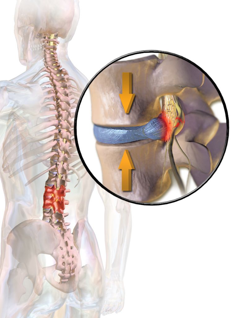
The language of fractures
A fracture (charted as Fx) is a break in bone continuity, and the descriptors fall into two families. The first describes whether skin is broken: a closed or simple fracture leaves skin intact; an open or compound fracture has bone communicating with the outside, which converts a bone injury into an infection risk and makes osteomyelitis a real concern. The second family describes the fracture line. A transverse fracture runs straight across; an oblique one runs at an angle; a spiral fracture twists around the shaft and results from a rotational force. A comminuted (KOM-ih-noo-ted) fracture shatters the bone into three or more pieces. An impacted fracture drives one fragment into another. A compression fracture collapses a vertebral body, and in an older adult it is often the first visible sign of osteoporosis.
Two more categories name a cause rather than a shape. A greenstick fracture is an incomplete break in the soft, pliable bone of a child — the bone bends and cracks on one side like a green twig. A pathologic fracture occurs through bone already weakened by disease such as osteoporosis, metastatic cancer or multiple myeloma, so a minimal force produces a major break. A stress fracture is a hairline break from repetitive loading, common in runners and new military recruits, and often invisible on the first x-ray.
Eponyms persist in fracture language even as the field moves toward descriptive names. A Colles (KOL-eez) fracture is a distal radius fracture with dorsal displacement, the classic result of falling on an outstretched hand. A Pott fracture involves the distal fibula at the ankle. Both remain in daily use, but modern radiology reports increasingly write “distal radius fracture with dorsal angulation” instead — and note that current style drops the possessive, so it is Colles fracture, not Colles’s.
Treatment vocabulary follows directly. Reduction means restoring the bone to alignment: closed reduction is done by manipulation without opening the skin, open reduction requires a surgical incision. Adding hardware gives internal fixation (plates, screws, rods) or external fixation (a frame outside the body), which is why the single most common orthopedic abbreviation on a surgical schedule is ORIF — open reduction with internal fixation.
Word parts that name skeletal disease
| Word part | Meaning | Example term | What the example means |
|---|---|---|---|
| -algia, -dynia | pain | myalgia, coccygodynia | muscle pain; pain in the tailbone |
| -itis | inflammation | bursitis | inflammation of a bursa |
| -osis | abnormal condition | spondylosis | degenerative condition of the vertebrae |
| -listhesis | slipping | spondylolisthesis | forward slipping of one vertebra over another |
| -schisis | splitting, cleft | rachischisis | congenital cleft of the vertebral column |
| -oma | tumor, mass (usually benign) | osteoma | benign bone tumor |
| -sarcoma | malignant connective tissue tumor | osteosarcoma | malignant bone tumor, most often in adolescents |
| -pathy | disease | arthropathy | any disease of a joint |
| -ptosis | drooping, sagging | visceroptosis | downward displacement of organs |
| kyph/o | hump | kyphosis | exaggerated outward curve of the thoracic spine |
| lord/o | bent backward, swayback | lordosis | exaggerated inward curve of the lumbar spine |
| scoli/o | crooked, bent sideways | scoliosis | lateral curvature of the spine |
The three spinal curvature terms reward a moment of care because they are frequently swapped. Kyphosis (kye-FOH-sis) is the exaggerated forward rounding of the upper back, the “dowager’s hump” of vertebral compression fractures. Lordosis (lor-DOH-sis) is the exaggerated inward sway of the lower back, seen in late pregnancy and in obesity. Scoliosis (skoh-lee-OH-sis) is the sideways S- or C-shaped curve screened for in adolescents. Kyphosis and lordosis exaggerate curves the spine normally has; scoliosis creates a curve that should not exist at all.
The arthritides and other joint disease
Osteoarthritis (OA), also charted as degenerative joint disease (DJD), is mechanical wear of articular cartilage. It favors weight-bearing joints and the distal finger joints, is typically asymmetric, produces morning stiffness that eases within about thirty minutes, and generates bony nodes at the finger joints. Rheumatoid arthritis (RA) is an autoimmune synovitis: symmetric, favoring the small joints of the hands and wrists, with morning stiffness lasting an hour or more and systemic features such as fatigue and low-grade fever. The distinction matters at the bedside because OA responds to activity modification and analgesia while RA needs disease-modifying drugs to prevent joint destruction.
Gout is arthritis caused by monosodium urate crystals depositing in a joint, classically the great toe — an attack there is called podagra (poh-DAG-rah), from pod/o meaning foot. Ankylosing spondylitis (ang-kih-LOH-sing spon-dih-LYE-tis) decodes as stiffening + vertebra + inflammation: a chronic inflammatory arthritis that begins at the sacroiliac joints and can fuse the spine into the rigid “bamboo” appearance seen on x-ray. Spondylolisthesis (spon-dih-loh-lis-THEE-sis) is a vertebra slipping forward on the one below it, most often at L5-S1, and it produces back pain with radiating nerve symptoms.
Soft-tissue diagnoses fill clinic schedules. Carpal tunnel syndrome (CTS) is compression of the median nerve as it passes beneath the transverse carpal ligament, causing numbness in the thumb, index and middle fingers. Plantar fasciitis (PLAN-tar fash-ee-EYE-tis) is inflammation of the fascia along the sole, producing sharp heel pain with the first steps of the morning. Lateral epicondylitis is tennis elbow; medial epicondylitis is golfer’s elbow. Herniated nucleus pulposus (HNP) is protrusion of the gel-like disc center through its outer ring, pressing on a nerve root.
Muscle and bone disease
Muscular dystrophy is a group of inherited disorders of progressive muscle degeneration; the Duchenne form is X-linked, appears in young boys and is caused by absent dystrophin protein. Myasthenia gravis (my-as-THEE-nee-ah GRAV-is) — literally grave muscle weakness — is an autoimmune attack on acetylcholine receptors at the neuromuscular junction, producing weakness that worsens with use and improves with rest, often first noticed as drooping eyelids and double vision. Fibromyalgia (fye-broh-my-AL-jee-ah) decodes as fiber + muscle + pain and describes widespread musculoskeletal pain with fatigue and sleep disturbance. Polymyositis (pol-ee-my-oh-SYE-tis) is inflammation of many muscles, producing symmetric proximal weakness.
On the bone side, osteogenesis imperfecta is a collagen defect producing bones that fracture with trivial force — the name translates as imperfect bone formation. Paget disease of bone, historically called osteitis deformans, is disorganized bone remodeling that leaves bone enlarged but structurally weak, with a characteristically elevated alkaline phosphatase. Osteomyelitis is bone and marrow infection, frequently seen with diabetic foot ulcers and open fractures. Multiple myeloma is a marrow plasma cell malignancy that produces punched-out lytic lesions and pathologic fractures.
Musculoskeletal pathology names either the shape of an injury, the tissue that is inflamed, or the direction a structure has moved — so the suffix almost always tells you which kind of problem you are reading about.
Section 5Diagnosis, Surgery and the Chart
Imaging and testing
Plain radiography remains the first study for bone, because calcium is dense and shows beautifully on film. Arthrography is imaging of a joint after contrast is injected into it. Arthroscopy (ar-THROS-koh-pee) uses a fiberoptic scope inserted through a small incision to look directly inside a joint, and because the scope carries instruments, the same procedure is often diagnostic and therapeutic in one sitting. Computed tomography shows bone detail in cross-section; magnetic resonance imaging shows the soft tissue — ligaments, menisci, discs, marrow edema — that x-ray cannot.
Dual-energy x-ray absorptiometry (DXA, sometimes written DEXA) measures bone mineral density and reports a T-score comparing the patient to a healthy young adult. A T-score of -1.0 to -2.5 is osteopenia; -2.5 or lower is osteoporosis. A bone scan, or skeletal scintigraphy, injects a radioactive tracer that concentrates wherever bone turnover is high, making it sensitive for metastases, occult fractures and osteomyelitis. Electromyography (ee-lek-troh-my-OG-rah-fee), abbreviated EMG, records the electrical activity of muscle and distinguishes a muscle problem from a nerve problem. Goniometry uses a goniometer (goh-nee-OM-eh-ter) to measure joint angles in degrees, and it is how a therapist documents range of motion objectively rather than as “improved.”
Laboratory values complete the picture. Creatine kinase (CK) rises when muscle is damaged, dramatically so in rhabdomyolysis. Uric acid supports a diagnosis of gout, though the definitive test is finding urate crystals in fluid obtained by arthrocentesis (ar-throh-sen-TEE-sis) — joint + surgical puncture to withdraw fluid. Rheumatoid factor, anti-CCP antibody, antinuclear antibody (ANA), erythrocyte sedimentation rate (ESR) and C-reactive protein help sort inflammatory from degenerative arthritis. Alkaline phosphatase climbs in Paget disease and in healing fracture; serum calcium and phosphorus anchor the metabolic bone work-up.
Surgical suffixes in orthopedic context
| Suffix | Meaning | Example term | What the example means |
|---|---|---|---|
| -otomy | surgical incision, cutting into | osteotomy | cutting bone, usually to realign it |
| -ectomy | surgical removal, excision | meniscectomy | removal of a torn knee meniscus |
| -ostomy | creation of a new opening | colostomy | a new opening from colon to abdominal wall |
| -plasty | surgical repair or reshaping | arthroplasty | joint repair or total joint replacement |
| -desis | surgical fusion or binding | arthrodesis | deliberate fusion of a joint to stop painful motion |
| -clasis, -clasia | intentional breaking | osteoclasis | surgically breaking a bone to correct a deformity |
| -centesis | surgical puncture to remove fluid | arthrocentesis | aspiration of fluid from a joint |
| -rrhaphy | suture, sewing together | tenorrhaphy | surgical suture of a torn tendon |
| -pexy | surgical fixation in place | myopexy | surgical fixation of a muscle to a new position |
| -scopy | visual examination with a scope | arthroscopy | looking inside a joint with a fiberoptic scope |
| -graphy | process of recording an image | arthrography | imaging a joint after contrast injection |
| -metry | process of measuring | densitometry | measurement of bone density |
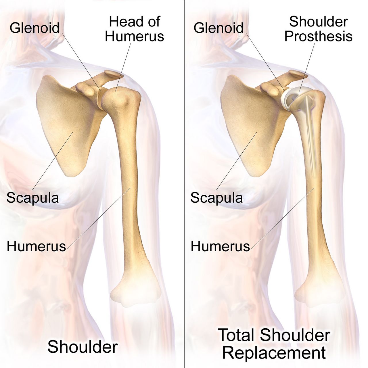
Common orthopedic operations fall out of these parts almost automatically. Total hip arthroplasty (THA) and total knee arthroplasty (TKA) replace the joint surfaces with prosthetic components. Spinal fusion joins vertebrae so a painful or unstable segment stops moving. Fasciotomy (fash-ee-OT-oh-mee) splits fascia to relieve the dangerous pressure of compartment syndrome, and it must be done urgently to save the limb. Carpal tunnel release divides the transverse carpal ligament to free the median nerve. Amputation removes a limb or part of one, charted as AKA or BKA depending on whether the level is above or below the knee.
Abbreviations you will meet on day one
| Abbreviation | Meaning | Where you will see it |
|---|---|---|
| Fx | fracture | Emergency triage, radiology reports |
| ROM | range of motion | Therapy notes, daily nursing assessment |
| AROM / PROM | active / passive range of motion | Physical and occupational therapy goals |
| ORIF | open reduction, internal fixation | Operative reports, surgical schedule |
| THA / TKA | total hip / total knee arthroplasty | Orthopedic surgery, post-op orders |
| OA | osteoarthritis | Problem lists |
| RA | rheumatoid arthritis | Problem lists, rheumatology notes |
| DJD | degenerative joint disease | Radiology impressions |
| HNP | herniated nucleus pulposus | Spine clinic, MRI reports |
| CTS | carpal tunnel syndrome | Occupational health, hand surgery |
| DXA (DEXA) | dual-energy x-ray absorptiometry | Osteoporosis screening |
| EMG | electromyography | Neurology and neuromuscular work-ups |
| ACL / PCL | anterior / posterior cruciate ligament | Knee injury and sports medicine notes |
| MCL / LCL | medial / lateral collateral ligament | Knee injury documentation |
| TMJ | temporomandibular joint | Dental and facial pain notes |
| C1-C7, T1-T12, L1-L5 | cervical, thoracic and lumbar vertebrae | Spine imaging and injury levels |
| NWB / PWB / WBAT | non-weight-bearing / partial weight-bearing / weight-bearing as tolerated | Post-op mobility orders |
| AKA / BKA | above-knee / below-knee amputation | Vascular and orthopedic surgery |
| NSAID | nonsteroidal anti-inflammatory drug | Medication lists |
| IM | intramuscular | Medication administration routes |
Ambiguous abbreviations are a genuine safety issue, and orthopedics has several. MD can mean muscular dystrophy or Doctor of Medicine. PT can mean physical therapy, prothrombin time or patient. OS means the left eye in ophthalmology but os means bone in anatomy. DC can mean discharge, discontinue or Doctor of Chiropractic. When an abbreviation could plausibly carry two meanings in the setting you are charting in, write the term out. Every institution also maintains a “do not use” list of abbreviations banned for safety reasons; learn yours on your first day.
Diagnostic and surgical terms in orthopedics are the same handful of roots wearing different suffixes, so identifying the suffix tells you immediately whether the note is describing a look, a measurement, a removal, a repair or a fusion.
The chapter in one breath
The musculoskeletal system is the most word-part-friendly system in the body. Bone is oste/o in clinical terms and oss/e in anatomic ones; cartilage is chondr/o; marrow and spinal cord share myel/o, which is why my/o for muscle must never be blurred with it. The structural anatomy of a long bone gives you the suffix -physis and the prefixes epi-, dia-, meta-, peri- and endo-, all of which reappear throughout medicine. Every named bone lends its Latin name to a combining form, so an unfamiliar orthopedic word usually resolves the moment you spot the bone inside it — and its plural obeys the Latin or Greek ending, not English. Joints belong to arthr/o and articul/o, with synovi/o, burs/o, ten/o, ligament/o and fasci/o filling in the soft tissue. Muscle takes my/o, rhabdomy/o and leiomy/o, and the suffixes -trophy, -asthenia, -plegia and -paresis convert those roots into statements about function. Pathology names either a shape, an inflamed tissue or a direction of movement; procedures name a look, a measurement, a removal, a repair or a fusion. Learn the parts and the chart reads itself.
Words worth knowing
| Term | What it means |
|---|---|
| Arthrocentesis (ar-throh-sen-TEE-sis) | Surgical puncture of a joint to withdraw fluid for analysis or relief of pressure. |
| Arthrodesis (ar-throh-DEE-sis) | Surgical fusion of a joint, deliberately eliminating motion to relieve pain. |
| Arthroplasty (AR-throh-plas-tee) | Surgical repair or replacement of a joint, as in total hip or knee arthroplasty. |
| Bursa (BER-sah), plural bursae | Fluid-filled sac that cushions tendon against bone near a joint. |
| Comminuted fracture | A fracture in which the bone is broken into three or more fragments. |
| Contracture (kon-TRAK-cher) | Permanent shortening of muscle, tendon or skin that fixes a joint in one position. |
| Crepitus (KREP-ih-tus) | Grating sound or sensation from bone or roughened cartilage rubbing together. |
| Diaphysis (dye-AF-ih-sis) | The shaft of a long bone; dia- (through) plus -physis (growth). |
| Epiphysis (eh-PIF-ih-sis) | The widened end of a long bone; epi- (upon) plus -physis (growth). |
| Kyphosis (kye-FOH-sis) | Exaggerated outward curvature of the thoracic spine. |
| Osteomyelitis (os-tee-oh-my-eh-LYE-tis) | Infection and inflammation of bone and its marrow. |
| Osteoporosis (os-tee-oh-por-OH-sis) | Loss of bone mass producing porous, fracture-prone bone; DXA T-score of -2.5 or lower. |
| Periosteum (per-ee-OS-tee-um) | Fibrous membrane covering the outer surface of bone. |
| Rhabdomyolysis (rab-doh-my-OL-ih-sis) | Breakdown of skeletal muscle releasing myoglobin, threatening the kidneys. |
| Sprain | Injury to a ligament from overstretching or tearing. |
| Strain | Injury to a muscle or tendon from overuse or overstretching. |
| Spondylolisthesis (spon-dih-loh-lis-THEE-sis) | Forward slipping of one vertebra over the vertebra beneath it. |
| Subluxation (sub-luks-AY-shun) | Partial dislocation in which joint surfaces remain partly in contact. |
| Synovial membrane (sih-NOH-vee-al) | Lining of a freely movable joint that secretes lubricating synovial fluid. |
| Tendinitis | Inflammation of a tendon; built on the Latin stem tendin- with the combining vowel dropped before -itis, rather than the common spelling tendonitis. |