Respiratory Disorders: Airway and Obstructive Disease
Obstructive lung disease is the quiet backbone of every med-surg floor. The patient in 412 with a COPD exacerbation, the twenty-two-year-old in 408 whose asthma flared after a viral illness, the man in 415 with a new tracheostomy and a mucus plug he cannot clear — all three are failing at the same physiologic step, which is moving air out. In obstructive disease the problem is not getting air in; it is emptying the chest before the next breath arrives. Air stacks, the chest hyperinflates, the diaphragm flattens, the work of breathing climbs, and eventually carbon dioxide rises. Everything you will do — positioning, bronchodilators, pursed-lip breathing, oxygen titration, airway clearance, teaching — is aimed at that single mechanical problem. This chapter builds the mechanism first, then the assessment, the diagnostics with real numbers, the drug classes by what they actually do, and the priority logic the NCLEX and your charge nurse will both demand of you.
By the end of this chapter you can
- Explain how airflow obstruction, air trapping and hyperinflation produce the classic findings of asthma and COPD, including the prolonged expiratory phase and barrel chest.
- Perform and document a focused respiratory assessment, distinguishing wheezes, rhonchi, crackles and a silent chest, and recognise accessory muscle use and tripod positioning as compensation.
- Interpret an arterial blood gas, pulse oximetry, capnography and spirometry using stated reference ranges, and identify when a “normalising” PaCO2 signals impending failure.
- Differentiate asthma from COPD by reversibility, and apply GOLD staging and peak flow zones to plan care.
- Compare bronchodilator and anti-inflammatory drug classes — SABA, SAMA, LABA, LAMA, inhaled corticosteroids, leukotriene modifiers, biologics, methylxanthines, roflumilast — by mechanism, nursing implications and monitoring.
- Manage bronchiectasis, cystic fibrosis and obstructive sleep apnea, including the correct sequence of airway clearance therapies.
- Select and titrate oxygen delivery devices safely, including the 88–92 percent target in chronic hypercapnia, and deliver competent tracheostomy care and suctioning.
- Apply ABC, acute-over-chronic and unstable-over-stable reasoning to decide what you do first, and defend delegation decisions to LPN and UAP.
- Recognise red flags that require immediate escalation and communicate them using SBAR, including the atypical presentation of hypoxemia in the older adult.
Section 1Gas Exchange, Focused Assessment and the Tests That Matter
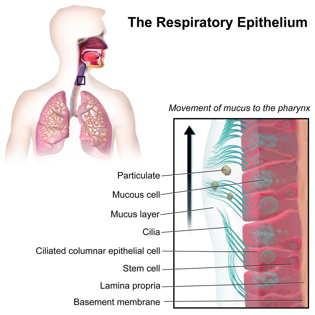
Ventilation, perfusion and why obstruction hurts on the way out
Two things must happen for a patient to oxygenate: air must reach the alveoli (ventilation) and blood must reach those same alveoli (perfusion). When the two are matched, gas exchange is efficient. When they are mismatched — ventilated alveoli with no blood flow, as in pulmonary embolism, or perfused alveoli with no air, as in a mucus-plugged lobe — oxygenation falls. Obstructive disease is fundamentally a ventilation problem, and the specific place it fails is exhalation.
Inhalation is active. The diaphragm contracts, the thorax enlarges, intrathoracic pressure drops and air is pulled in; the negative pressure around the airways actually helps hold them open. Exhalation in a healthy person is passive, driven by the elastic recoil of lung tissue. In obstructive disease that recoil is lost (emphysema destroys the elastin scaffolding) or the lumen is narrowed by smooth-muscle spasm, mucosal oedema and thick secretions (asthma, chronic bronchitis). Now the patient must push air out, and pushing raises the pressure around the small airways, which collapses them before the alveoli have emptied. Air stacks behind the closed airway. This is air trapping, and it is the single mechanism that explains nearly every finding in this chapter.
Follow the consequences. Trapped air means the lungs sit at a larger resting volume, so the chest becomes hyperinflated and the anteroposterior diameter approaches the lateral diameter — the barrel chest. Hyperinflation flattens the diaphragm; a flat diaphragm is a mechanically inefficient muscle, so the patient recruits sternocleidomastoid, scalene and intercostal muscles and leans forward onto their elbows to fix the shoulder girdle and give those accessory muscles something to pull against. That is the tripod position, and it is not a comfort preference — it is a compensation. Trapped air also means each new breath begins against a positive alveolar pressure the patient must overcome before any air moves, which is the intrinsic PEEP that makes these patients so exhausting to breathe for. Finally, because carbon dioxide is cleared by ventilation, failure to empty the chest eventually raises PaCO2.
The focused respiratory assessment
Start before you touch the patient. From the doorway, count the respiratory rate for a full minute (normal 12–20 breaths per minute in an adult) and watch the ratio of inspiration to expiration. Normal is roughly 1 to 2. In obstruction it stretches to 1 to 3 or 1 to 4, and a prolonged expiratory phase is one of the most reliable and most under-documented findings on a med-surg floor. Note the position, the ability to speak in full sentences, the use of accessory muscles, nasal flaring, and whether the skin at the suprasternal notch or between the ribs pulls in with each breath. A patient who can speak only three or four words between breaths is in significant distress regardless of what the pulse oximeter reads.
Auscultate systematically, comparing side to side, apex to base, anterior and posterior, over bare skin. Wheezes are continuous musical sounds produced by air moving through narrowed airways, classically expiratory in obstruction; inspiratory and expiratory wheezing means more severe narrowing. Rhonchi are low-pitched, snoring or gurgling sounds from secretions in larger airways and characteristically change or clear with coughing. Crackles are discontinuous popping sounds from the opening of collapsed alveoli or from fluid, and they point you toward pneumonia, heart failure or atelectasis rather than pure obstruction. Stridor is a high-pitched inspiratory crow heard without a stethoscope and means upper airway obstruction until proven otherwise — it is an emergency.
The finding students most often mis-read is the silent chest. Wheezing requires airflow. A patient who was wheezing loudly an hour ago and is now quiet, with a rising respiratory rate, rising heart rate, a drowsy affect and a falling ability to speak, has not improved — they have stopped moving enough air to make noise. That is impending respiratory arrest, and the correct action is to call for help, apply oxygen, prepare for continuous nebulised bronchodilator and anticipate intubation, not to chart “lungs clear.”
Round out the exam with percussion (hyperresonance over hyperinflated lung), a look at the fingernails for clubbing, and the sputum. Clubbing is worth memorising because it is not a feature of uncomplicated COPD or asthma. When you see it, think bronchiectasis, cystic fibrosis, lung cancer or interstitial disease. Document sputum by colour, consistency, volume and whether it changed — a change from clear-white to thick yellow-green with increased volume is one of the three cardinal signs of a COPD exacerbation.
Pearl. Pulse oximetry measures how saturated the haemoglobin is, not how much haemoglobin there is and not how well carbon dioxide is being cleared. A patient with a haemoglobin of 6 g/dL can read 99 percent and still be tissue-hypoxic, and a patient in early respiratory failure can read 96 percent while the PaCO2 climbs. Carbon monoxide poisoning falsely elevates the reading outright. Trend the number, but treat the patient in front of you.
Arterial blood gases, oximetry and capnography
Learn the reference values cold, because you will be asked to interpret gases at three in the morning: pH 7.35–7.45, PaCO2 35–45 mmHg, HCO3− 22–26 mEq/L, PaO2 80–100 mmHg and SaO2 95–100 percent on room air. Read a gas in three steps. First, is the pH acidotic, alkalotic or normal? Second, which value moves in the direction that would explain the pH — a high PaCO2 explains an acidosis (respiratory), a low HCO3− also explains an acidosis (metabolic). Third, has the other system compensated? If the pH has returned into range while both PaCO2 and HCO3− are abnormal, compensation is complete.
Apply that to obstruction. A patient in an early, frightening asthma attack hyperventilates: pH 7.48, PaCO2 28, HCO3− 23 — an acute respiratory alkalosis, which is the expected gas. If that same patient is rechecked an hour later and the gas reads pH 7.36, PaCO2 41, the temptation is to relax. Do not. A tachypneic, struggling asthmatic should be blowing off carbon dioxide. A normalising PaCO2 in that setting means the patient is tiring and can no longer sustain the minute ventilation. It is the gas that precedes intubation.
Contrast that with the patient who has lived with COPD for fifteen years: pH 7.36, PaCO2 58, HCO3− 32, PaO2 62 on 2 L nasal cannula. That is a compensated respiratory acidosis and it may be this patient’s baseline. The kidney has retained bicarbonate over months to buffer chronic hypercapnia. You do not treat that number; you treat the change from it. If the same patient presents with pH 7.24 and PaCO2 78, that is acute-on-chronic respiratory acidosis and it earns a rapid response call, non-invasive ventilation and an intensivist.
Two bedside adjuncts deserve mention. Continuous waveform capnography reports end-tidal CO2, normally 35–45 mmHg, and in obstruction the waveform loses its square shape and takes on a “shark-fin” upslope that visibly improves as bronchodilators work — it is the earliest objective sign of response. Arterial line draws and radial punctures require you to hold pressure for five minutes, or twenty minutes if the patient is anticoagulated. Then expel any air bubbles, cap the syringe, mix it and have it run at room temperature within thirty minutes. A modern plastic syringe is not routinely iced: chilling plastic accelerates oxygen diffusion through the barrel wall and falsely raises the PaO2, and prolonged cooling leaks potassium out of the cells. Ice only a glass syringe, or a plastic syringe whose analysis will be delayed well beyond thirty minutes, and note on the requisition that the specimen was chilled.
| Gas pattern | Typical values | What it means at the bedside |
|---|---|---|
| Acute respiratory alkalosis | pH 7.50, PaCO2 28, HCO3− 23 | Early asthma attack, anxiety, pain, PE. Expected early; reassuring only in context. |
| Acute respiratory acidosis | pH 7.22, PaCO2 68, HCO3− 25 | Ventilatory failure. Bicarbonate has not had time to rise. Escalate now. |
| Compensated respiratory acidosis | pH 7.36, PaCO2 58, HCO3− 32 | Chronic CO2 retention, common baseline in advanced COPD. Compare to prior gases. |
| Acute-on-chronic | pH 7.25, PaCO2 78, HCO3− 33 | Exacerbation on top of chronic retention. Non-invasive ventilation is the usual answer. |
| “Normal” gas in a distressed asthmatic | pH 7.37, PaCO2 42, RR 34 | Ominous. Fatigue, not improvement. Prepare for intubation. |
Spirometry, peak flow and imaging
Spirometry is the diagnostic gold standard for obstructive disease. Two numbers matter. FVC is the total volume forcibly exhaled after a maximal inhalation. FEV1 is the volume exhaled in the first second. In obstruction the FEV1 falls further and faster than the FVC, so the ratio drops. A post-bronchodilator FEV1/FVC ratio below 0.70 confirms persistent, incompletely reversible airflow limitation and is the diagnostic threshold for COPD. In restrictive disease the ratio is normal or high while both volumes shrink — a useful contrast to keep straight.
Reversibility separates asthma from COPD. Repeat spirometry ten to fifteen minutes after a short-acting bronchodilator; an increase in FEV1 of at least 12 percent and at least 200 mL is a significant bronchodilator response and supports asthma. When baseline spirometry is normal but the history is suggestive, a methacholine challenge is used; a fall in FEV1 of 20 percent or more is positive for airway hyperresponsiveness. Fractional exhaled nitric oxide (FeNO) of 25 ppb or higher in an adult suggests eosinophilic airway inflammation likely to respond to inhaled corticosteroids.
Peak expiratory flow is the home version — less precise, but the number the patient will actually use. Teach the patient to establish a personal best over two weeks of good control and then to act on zones: green is 80–100 percent of personal best (continue current plan), yellow is 50–79 percent (use the reliever, follow the written action plan, contact the provider if not back to green), red is below 50 percent (take the reliever and seek emergency care now). Nursing pointers: stand up, seal the lips tightly, blow one hard fast blast, do three attempts and record the highest, and measure at the same time each day before bronchodilators.
Imaging is supportive rather than diagnostic in obstruction. A chest radiograph in advanced COPD shows hyperinflation, flattened hemidiaphragms, a narrow vertical heart and increased retrosternal air space; its real job is to rule out pneumonia, pneumothorax, effusion or a mass. High-resolution CT is the test of choice for bronchiectasis, where it shows dilated, thickened airways with “tram-track” and “signet-ring” signs. Supporting labs include a CBC (white cells 5,000–10,000/mm3; haematocrit above 55 percent suggests chronic hypoxemia), a blood eosinophil count (300 cells/µL or more predicts steroid responsiveness), alpha-1 antitrypsin level in emphysema under age 45 or without smoking history, and BNP, where a value below 100 pg/mL makes heart failure an unlikely cause of the dyspnoea.
Obstructive disease is a failure of exhalation, so the assessment findings, the gases and the spirometry all trace back to trapped air — and a silent chest or a normalising PaCO2 in a struggling patient means exhaustion, not improvement.
Section 2Asthma: Reversible Obstruction and the Rescue Plan
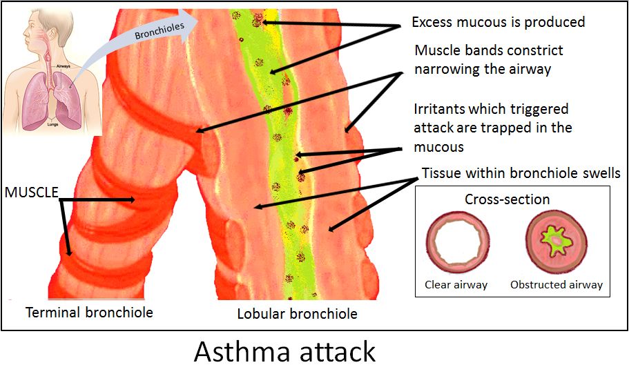
Pathophysiology: three narrowings, not one
Asthma is a chronic inflammatory disorder of the airways with variable, largely reversible obstruction. An inhaled trigger — allergen, cold air, exercise, viral infection, smoke, aspirin or an NSAID, a beta-blocker, gastro-oesophageal reflux, emotional stress — activates mast cells and eosinophils in a sensitised airway. Mediators including histamine, leukotrienes and prostaglandins are released, and three things happen at once: bronchial smooth muscle contracts (bronchospasm), the mucosa becomes oedematous, and goblet cells pour out thick tenacious mucus. Each narrowing is additive. This is why a bronchodilator alone opens the airway only partially and why the inflammation must be treated as well.
There are two phases. The early phase begins within minutes and is dominated by bronchospasm; it responds beautifully to a short-acting beta2-agonist. The late phase arrives four to twelve hours later, is dominated by eosinophilic inflammation and mucosal swelling, and does not respond well to bronchodilators at all. That biphasic pattern is the reason a patient who feels better in the emergency department can deteriorate at home overnight, and the reason systemic corticosteroids are given even when the patient improves acutely.
Over years, repeated inflammation produces airway remodelling — subepithelial fibrosis, smooth-muscle hypertrophy and mucous gland hyperplasia — which converts some of the reversible obstruction into fixed obstruction. Preventing remodelling is the entire argument for adherence to controller therapy in a patient who feels perfectly well between attacks, and it is the single most important thing you will teach.
Assessment, severity and diagnostics
The classic triad is wheezing, cough (often nocturnal or early morning) and chest tightness with dyspnoea. Ask about the pattern: what triggers it, how often the reliever inhaler is used, whether symptoms wake the patient at night, whether school or work is missed, and — the highest-yield question — whether the patient has ever been intubated or admitted to intensive care for asthma. A history of intubation identifies a patient at high risk of a fatal attack and changes your threshold for escalation permanently.
Grade severity at the bedside using speech, posture, accessory muscles, mental status and peak flow rather than a single number. In a mild-to-moderate exacerbation the patient speaks in phrases, wheezes on expiration, has a peak flow of 50–79 percent of personal best and an oxygen saturation above 92 percent. In a severe exacerbation the patient speaks in single words, sits upright or tripods, uses accessory muscles, wheezes throughout inspiration and expiration, has a peak flow below 50 percent and a saturation below 90 percent. In life-threatening asthma the chest is silent, the patient is drowsy or confused, the effort is feeble, and cyanosis, bradycardia or hypotension appear.
Diagnostics: spirometry with a bronchodilator response of at least 12 percent and 200 mL, peak flow variability, FeNO, blood eosinophils, and allergy testing when triggers are unclear. During an acute attack, obtain an ABG only if the patient is severe or deteriorating — and interpret it against the effort, as above. Serum potassium (normal 3.5–5.0 mEq/L) matters more than students expect, because repeated beta2-agonist dosing drives potassium into cells and a patient on continuous albuterol can become genuinely hypokalaemic.
Pharmacology: relievers, controllers and the ones that are never rescue
Short-acting beta2-agonists (SABA) — albuterol, levalbuterol — stimulate beta2 receptors on bronchial smooth muscle, relaxing it within five minutes and lasting four to six hours. This is the rescue drug. Expect tachycardia, tremor, nervousness and hypokalaemia; teach the patient that these effects are common and not an allergy. Short-acting muscarinic antagonists (SAMA) — ipratropium — block acetylcholine-mediated bronchoconstriction, take about fifteen minutes to work, and are added to albuterol in moderate-to-severe exacerbations but are not a rescue drug on their own. Anticholinergic effects are dry mouth, urinary retention and blurred vision, and the drug is used cautiously in narrow-angle glaucoma and benign prostatic hyperplasia.
Inhaled corticosteroids (ICS) — fluticasone, budesonide, beclomethasone — are the cornerstone controller. They suppress airway inflammation over days to weeks and have no acute effect whatsoever, which is why a patient must be told explicitly, in plain language, that this inhaler will not help during an attack and must still be taken every day. Teach rinsing and spitting after each use to prevent oral candidiasis and hoarseness, and use of a valved holding chamber or spacer to reduce oropharyngeal deposition. Long-acting beta2-agonists (LABA) — salmeterol, formoterol — provide twelve hours of bronchodilation and must never be used as monotherapy in asthma; they are always combined with an inhaled corticosteroid. Current guidance increasingly favours an ICS–formoterol combination used as both maintenance and reliever, because it treats the inflammation every time the patient reaches for relief.
Leukotriene receptor antagonists — montelukast, zafirlukast — block leukotriene-mediated bronchoconstriction and inflammation, are oral, and are useful in exercise-induced and aspirin-sensitive asthma. The two are not interchangeable at the bedside, and the difference is entirely a nursing-teaching difference. Montelukast is taken once daily in the evening, with or without food, and it is montelukast — not the class — that carries a boxed warning for serious neuropsychiatric events including agitation, depression, sleep disturbance and suicidal thinking; ask about mood at every encounter and tell families what to watch for. Zafirlukast is 20 mg twice daily and must be taken on an empty stomach, at least one hour before or two hours after a meal, because food reduces its absorption by roughly 40 percent. Zafirlukast also needs liver-enzyme monitoring, since hepatic injury has been reported — teach the patient to report anorexia, nausea, right upper quadrant pain, dark urine or jaundice — and it raises warfarin levels, so the INR is watched closely when the two are given together. Biologics — omalizumab (anti-IgE), mepolizumab and benralizumab (anti-IL-5), dupilumab (anti-IL-4/13) — are reserved for severe eosinophilic or allergic asthma; omalizumab carries a risk of anaphylaxis, so observe the patient after injection and ensure epinephrine is available. Methylxanthines — theophylline — are rarely used now because of a narrow therapeutic index; the therapeutic serum level is roughly 10–20 mcg/mL and many clinicians target 5–15 mcg/mL. Toxicity begins with nausea, vomiting, insomnia and tachycardia and progresses to dysrhythmias and seizures. Caffeine potentiates it; smoking accelerates its metabolism.
Pearl. If a patient uses a short-acting rescue inhaler more than twice a week for symptoms, or empties more than one canister a month, their asthma is not controlled no matter what they tell you. Reliever overuse is the most sensitive early marker of a patient heading for a severe attack, and it is a nursing assessment finding you can collect in one question.
Status asthmaticus, priorities and teaching
Status asthmaticus is a severe exacerbation that does not respond to standard bronchodilator and corticosteroid therapy. Priorities in order: place the patient in high Fowler or tripod position, apply oxygen titrated to a saturation of 93–95 percent, give continuous or back-to-back nebulised albuterol with ipratropium, establish IV access, give systemic corticosteroids (methylprednisolone IV or prednisone orally — onset four to six hours, so give early), and stay with the patient. Add magnesium sulfate, typically 2 g IV over twenty minutes, for severe refractory bronchospasm; it relaxes smooth muscle, and you monitor for hypotension, flushing and loss of deep tendon reflexes. Subcutaneous or intramuscular epinephrine is used when anaphylaxis is part of the picture. Heliox and non-invasive ventilation are bridges; intubation is the endpoint if the patient tires.
Sedatives and opioids are avoided in an unstable asthmatic outside of controlled intubation because they blunt respiratory drive. Do not give a beta-blocker — even an ophthalmic one such as timolol — and check the home list for it. Hydrate the patient adequately to thin secretions, but remember that a struggling asthmatic often cannot drink; IV fluids may be needed.
Teaching is where nursing changes outcomes. Every asthmatic goes home with a written asthma action plan in zones tied to their own peak flow numbers and symptoms. Demonstrate inhaler technique and have the patient teach it back — studies consistently show most patients use inhalers incorrectly, and a mis-used inhaler is an untaken drug. Cover trigger avoidance concretely (dust-mite covers, no indoor smoking, pet management, humidity control, pre-medicating with a SABA fifteen minutes before exercise), annual influenza vaccination and current COVID-19 and pneumococcal vaccination as indicated, and the specific circumstances that mean go to the emergency department now: peak flow below 50 percent of personal best, no relief from the reliever, inability to speak a full sentence, or lips and nail beds turning blue.
Asthma is reversible obstruction driven by bronchospasm, oedema and mucus, so relievers open the airway while controllers — taken daily even when the patient feels well — treat the inflammation that causes attacks and remodelling.
Section 3COPD: Emphysema, Chronic Bronchitis and the Exacerbation
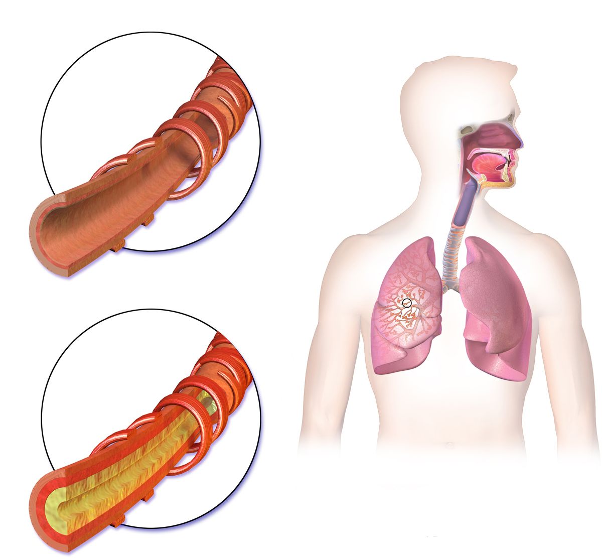
Two mechanisms under one name
Chronic obstructive pulmonary disease is persistent, largely irreversible airflow limitation caused by an abnormal inflammatory response to noxious particles — cigarette smoke above all, plus occupational dust, biomass cooking fuel and air pollution. It is conventionally described as two overlapping processes, and understanding both explains the whole clinical picture.
In emphysema, proteolytic enzymes released by inflammatory cells digest the alveolar walls. Alveoli merge into large floppy air sacs called bullae. Two things are lost: surface area for gas exchange, and the elastic recoil that drives passive exhalation and tethers the small airways open. The result is severe air trapping, hyperinflation, a barrel chest, marked dyspnoea, minimal cough, weight loss from the caloric cost of breathing, and pursed-lip breathing adopted spontaneously because it creates back-pressure that splints the airways open. Because ventilation and perfusion are lost together in the same destroyed unit, hypoxemia is relatively mild until late — this is the patient older texts called the “pink puffer.”
In chronic bronchitis, defined clinically as a productive cough for at least three months in each of two consecutive years, the insult is to the airway lining. Goblet cells and mucous glands hypertrophy, cilia are destroyed, and the airway fills with mucus it can no longer clear. Perfused alveoli sit behind plugged airways, so shunt physiology produces earlier and more severe hypoxemia and hypercapnia, with cyanosis, polycythaemia, right-sided heart strain and peripheral oedema — the “blue bloater.” Most real patients sit somewhere between these two portraits. A separate cause worth knowing is alpha-1 antitrypsin deficiency. Alpha-1 antitrypsin is not an enzyme; it is an anti-enzyme — a protease inhibitor made by the liver whose job is to neutralise the neutrophil elastase that inflammatory cells release into the airway. When it is hereditarily deficient that elastase is left unopposed and digests the alveolar walls. Suspect it in emphysema before age 45, in a never-smoker, or with a strong family history, and note that it produces lower-lobe rather than upper-lobe disease.
Assessment, staging and diagnostics
Expect a barrel chest with an anteroposterior-to-lateral ratio approaching 1 to 1, prolonged expiration, diminished breath sounds (the destroyed lung transmits sound poorly — quiet is bad here too), hyperresonance to percussion, distant heart sounds, use of accessory muscles, and often a thin, wasted body habitus with a large chest. The patient may sleep in a recliner. Ask about the pack-year history: packs per day multiplied by years smoked. Grade functional dyspnoea by what the patient can no longer do — one flight of stairs, walking to the mailbox, dressing.
Diagnosis requires post-bronchodilator spirometry showing an FEV1/FVC ratio below 0.70. Severity is then graded by FEV1 as a percentage of predicted: GOLD 1 (mild) FEV1 80 percent or more, GOLD 2 (moderate) 50–79 percent, GOLD 3 (severe) 30–49 percent, GOLD 4 (very severe) below 30 percent. Additional expected findings: an ABG showing compensated respiratory acidosis, a haematocrit above 55 percent from chronic hypoxemic stimulation of erythropoietin, a chest film with flattened diaphragms and increased retrosternal air space, and an ECG that may show right axis deviation or P pulmonale from right heart strain.
| Feature | Asthma | COPD |
|---|---|---|
| Onset | Often childhood or young adult | Usually after age 40, after long exposure |
| Reversibility | Largely reversible; FEV1 rises 12 percent and 200 mL post-bronchodilator | Persistent; ratio stays below 0.70 after bronchodilator |
| Symptom pattern | Episodic, often nocturnal, symptom-free between attacks | Progressive, daily, worst in the morning |
| Typical trigger | Allergen, exercise, cold air, virus | Tobacco or occupational exposure; exacerbated by infection |
| Sputum | Scant, tenacious, often clear | Chronic, often copious, changes colour with exacerbation |
| Oxygen target | 93–95 percent | 88–92 percent if chronically hypercapnic |
| Cornerstone drug | Inhaled corticosteroid (controller) | Long-acting bronchodilator (LAMA and/or LABA) |
Management: what actually changes the trajectory
Only three interventions are proven to prolong survival in COPD: smoking cessation, long-term oxygen therapy in patients with resting hypoxemia, and lung volume reduction surgery or transplantation in carefully selected patients. Everything else improves symptoms and reduces exacerbations, which matters enormously for quality of life but does not change the slope of decline. Smoking cessation is the only intervention that slows the annual loss of FEV1, and it works at any age and any stage — say so, every admission, without lecturing. Offer nicotine replacement, varenicline or bupropion and a referral.
Bronchodilators are the pharmacologic backbone. Long-acting muscarinic antagonists (LAMA) — tiotropium, umeclidinium, aclidinium — are first-line and reduce exacerbations; tiotropium is a once-daily maintenance drug and is never a rescue inhaler. If it comes as a capsule for a dry-powder device, teach the patient that the capsule is placed in the inhaler and is never swallowed. LABAs are added or combined. Inhaled corticosteroids are added in COPD only for frequent exacerbators, particularly those with a blood eosinophil count of 300 cells/µL or more, because in COPD they raise the risk of pneumonia. Roflumilast, a phosphodiesterase-4 inhibitor, reduces exacerbations in severe chronic-bronchitis-predominant disease with FEV1 below 50 percent; monitor for weight loss, diarrhoea and psychiatric symptoms. Chronic low-dose azithromycin reduces exacerbations in selected patients but requires baseline ECG for QT and hearing monitoring.
Long-term oxygen therapy is indicated when resting PaO2 is 55 mmHg or less, or SpO2 is 88 percent or less; or when PaO2 is 56–59 mmHg in the presence of cor pulmonale, right heart failure or a haematocrit above 55 percent. To improve survival it must be worn at least fifteen hours a day, and ideally continuously. This is a teaching problem more than a prescribing problem: patients take it off to shower, to eat, to go out, and the survival benefit evaporates. Set the target saturation at 88–92 percent in a chronically hypercapnic patient. Titrating far above that risks worsening hypercapnia through several mechanisms — release of hypoxic pulmonary vasoconstriction worsening ventilation–perfusion matching, the Haldane effect, and to a lesser degree reduced respiratory drive. That said, never withhold oxygen from a hypoxemic patient out of fear of CO2 retention. Give what is needed, titrate down, and watch.
Non-pharmacologic care is where nurses do the most good. Teach pursed-lip breathing (inhale through the nose for a count of two, exhale slowly through pursed lips for a count of four to six) to prevent airway collapse and reduce air trapping, and diaphragmatic breathing with a hand on the abdomen. Refer to pulmonary rehabilitation, which reliably improves exercise capacity and dyspnoea. Address nutrition directly: these patients burn extra calories to breathe and become breathless while eating, so offer small frequent calorie-dense high-protein meals, rest before eating, use bronchodilators before meals, avoid gas-forming foods that splint the diaphragm, and monitor weight weekly. Ensure influenza vaccination annually plus pneumococcal, COVID-19, RSV and Tdap vaccination as indicated. Assess mood; anxiety and depression are extremely common and worsen dyspnoea.
Pearl. A COPD patient with a saturation of 89 percent who is comfortable, alert and at their documented baseline does not need the oxygen turned up. A COPD patient with a saturation of 94 percent who is newly confused, tachypneic and drowsy is in trouble. In chronic lung disease, the mental status and the work of breathing are more sensitive than the pulse oximeter.
Exacerbations and complications
A COPD exacerbation is diagnosed by the three cardinal signs: increased dyspnoea, increased sputum volume, and increased sputum purulence. Viral or bacterial infection is the most common trigger. Management is bronchodilators (nebulised albuterol with ipratropium), systemic corticosteroids for roughly five days, antibiotics when all three cardinal signs are present, or when two are present and one of them is increased sputum purulence, or when the patient needs invasive or non-invasive ventilatory support, controlled oxygen to a target of 88–92 percent, and non-invasive positive pressure ventilation (BiPAP) for acute hypercapnic respiratory acidosis. BiPAP in this setting reduces intubation rates and mortality; it is one of the highest-value interventions in respiratory medicine, and the nursing work is skin protection at the mask, monitoring for gastric distension and aspiration risk, and reassessing the gas in one to two hours.
Complications to anticipate: cor pulmonale, right ventricular failure secondary to chronic hypoxic pulmonary vasoconstriction, presenting with jugular venous distension, dependent oedema, hepatomegaly, ascites and a loud pulmonic second heart sound — treated by correcting the hypoxemia, with diuretics for congestion. Secondary polycythaemia raises viscosity and thrombotic risk. Spontaneous pneumothorax occurs when a bulla ruptures; suspect it with sudden unilateral pleuritic pain, absent breath sounds on that side and tracheal deviation away from the affected side in tension physiology. Respiratory failure, pneumonia, venous thromboembolism, osteoporosis from chronic steroids and inactivity, and skeletal muscle wasting round out the list.
Escalate immediately — and use SBAR — for a change in mental status, a respiratory rate above 30 or falling toward exhaustion, saturation that will not rise despite appropriate oxygen, accessory muscle use with paradoxical abdominal motion, a pH below 7.30, or new haemodynamic instability. Situation: “This is Maria, RN, on 4 West. Mr. Alvarez in 412 with a COPD exacerbation has become confused in the last thirty minutes.” Background: “He is day two, on 2 L nasal cannula, baseline gas last night pH 7.36, PaCO2 56.” Assessment: “Respiratory rate 32, saturation 86 percent on 2 L, using accessory muscles, oriented to person only, breath sounds markedly diminished.” Recommendation: “I have him upright and I have increased oxygen while I call. I need you at the bedside now, and I would like an ABG and an order for BiPAP.”
COPD is irreversible airflow limitation in which smoking cessation, long-term oxygen for resting hypoxemia and pulmonary rehabilitation do more for the patient than any inhaler — and in an exacerbation the target saturation is 88–92 percent with BiPAP for hypercapnic acidosis.
Section 4Bronchiectasis, Cystic Fibrosis and Sleep-Disordered Breathing
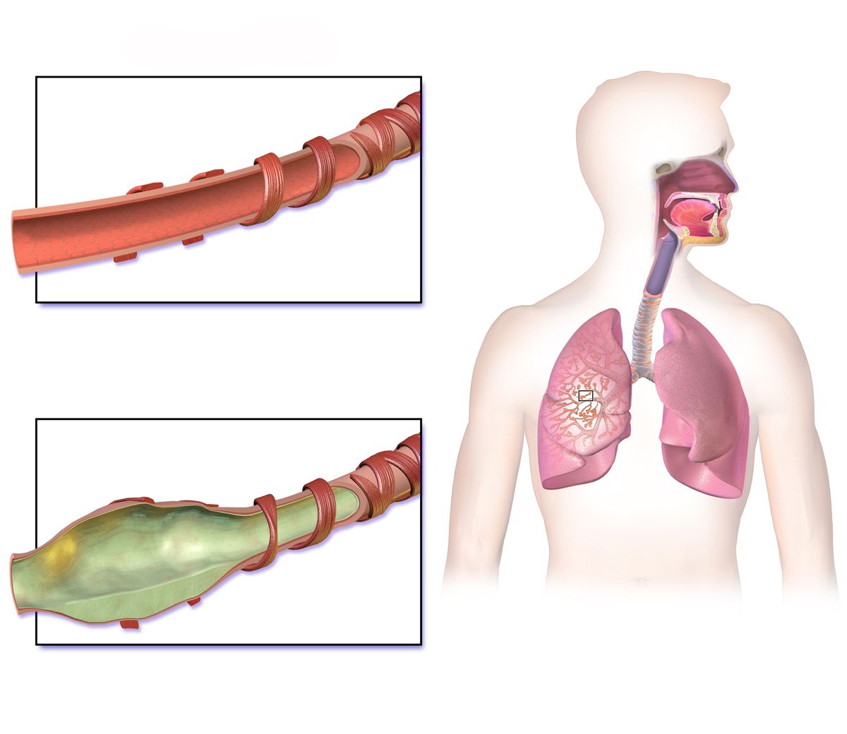
Bronchiectasis: the airway that cannot clear itself
Bronchiectasis is permanent, abnormal dilation and destruction of bronchial walls, usually the consequence of a vicious cycle: an initial insult (severe pneumonia, tuberculosis, aspiration, an inhaled foreign body, immunodeficiency, cystic fibrosis) impairs mucociliary clearance; retained secretions become colonised; chronic infection releases enzymes that destroy the elastic and muscular wall; the floppy dilated airway clears even less well; and the cycle accelerates.
The hallmark is a chronic productive cough with copious purulent sputum — often described in cupfuls, worst on rising, and layering into three levels when left standing. Expect crackles, wheezes, recurrent respiratory infections, fatigue, weight loss and digital clubbing. Haemoptysis is common because the dilated airway wall erodes into bronchial arteries; small streaks are frequent, but massive haemoptysis is a genuine emergency in which the patient is positioned with the bleeding lung down to protect the good lung, oxygen is applied and interventional bronchial artery embolisation is arranged.
Diagnosis is by high-resolution CT. Management is chronic and unglamorous: daily airway clearance (active cycle of breathing, oscillating positive expiratory pressure device such as a flutter or Acapella, high-frequency chest wall oscillation vest, postural drainage), hydration, bronchodilators, sputum cultures to guide antibiotics, treatment of exacerbations with a longer antibiotic course of ten to fourteen days, vaccination, and pulmonary rehabilitation. Nursing role: schedule airway clearance before meals to avoid vomiting, document sputum volume and character every shift as an objective outcome measure, and reinforce that clearance is done every day, not only when sick.
Cystic fibrosis: one gene, many organs
Cystic fibrosis is an autosomal recessive disorder of the CFTR chloride channel. Defective chloride transport at epithelial surfaces means water is not drawn into secretions, so mucus everywhere in the body becomes thick and sticky. In the lungs this produces obstruction, chronic infection (Staphylococcus aureus early, Pseudomonas aeruginosa later) and progressive bronchiectasis. In the pancreas, thick secretions plug the ducts and cause exocrine insufficiency with malabsorption, steatorrhoea and failure to thrive, and eventually CF-related diabetes. Sweat glands cannot reabsorb chloride, so the sweat is salty — the parent’s report that the baby tastes salty is a genuine clinical clue.
Diagnosis is by newborn screening followed by a sweat chloride test: a value of 60 mmol/L or greater is diagnostic, 30–59 mmol/L is intermediate and requires further testing, and below 30 mmol/L is normal. Genetic testing identifies the specific CFTR variant, which now determines therapy.
Management has been transformed by CFTR modulators such as elexacaftor–tezacaftor–ivacaftor, which partially correct the defective protein in patients with eligible variants; monitor liver enzymes and for cataracts. Respiratory care follows a specific and testable order: bronchodilator first to open the airway, then mucolytics — inhaled dornase alfa, which cleaves the DNA released by dead neutrophils, and hypertonic saline 7 percent, which draws water onto the airway surface — then airway clearance to move the loosened secretions, and finally inhaled antibiotics such as tobramycin so the drug is deposited in a cleared airway. Nutritional care is equally essential: pancreatic enzyme replacement with every meal and every snack, taken with the food and never crushed or chewed if enteric-coated; fat-soluble vitamins A, D, E and K; a high-calorie, high-protein diet; and added salt, especially in hot weather or with exercise, because salt losses are large. Teach strict infection prevention and segregation from other CF patients to prevent cross-infection.
Pearl. Sequence is a nursing intervention. Bronchodilator, then hypertonic saline or dornase alfa, then airway clearance, then inhaled antibiotic. Give the antibiotic first and you have deposited an expensive drug onto a wall of mucus, then knocked it right back out with the vest. Students lose this question constantly, and patients lose therapeutic benefit when the order is wrong.
Obstructive sleep apnea and upper airway obstruction
Obstructive sleep apnea is repetitive collapse of the pharyngeal airway during sleep despite continued respiratory effort. Each apnoea produces desaturation and a surge of sympathetic activity that arouses the patient briefly; the arousals fragment sleep and the desaturations plus sympathetic surges drive hypertension, atrial fibrillation, pulmonary hypertension, stroke risk, insulin resistance and daytime somnolence. Risk factors are obesity, a large neck circumference, male sex, older age, retrognathia, tonsillar hypertrophy, alcohol and sedatives.
Screen with STOP-BANG (Snoring, Tiredness, Observed apnoea, Pressure, BMI above 35, Age above 50, Neck circumference above 40 cm, Gender male); a score of three or more indicates elevated risk. Diagnosis is by polysomnography, reported as the apnea–hypopnea index: 5–15 events per hour is mild, 15–30 moderate, and above 30 severe. Treatment is CPAP, plus weight loss, positional therapy, avoidance of alcohol and sedating drugs, and in selected patients an oral appliance or surgery.
The med-surg relevance is perioperative and pharmacologic. A patient with untreated or unrecognised OSA who receives opioids, benzodiazepines or anaesthesia is at high risk of postoperative airway obstruction and respiratory arrest, particularly in the first 24 hours and particularly during the first uninterrupted sleep. Bring the patient’s own CPAP machine into the hospital and use it whenever they sleep, including naps; nurse them with the head of bed elevated and in a lateral rather than supine position; use continuous pulse oximetry and, when opioids are given, capnography; use multimodal non-opioid analgesia; and never assume a quiet, sleeping postoperative patient is a comfortable one.
Acute upper airway obstruction — from a foreign body, angioedema, anaphylaxis, epiglottitis, tumour, trauma or post-extubation laryngeal oedema — announces itself with stridor, drooling, an inability to speak, severe anxiety and rapidly progressive distress. This is the most time-critical scenario in the chapter. Do not leave the patient. Call for emergency help, keep the patient upright and calm, give high-flow oxygen, avoid anything that agitates them, and prepare for a difficult airway with intubation or surgical airway equipment at the bedside. For a conscious choking adult with a complete obstruction, perform abdominal thrusts; if the patient becomes unresponsive, begin CPR and look in the mouth for a visible object before each set of ventilations. In anaphylaxis, intramuscular epinephrine into the lateral thigh is the first drug and antihistamines and steroids are adjuncts, never substitutes.
Bronchiectasis and cystic fibrosis are diseases of failed secretion clearance managed by daily airway clearance in the correct sequence, while obstructive sleep apnea turns dangerous the moment a patient receives sedation or opioids.
Section 5Airway Management, Oxygen Delivery and Setting Priorities
Oxygen delivery devices and oxygen safety
Oxygen is a drug. It requires an order (or protocol), a dose, a route, a target and reassessment, and it carries real toxicity: prolonged high concentrations cause absorption atelectasis and, over days, oxidative alveolar injury. Choose the device by how much oxygen the patient needs and how precisely you need to control it, and remember that with any low-flow device the actual inspired concentration falls as the patient’s minute ventilation rises — a tachypneic patient entrains more room air and gets a lower effective dose.
| Device | Flow rate | Approximate FiO2 | Nursing notes |
|---|---|---|---|
| Nasal cannula | 1–6 L/min | 24–44 percent | Patient can eat and talk; humidify above 4 L; check ears and nares for pressure injury. |
| Simple face mask | 6–10 L/min | 35–50 percent | Never run below 5 L/min — the patient rebreathes exhaled CO2. |
| Partial rebreather | 6–11 L/min | 60–75 percent | Reservoir bag must stay at least one-third inflated on inspiration. |
| Non-rebreather | 10–15 L/min | Up to about 90 percent | Short-term emergency use. Inflate the bag before applying; never let it collapse. |
| Venturi mask | Per colour-coded adapter | 24–60 percent, precise | Device of choice when exact FiO2 matters, as in a CO2-retaining COPD patient. |
| High-flow nasal cannula | Up to 60 L/min | 21–100 percent | Heated and humidified; washes out dead space and gives modest positive pressure. |
| BiPAP / CPAP | Set pressures | Titrated | BiPAP for hypercapnic COPD exacerbation; watch skin, gastric distension and aspiration risk. |
Safety teaching for home oxygen is a board favourite and a genuine fire-prevention issue: no smoking by the patient or anyone in the home, no open flames or gas stoves near the tubing, keep cylinders upright and secured at least six to ten feet from heat sources, use cotton rather than wool or synthetic blankets to limit static, avoid petroleum-based products on the face (use water-soluble lubricant for dry nares), post “oxygen in use” signage, and keep a working fire extinguisher and smoke alarms. Never adjust the litre flow without an order or a protocol, and always re-check saturation and work of breathing after any change.
Tracheostomy care and suctioning
Keep at the bedside of every tracheostomy patient: the obturator for the current tube, a spare tube of the same size, a spare tube one size smaller, a suction setup, an Ambu bag, and sterile saline and gloves. If the tube becomes dislodged in a mature tract, hyperextend the neck, spread the stoma with a curved haemostat and reinsert the spare tube with the obturator in place, then immediately remove the obturator so the patient can breathe. If the tract is fresh — less than seven days — do not attempt blind reinsertion; call for emergency help, occlude the stoma and ventilate over the mouth and nose with a bag-valve-mask.
Routine care: assess the site every shift for redness, drainage, subcutaneous emphysema and skin breakdown; clean the inner cannula or replace a disposable one per policy; use pre-cut or manufactured tracheostomy dressings and never cut gauze, because loose fibres can be aspirated; secure ties with one to two fingers’ space beneath them; and always have a second person hold the tube while ties are changed. Maintain cuff pressure at 20–25 cmH2O — higher pressures compress tracheal mucosal capillaries and cause ischaemia, stenosis and fistula, while lower pressures allow aspiration. A cuffed tube prevents aspiration and permits positive-pressure ventilation but blocks airflow through the larynx, so the patient cannot speak; a speaking valve is used only with the cuff fully deflated, and placing one on an inflated cuff is a fatal error.
Suction only when indicated — audible or visible secretions, coarse rhonchi, a rise in peak airway pressure, a fall in saturation, or an ineffective cough — not on a schedule. Technique: explain the procedure, hyperoxygenate with 100 percent oxygen for thirty to sixty seconds, use sterile technique for tracheal suctioning, choose a catheter no larger than half the internal diameter of the airway, set wall suction to 80–120 mmHg for an adult, insert without suction until resistance is met and then withdraw about 1 cm, apply intermittent suction for no more than 10–15 seconds while withdrawing in a rotating motion, allow the patient to recover and re-oxygenate between passes, and limit to two or three passes. Do not instil normal saline before suctioning — it does not thin secretions, it does dislodge bacteria from the tube into the lower airway, and it causes desaturation. Watch for hypoxemia, dysrhythmias from vagal stimulation, bronchospasm, bleeding and raised intracranial pressure. Stop and re-oxygenate for bradycardia, a drop in saturation, or new dysrhythmia.
Inhaler and nebuliser technique — the teaching that actually changes outcomes
For a metered-dose inhaler: shake for five seconds, attach a spacer whenever one is available, exhale fully away from the device, seal the lips around the mouthpiece, begin a slow deep inhalation while actuating one puff, continue inhaling over three to five seconds, hold the breath for ten seconds, exhale slowly, and wait about one minute between puffs of the same drug. When two different inhalers are due, give the bronchodilator first and wait five minutes before the corticosteroid, so the second drug reaches an opened airway. Rinse and spit after any inhaled corticosteroid.
For a dry-powder inhaler, the technique reverses in two important ways: do not shake, do not exhale into the device (humidity clumps the powder), and inhale quickly and forcefully rather than slowly, because the patient’s own effort disaggregates the powder. For a small-volume nebuliser, the patient sits upright and breathes normally with occasional deep breaths through the mouthpiece until the mist stops, typically eight to twelve minutes; tapping the reservoir recovers the last of the dose. Assess heart rate before and after a beta-agonist, and reassess lung sounds, respiratory rate, saturation and the patient’s subjective breathing afterward — that reassessment is the nursing evaluation step and it belongs in your note.
Priorities, delegation and the older adult
When two respiratory patients need you at once, sort with the same three filters every time. First, ABC: airway before breathing before circulation, and within breathing, the patient with the least effective air movement wins. Second, acute over chronic: a new finding beats a chronic one, so a COPD patient at their documented baseline of 89 percent waits while the post-operative patient who has dropped from 97 to 91 percent gets seen. Third, unstable over stable and actual over potential problems. Applying that: a patient with stridor outranks a patient with a silent chest, who outranks a patient wheezing loudly, who outranks a patient asking for a scheduled inhaler. A drowsy patient with a “good” saturation on a non-rebreather outranks nearly everyone, because that combination means CO2 is climbing.
| Task | Who | Why |
|---|---|---|
| Initial and ongoing respiratory assessment, lung sounds, evaluation of response to therapy | RN only | Assessment and evaluation are not delegable. |
| Patient teaching (inhaler technique, action plan, oxygen safety) and the first dose of a new drug | RN only | Teaching and initial evaluation require the nursing process. |
| Titrating oxygen, admitting an unstable patient, care of a fresh tracheostomy | RN only | Unstable and unpredictable outcomes. |
| Administering routine nebulised or oral drugs, tracheostomy care on a stable established stoma, sterile suctioning of a stable patient, reinforcing teaching | LPN/LVN (per state scope and facility policy) | Stable, predictable patient with an established plan of care. |
| Measuring vital signs and pulse oximetry, positioning, assisting with ambulation and ADLs, recording intake and output, encouraging incentive spirometry the RN has already taught, oral care, weights | UAP | Standardised tasks with predictable outcomes; the UAP reports the number, the RN interprets it. |
Older adults change the picture in ways that catch students out. Ageing reduces chest wall compliance and elastic recoil, weakens respiratory muscles, blunts the cough and ciliary clearance, and dulls the ventilatory response to both hypoxia and hypercapnia — so an older patient can be significantly hypoxemic and neither look nor feel distressed. The first sign of hypoxemia or pneumonia in an older adult is frequently new confusion, restlessness or a fall, sometimes with a normal temperature and no cough at all. Treat acute change in mental status in an older patient as hypoxia or infection until proven otherwise, and check a saturation before you reach for anything else. Anticholinergic inhalers may precipitate urinary retention in a man with prostatic enlargement or worsen narrow-angle glaucoma; beta-agonists cause more tremor and tachycardia; theophylline clearance falls, so toxicity appears at doses that were previously safe; and arthritic hands and reduced inspiratory flow may make a metered-dose inhaler or dry-powder device unusable, in which case a spacer or nebuliser is the right accommodation, not a compliance problem.
Pearl. Before you conclude that an older patient is “non-adherent” with inhaled therapy, hand them the device and watch. The most common reasons are a canister that has been empty for weeks, a dry-powder device they cannot generate enough flow to trigger, hands that cannot press the canister and inhale simultaneously, or nobody ever telling them the steroid inhaler will not help during an attack. All four are fixed by nursing, not by prescribing.
Oxygen is a drug with a target, suctioning is done for indication rather than by schedule, and priority in respiratory care follows airway before breathing, acute before chronic and unstable before stable — with new confusion in an older adult treated as hypoxia until proven otherwise.
The chapter in one breath
Obstructive lung disease is a failure of exhalation. Narrowed or collapsible airways trap air, hyperinflate the chest, flatten the diaphragm and drive up the work of breathing until carbon dioxide rises. Everything visible at the bedside — prolonged expiration, barrel chest, tripod position, accessory muscle use, pursed-lip breathing — is downstream of that one mechanism. Asthma is reversible obstruction from bronchospasm, mucosal oedema and mucus; relievers open the airway and daily inhaled corticosteroids treat the inflammation that causes attacks and long-term remodelling, and a silent chest or a normalising PaCO2 in a struggling asthmatic means impending failure. COPD is fixed obstruction from alveolar destruction and mucus hypersecretion, staged by an FEV1/FVC ratio below 0.70; only smoking cessation, long-term oxygen for resting hypoxemia and selected surgery change survival, the saturation target in chronic hypercapnia is 88–92 percent, and BiPAP is the answer to acute hypercapnic acidosis. Bronchiectasis and cystic fibrosis are managed by daily airway clearance performed in the right sequence, and sleep apnea becomes dangerous the moment sedation is given. Prioritise by airway, then breathing, acute over chronic, unstable over stable — and in an older adult, treat new confusion as hypoxia until proven otherwise.
Words worth knowing
| Term | What it means |
|---|---|
| Air trapping | Failure to fully exhale, so residual volume rises and each breath begins against positive alveolar pressure; the core mechanism of obstructive disease. |
| Barrel chest | Anteroposterior chest diameter approaching the lateral diameter (about 1 to 1) from chronic hyperinflation. |
| FEV1/FVC ratio | Fraction of forced vital capacity exhaled in the first second; a post-bronchodilator value below 0.70 defines persistent airflow obstruction. |
| Bronchodilator reversibility | An FEV1 increase of at least 12 percent and 200 mL after a short-acting bronchodilator, supporting asthma. |
| Peak expiratory flow zones | Green 80–100 percent, yellow 50–79 percent, red below 50 percent of the patient’s personal best. |
| Silent chest | Absence of wheezing in a severely obstructed patient because airflow is too low to generate sound; a pre-arrest finding. |
| Status asthmaticus | A severe asthma exacerbation unresponsive to standard bronchodilator and corticosteroid therapy. |
| Airway remodelling | Fibrosis, smooth-muscle hypertrophy and mucous gland hyperplasia from chronic inflammation, converting reversible obstruction to fixed obstruction. |
| GOLD stages | COPD severity by FEV1 percent predicted: 1 at 80 percent or more, 2 at 50–79, 3 at 30–49, 4 below 30. |
| Compensated respiratory acidosis | Chronically elevated PaCO2 with a compensating rise in bicarbonate and a near-normal pH; a common COPD baseline. |
| Cor pulmonale | Right ventricular hypertrophy and failure caused by chronic pulmonary hypertension from lung disease. |
| Pursed-lip breathing | Slow exhalation through pursed lips that creates back-pressure to splint small airways open and reduce air trapping. |
| Long-term oxygen therapy | Oxygen for resting PaO2 of 55 mmHg or less or SpO2 of 88 percent or less, worn at least fifteen hours daily to improve survival. |
| Bronchiectasis | Permanent bronchial dilation and wall destruction with copious purulent sputum, clubbing and haemoptysis; diagnosed on high-resolution CT. |
| Sweat chloride test | Diagnostic test for cystic fibrosis; 60 mmol/L or greater is diagnostic, 30–59 intermediate, below 30 normal. |
| CFTR modulator | Drug that partially restores function of the defective chloride channel in patients with eligible cystic fibrosis variants. |
| Apnea–hypopnea index | Events per hour on polysomnography: 5–15 mild, 15–30 moderate, above 30 severe obstructive sleep apnea. |
| Venturi mask | High-flow device delivering a precise FiO2 of 24–60 percent regardless of the patient’s breathing pattern. |
| Cuff pressure | Pressure in a tracheostomy or endotracheal tube cuff, maintained at 20–25 cmH2O to seal the airway without causing mucosal ischaemia. |
| Obturator | The rounded guide inserted inside a tracheostomy tube during placement; kept at the bedside and removed immediately after insertion. |