The Child in Crisis: Emergencies, Safeguarding and the Family
Almost no child arrests without warning. The warning is usually written in the minutes before — a child who has stopped fighting the oxygen mask, a fontanelle that has sunk, a heart rate that has been climbing since the last set of observations, a bruise on an infant who cannot yet roll over. This chapter is about seeing that writing and acting on it in the right order, because in paediatric emergencies the correct first action is frequently not the intuitive one. You will learn to read a child across a room using the Paediatric Assessment Triangle, to grade dehydration by percentage and match it to fluid orders, to calculate every dose per kilogram and check it against an adult ceiling, to run the fixed sequences that save a child in anaphylaxis, status epilepticus, shock or ketoacidosis, and to recognise injuries that do not fit the story or the child’s developmental stage. Then you will learn to stay in the room with the family afterwards — through resuscitation, through bad news, and through the death of a baby or a child.
By the end of this chapter you can
- Apply the Paediatric Assessment Triangle and its TICLS appearance cues to name a child’s physiological state within sixty seconds of entering the room.
- State normal heart rate, respiratory rate and the hypotension threshold for each paediatric age band, and use the formula systolic BP less than 70 + (2 × age in years) for children 1 to 10 years.
- Distinguish compensated from decompensated shock and explain why hypotension and bradycardia are late, ominous findings in a child, and state the compression rate, depth, ratios and defibrillation energies used in paediatric life support.
- Grade dehydration as mild, moderate or severe by percentage weight loss, match each grade to its clinical signs, and calculate the fluid deficit in millilitres per kilogram.
- Calculate maintenance fluids using the 4–2–1 rule and calculate weight-based doses showing the arithmetic, always checking the result against the maximum adult dose.
- Perform the correct first action in anaphylaxis, status epilepticus, paediatric septic shock, diabetic ketoacidosis, poisoning and foreign body airway obstruction, and name the drug, manoeuvre or delay that is contraindicated in each.
- Identify injuries inconsistent with a child’s developmental stage using TEN-4-FACESp and the sentinel injury concept, and describe the nurse’s personal mandatory-reporting duty and documentation standard.
- Escalate deterioration using SBAR, and apply delegation limits that keep assessment, teaching, evaluation and the unstable patient with the registered nurse.
- Support a family through resuscitation, bad news and perinatal or paediatric loss using family-centred, non-blaming language and developmentally appropriate explanations of death, and explain the investigation that follows a sudden unexpected infant death, including the ABCs of safe sleep the investigation asks about.
Section 1The First Sixty Seconds: Reading the Deteriorating Child
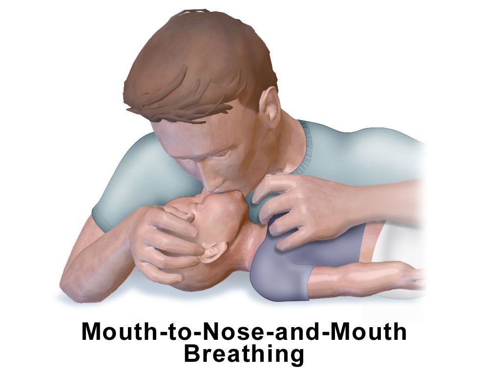
The Paediatric Assessment Triangle
The Paediatric Assessment Triangle (PAT) is a structured way of doing what experienced paediatric nurses do instinctively: forming an impression before touching the child. It uses only what you can see and hear from the end of the bed, takes under a minute, requires no equipment, and works on a child who is asleep, terrified or hostile. Its three legs are appearance, work of breathing and circulation to the skin. It does not replace the primary survey; it tells you how fast the primary survey needs to happen and whether you should be calling for help before you start it.
Appearance is the leg students under-weight, and it is the most important. It is the outward sign of brain perfusion, oxygenation and substrate delivery, and it is summarised by the mnemonic TICLS: Tone, Interactiveness, Consolability, Look or gaze, and Speech or cry. A well child has flexed limbs and resists you; a sick child is limp or has lost the normal flexed posture. A well infant tracks a face and reaches for a bright object; a sick infant stares through you. A well child is comforted by a caregiver; the inconsolable child, and equally the child who has stopped protesting when a stranger approaches, are both worrying. A vigorous angry cry is reassuring. A weak, whimpering or grunting cry is not.
Work of breathing is assessed by listening for abnormal airway sounds and looking for the visible effort of moving air. Stridor is upper airway, wheeze is lower airway, grunting is alveolar — a child grunting is generating their own positive end-expiratory pressure against collapsing alveoli, and grunting is one of the most reliable single markers of serious illness in an infant. Look for nasal flaring, head bobbing, tracheal tug and retractions, naming the level (suprasternal, intercostal, subcostal). Look at the position the child has chosen: the tripod position, the sniffing position, or an infant who will only settle sitting forward. Circulation to skin is the third leg and is judged by colour: pallor, mottling and cyanosis, in that order of progression. Cyanosis in a child is late.
The value of the triangle lies in the combinations. Which legs are abnormal names the physiological problem, and the physiological problem determines the first intervention.
| Appearance | Work of breathing | Circulation to skin | Physiological state | First priority |
|---|---|---|---|---|
| Normal | Normal | Normal | Stable | Complete assessment at a normal pace |
| Normal | Abnormal | Normal | Respiratory distress | Oxygen, position of comfort, keep the child calm |
| Abnormal | Abnormal | Normal | Respiratory failure | Assisted ventilation with bag-mask; call for help now |
| Normal | Normal | Abnormal | Compensated shock | Vascular access and a fluid bolus before the blood pressure falls |
| Abnormal | Normal | Abnormal | Shock that has reached the brain — decompensated once the systolic pressure falls below the age threshold | Immediate bolus, escalate, prepare for deterioration |
| Abnormal | Normal | Normal | Primary brain or metabolic problem | Check glucose, consider seizure, poisoning, sepsis, trauma |
| Abnormal | Abnormal | Abnormal | Cardiopulmonary failure | Resuscitation team; arrest is minutes away |
Check a glucose in every child with an abnormal appearance. Hypoglycaemia mimics sepsis, head injury, poisoning and seizure, and it is the one cause on that list you can correct in ninety seconds. Infants have small glycogen stores and a high metabolic rate; a child who has been vomiting for a day may be profoundly hypoglycaemic with a normal-looking chest and normal skin colour. Treat a blood glucose below about 2.6 mmol/L (47 mg/dL) in a neonate, or below 3.3 mmol/L (60 mg/dL) in an infant or child, and treat any symptomatic child regardless of the number: give dextrose 0.5 to 1 g/kg — in practice 5 to 10 mL/kg of 10% dextrose for an infant, or 2 to 4 mL/kg of 25% dextrose for an older child — and then recheck.
The numbers, by age band
A single set of adult ranges is useless in paediatrics and actively dangerous. A heart rate of 150 is normal in a two-month-old and a resuscitation call in a twelve-year-old. A respiratory rate of 20 is normal in a school-age child and a pre-terminal sign in a neonate. Learn the bands, and learn that heart rate and respiratory rate are the sensitive early markers while blood pressure is the last thing to change.
| Age band | Heart rate, awake (beats/min) | Respiratory rate (breaths/min) | Hypotension if systolic BP is |
|---|---|---|---|
| Neonate, birth to under 1 month | 100–205 | 30–60 | less than 60 mmHg |
| Infant, 1 month to under 1 year | 100–190 | 30–53 | less than 70 mmHg |
| Toddler, 1 year to under 3 years | 98–140 | 22–37 | less than 70 + (2 × age in years) |
| Preschool, 3 years to under 6 years | 80–120 | 20–28 | less than 70 + (2 × age in years) |
| School-age, 6 years to under 12 years | 75–118 | 18–25 | less than 70 + (2 × age in years) up to age 10; less than 90 mmHg at 11 years |
| Adolescent, 12 years and over | 60–100 | 12–20 | less than 90 mmHg |
Work the formula until it is automatic. A four-year-old is hypotensive below 70 + (2 × 4) = 78 mmHg systolic. A seven-year-old is hypotensive below 70 + (2 × 7) = 84 mmHg. A recorded systolic of 82 in that seven-year-old is not “a bit soft”; it is decompensated shock in a child who has already exhausted their compensatory reserve, and it demands an immediate bolus and a call, not a repeat cuff in fifteen minutes. Sleeping heart rates run roughly 20 to 30 beats/min lower than the awake ranges above, so a sleeping infant at 175 is tachycardic even though the number sits comfortably inside the awake band of 100 to 190.
Two other numbers belong here. Temperature: a rectal temperature of 38.0°C or higher in an infant under 28 days is a medical emergency requiring full evaluation for serious bacterial infection and parenteral antibiotics, regardless of how well the baby looks, because neonates mount almost no localising signs. Weight: if the child cannot be weighed, estimate with (age in years × 2) + 8 kg for children 1 to 5 years and (age in years × 3) + 7 kg for children 6 to 12 years — or better, use a length-based resuscitation tape. Every drug and every fluid volume in this chapter depends on that number being right.
From compensation to collapse, and the escalation that prevents it
Children compensate for hypovolaemia and sepsis extremely well and then fail extremely fast. Cardiac output in a small child is largely rate-dependent because stroke volume is relatively fixed, so the first response to any fall in preload is tachycardia, followed by intense peripheral vasoconstriction. That vasoconstriction is what you are seeing in prolonged capillary refill, cool mottled peripheries, a narrowed pulse pressure and weak peripheral pulses with a preserved central pulse — and it is what keeps the blood pressure normal. This is compensated shock, and it is entirely treatable. Once the blood pressure falls, the child has run out of reserve; this is decompensated shock, and mortality climbs steeply.
Bradycardia in a sick child is not a rhythm problem to be observed. It is hypoxia or acidosis reaching the myocardium and it immediately precedes arrest. In paediatric advanced life support, a heart rate under 60 beats/min with signs of poor perfusion despite adequate oxygenation and ventilation is an indication to start chest compressions — you do not wait for pulselessness. Most paediatric arrests are respiratory in origin and hypoxic in mechanism, which is why effective bag-mask ventilation, not defibrillation, is the intervention that saves the majority of these children. Compress at 100 to 120 per minute to a depth of at least one third of the anteroposterior chest diameter (about 4 cm in an infant, 5 cm in a child), at 30:2 for a single rescuer and 15:2 for two rescuers in infants and children. Defibrillation for a shockable rhythm starts at 2 J/kg, then 4 J/kg, then 4 J/kg or more without exceeding 10 J/kg or the adult dose. Adrenaline is 0.01 mg/kg intravenously or intraosseously (0.1 mL/kg of the 0.1 mg/mL concentration), maximum 1 mg, every 3 to 5 minutes.
The purpose of every observation set is escalation, and escalation has a script. SBAR converts a vague worry into a clinical demand. Situation: “I am calling about Maya Okoye, 4 years old, bed 6. Her heart rate is 172 and her capillary refill is 4 seconds.” Background: “She was admitted eight hours ago with gastroenteritis, has had six large watery stools and has not passed urine since arrival.” Assessment: “I think she is in shock. Her blood pressure is 84 systolic, which is still above her hypotension threshold of 78, so by pressure she is compensated — but her appearance has changed and she is now difficult to rouse, so the shock has already reached her brain and she is about to decompensate.” Recommendation: “I need you to review her now and I would like an order for a 20 mL/kg isotonic bolus and a bedside glucose.” Say the words “I need you to come now” when you mean them, and if the response does not match the child, use the chain of command or the rapid response criteria — both of which parents may also activate in many facilities.
Delegation limits tighten as the child destabilises. Unlicensed assistive personnel may weigh a child, record vital signs on a stable patient, apply a pulse oximeter probe, measure intake and output, and stay with a child for comfort. They may not perform the Paediatric Assessment Triangle, triage, interpret a change in condition, or take observations on an unstable child, because those acts are assessment. Teaching a caregiver, evaluating the response to a bolus, and the care of any child whose PAT is abnormal remain with the registered nurse.
| Red flag | Why it matters | Escalate as |
|---|---|---|
| Grunting, head bobbing or a silent chest | Alveolar collapse or exhaustion; imminent respiratory failure | Emergency call, prepare bag-mask |
| Capillary refill over 3 seconds with mottled cool skin | Compensated shock | Urgent review, bolus, glucose |
| Systolic BP below the age threshold | Decompensated shock | Resuscitation team |
| Heart rate under 60 with poor perfusion in an infant or child | Pre-arrest | Start CPR, call arrest team |
| Fever 38.0°C or higher in an infant under 28 days | Possible serious bacterial infection | Immediate medical review, blood cultures, antibiotics |
| A parent saying the child is “not themselves” | Caregiver concern independently predicts deterioration | Reassess in full; document and escalate |
Name the child’s physiological state from the Paediatric Assessment Triangle before you touch anything, and treat tachycardia and delayed capillary refill as shock, because a normal blood pressure in a child is the last reassurance you will get.
Section 2Fluids, Dehydration and the Arithmetic of a Small Body
Grading dehydration by percentage
Children dehydrate faster than adults for structural reasons: a higher proportion of body water is extracellular and therefore easily lost, the surface-area-to-mass ratio is larger so insensible losses are proportionally greater, the metabolic rate is higher, and the immature kidney concentrates urine poorly. An infant losing fluid through vomiting and diarrhoea can lose a tenth of their body mass in a day, which in an adult would be seven litres.
The gold standard for grading dehydration is an acute change in weight, because 1 kg of acute weight loss equals 1 litre of fluid. If a recent well-child weight exists, use it: a child weighing 12 kg who weighed 13.2 kg last week has lost 1.2 kg, which is 1200 mL, which is 9% of 13.2 kg — severe. When no recent weight exists, grade by clinical signs. Note that the same set of signs corresponds to different percentages in infants and older children, because infants have more water to lose. For the table below, use the infant column under 1 year of age and the older-child column from 1 year onwards, and convert percentage to volume with the rule that each 1% of dehydration is 10 mL/kg.
| Severity | Deficit (infant / older child) | Deficit in mL/kg (infant / older child) | Appearance and neurology | Circulation | Mucosa, eyes, fontanelle | Urine output |
|---|---|---|---|---|---|---|
| Mild | 5% / 3% | 50 / 30 mL/kg | Alert, thirsty, normal behaviour | Normal heart rate, capillary refill under 2 s, normal BP | Slightly dry mucosa, tears present, flat fontanelle, normal turgor | Slightly reduced |
| Moderate | 10% / 6% | 100 / 60 mL/kg | Irritable or listless, restless | Tachycardia, capillary refill 2–3 s, normal BP, weak peripheral pulses | Dry mucosa, few tears, sunken eyes and fontanelle, reduced turgor | Oliguria, under 1 mL/kg/h |
| Severe | 15% / 9% | 150 / 90 mL/kg | Lethargic, obtunded, may be unrousable | Marked tachycardia then bradycardia, capillary refill over 3 s, hypotension (late), thready or absent peripheral pulses, cool mottled limbs | Parched mucosa, no tears, deeply sunken eyes and fontanelle, tenting skin | Anuria |
Deep, sighing respirations in a dehydrated child are not respiratory distress — they are respiratory compensation for metabolic acidosis, and they mean the child is sicker than the chest examination suggests. Skin turgor and the fontanelle are useful but imperfect: turgor is falsely reassuring in a chubby infant and in hypernatraemic dehydration, where the skin feels doughy rather than tented. Capillary refill, heart rate, mental status and urine output remain the workhorses.
Maintenance, deficit and boluses: the 4–2–1 rule
Maintenance fluid replaces ongoing obligatory losses — urine, stool and insensible loss — in a child who is not currently depleted. The 4–2–1 rule gives the hourly rate: 4 mL/kg/h for the first 10 kg, plus 2 mL/kg/h for the next 10 kg, plus 1 mL/kg/h for every kilogram above 20 kg. Its daily equivalent is the 100–50–20 rule (100 mL/kg/day for the first 10 kg, 50 for the next 10, 20 thereafter).
Work an example. A child weighs 26 kg. First 10 kg: 10 × 4 = 40 mL/h. Next 10 kg: 10 × 2 = 20 mL/h. Remaining 6 kg: 6 × 1 = 6 mL/h. Total = 40 + 20 + 6 = 66 mL/h. Check it against the daily rule: (10 × 100) + (10 × 50) + (6 × 20) = 1000 + 500 + 120 = 1620 mL/day, which is 67.5 mL/h — close to the hourly answer but not identical. The gap is created in the first 20 kg: there the daily rule allows 1000 + 500 = 1500 mL/day, which is 62.5 mL/h, while the hourly rule allows only 40 + 20 = 60 mL/h, so the daily rule starts 2.5 mL/h ahead of the hourly one. Above 20 kg that reverses and the gap closes, because the hourly rule adds 1 mL/kg/h while the daily rule adds only 20 mL/kg/day, which is 0.83 mL/kg/h — the daily figure loses about 0.17 mL/h for every kilogram above 20. By 26 kg the 6 extra kilograms have clawed back 1 mL/h of the original 2.5, leaving the 1.5 mL/h difference you see. The two rules are therefore furthest apart at exactly 20 kg and converge as the child gets heavier, meeting at 35 kg where both give 75 mL/h. Use one of them consistently rather than mixing them, and treat the second as a sanity check rather than as proof. A second example: a 7 kg infant sits entirely inside the first band, so 7 × 4 = 28 mL/h. A third: a 15 kg toddler is (10 × 4) + (5 × 2) = 40 + 10 = 50 mL/h.
Maintenance is not resuscitation. A child in shock gets a bolus of 20 mL/kg of isotonic crystalloid over 5 to 20 minutes, then is reassessed — heart rate, capillary refill, mental status, urine output — before the next bolus is considered. For a 12 kg child that is 12 × 20 = 240 mL. Several conditions call for a 10 mL/kg bolus given more cautiously with reassessment between each: neonates, diabetic ketoacidosis (where guidelines advise even, unhurried rehydration), severe acute malnutrition, known or suspected cardiac failure, and suspected raised intracranial pressure. The exception is the child who is frankly shocked with hypotension or unrecordable pulses, who still needs 20 mL/kg rapidly — but that exception applies only beyond the newborn period, because three groups keep the cautious volume even in frank shock: neonates (10 mL/kg over 5 to 10 minutes), known cardiac disease and severe acute malnutrition, where 10 mL/kg aliquots with reassessment between each remain the rule. If a child needs more than 40 to 60 mL/kg, escalate for vasoactive support rather than continuing to pour in fluid.
Where dehydration is mild or moderate and the child can drink, oral rehydration solution is as effective as intravenous fluid and safer: give the estimated deficit from the table above over about 4 hours — that is 50 mL/kg for mild and 100 mL/kg for moderate dehydration in an infant under 1 year, and 30 mL/kg and 60 mL/kg respectively from 1 year onwards — given in small frequent volumes (5 to 10 mL by syringe or spoon every 1 to 2 minutes), plus about 10 mL/kg extra for every additional watery stool. Vomiting is not a contraindication — small volumes given slowly are usually tolerated. Plain water, fruit juice and sports drinks are not substitutes; juice and sports drinks are hyperosmolar and worsen diarrhoea, and free water alone risks hyponatraemia. When a deficit is replaced intravenously, the classic scheme gives the bolus first, then replaces the remaining deficit over 24 hours alongside maintenance, with half of the remainder in the first 8 hours. Isotonic maintenance fluid, generally with added dextrose, is now standard for most hospitalised children because hypotonic fluid caused hospital-acquired hyponatraemia.
Weight-based dosing and the adult ceiling
Children are not small adults, and paediatric dosing is not a scaled-down adult order — it is calculated from scratch, per kilogram, every time. Three habits prevent most of the harm. First, weigh in kilograms only and record only kilograms; a pounds-for-kilograms confusion produces a 2.2-fold overdose and it is the single commonest source of paediatric weight errors. Second, show the arithmetic in your own working before you draw anything up. Third, check the calculated dose against the maximum adult dose; a per-kilogram formula applied to a large adolescent can legitimately exceed the highest dose any adult should receive, and when it does, the adult maximum wins.
Work three examples. Paracetamol (acetaminophen) 15 mg/kg per dose for an 18 kg child: 15 × 18 = 270 mg, every 4 to 6 hours — but the interval alone does not finish the calculation, because the daily ceiling is 75 mg/kg, which here is 75 × 18 = 1350 mg. Divide the ceiling by the dose: 1350 ÷ 270 = 5, so this child may have no more than five doses in 24 hours. Six doses at the four-hourly end would be 1620 mg and would breach the ceiling, which is why 15 mg/kg is ordered four- to six-hourly to a maximum of five doses a day rather than around the clock. Now the same formula for a 60 kg adolescent: 15 × 60 = 900 mg per dose, which is under the 1000 mg adult single-dose ceiling and therefore acceptable — but the daily calculation 75 × 60 = 4500 mg exceeds the adult maximum of 4000 mg in 24 hours, so the daily total must be capped at the adult figure. Lorazepam for status epilepticus at 0.1 mg/kg for a 14 kg child: 0.1 × 14 = 1.4 mg; from a 2 mg/mL vial that is (1.4 ÷ 2) × 1 = 0.7 mL. The same formula in a 50 kg adolescent gives 5 mg, which is above the 4 mg maximum single dose, so 4 mg is what is given — the ceiling wins. Intramuscular adrenaline for anaphylaxis at 0.01 mg/kg of the 1 mg/mL concentration for a 22 kg child: 0.01 × 22 = 0.22 mg, which is 0.22 mL into the vastus lateralis, with a maximum single dose of 0.5 mg at any age — 0.3 mg is a widely used convention in small prepubertal children rather than an absolute ceiling. Auto-injector strengths and how to choose between them are set out with the rest of the anaphylaxis sequence in Section 3.
Some errors recur so predictably that they have names. A tenfold error follows a misplaced decimal, which is why you write 0.5 mg and never .5 mg, and 1 mg and never 1.0 mg. A concentration error follows confusing the 1 mg/mL and 0.1 mg/mL adrenaline presentations. A units error follows writing “u” for units in an insulin order. High-alert medicines in paediatrics — insulin, opioids, heparin, concentrated electrolytes, chemotherapy, magnesium sulfate and vasoactive infusions — require an independent double check, in which a second nurse calculates the dose from the original order without seeing your working, rather than simply agreeing with your number.
Three questions before every paediatric dose. Is the weight in kilograms, measured today, and is it this child’s weight? Does my per-kilogram arithmetic reproduce the ordered dose when I calculate it independently? Does the result exceed the maximum adult dose — and if so, have I stopped and queried it? A dose that is mathematically correct per kilogram but above the adult ceiling is still an overdose. Doses may also need adjusting for renal or hepatic impairment and for gestational age in neonates, where dosing intervals lengthen rather than doses shrinking.
Grade dehydration by percentage and match it to signs, calculate maintenance with 4–2–1 and resuscitation with 20 mL/kg (10 mL/kg in neonates, diabetic ketoacidosis, severe acute malnutrition, known or suspected cardiac failure and suspected raised intracranial pressure), and calculate every drug per kilogram while checking the answer against the maximum adult dose.
Section 3Paediatric Emergencies: When the Sequence Saves the Life
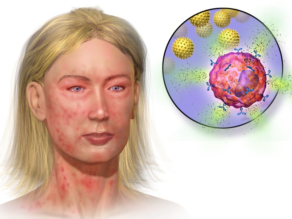
Anaphylaxis: adrenaline into the thigh before anything else
Every emergency in this section has a correct first action, and in most of them the intuitive action is the wrong one. The order is not a matter of tidiness. The order is the treatment.
Anaphylaxis is mast cell and basophil degranulation on a systemic scale, usually immunoglobulin E mediated and in children usually triggered by food — peanut, tree nut, cow’s milk, egg, sesame, shellfish — or by venom, latex or a drug. The mediators released do four things at once: they dilate vessels, open a capillary leak that can shift a third of the circulating plasma volume into the interstitium within minutes, constrict bronchi, and swell the larynx, tongue and pharynx. That combination is why a child can travel from itchy to unrecordable in the time it takes to find a set of observations.
Recognition rests on the speed of onset and the number of systems involved. Suspect anaphylaxis when a child develops, over minutes to a couple of hours, skin or mucosal signs — urticaria, flushing, angio-oedema — together with either respiratory or circulatory compromise; or when two or more systems are involved after exposure to a likely allergen; or when a child becomes hypotensive after exposure to a known allergen. In children the respiratory features dominate, and persistent vomiting or severe abdominal pain shortly after an allergen is a cardinal sign of a systemic reaction rather than a stomach upset. Crucially, something like one child in ten to one in five has no skin signs at all, so the absence of a rash does not exclude anaphylaxis in a child who is stridulous, wheezy, pale and floppy after eating.
The first action is intramuscular adrenaline into the anterolateral thigh, into the vastus lateralis — before an antihistamine, before a steroid, before intravenous access, and before anyone goes to look for the allergy plan. The dose is 0.01 mg/kg of the 1 mg/mL (1:1000) solution, to a maximum single dose of 0.5 mg at any age (0.3 mg is the conventional ceiling used in small prepubertal children), repeated after 5 to 15 minutes if the response is inadequate. The site matters, because intramuscular injection into the thigh produces a higher and faster peak plasma concentration than the deltoid or than any subcutaneous route. Adrenaline is also the only agent that reverses all four mechanisms at once: it constricts vessels, raises blood pressure, shrinks mucosal oedema, dilates bronchi and switches off further mediator release. Delay is what kills. In fatal anaphylaxis the interval from exposure to cardiorespiratory arrest is measured in minutes — shortest with intravenous drugs, then venom, then food — and the finding that runs through fatality reviews is that adrenaline was given late or not at all while an antihistamine was given early. There is no absolute contraindication to adrenaline in anaphylaxis.
Position the child according to what is failing, and change position gently. A child with circulatory compromise lies flat with the legs raised; a child whose breathing is the dominant problem may sit up; a child who is unconscious or vomiting goes into the recovery position. Never make a hypotensive child sit up abruptly, stand, or walk to a trolley. In a vasodilated, volume-depleted circulation venous return is posture-dependent, and suddenly emptying the right ventricle in that state has caused cardiac arrest — deaths are documented in patients who were sat or stood up. Alongside the adrenaline: call for help, remove the trigger where you can (stop the infusion, scrape out a stinger), give high-flow oxygen, obtain access and give a 20 mL/kg isotonic crystalloid bolus if there is any circulatory compromise, and add inhaled salbutamol only for bronchospasm that persists after adrenaline. Observe afterwards for a biphasic reaction, and no child goes home without an adrenaline auto-injector, training in its use, a written action plan and an allergy referral.
| Agent | Dose and route in a child | Role and cautions |
|---|---|---|
| Adrenaline (epinephrine) 1 mg/mL | 0.01 mg/kg intramuscularly into the anterolateral thigh; maximum single dose 0.5 mg at any age, with 0.3 mg the conventional ceiling in small prepubertal children; repeat after 5–15 min if needed | First-line and time-critical. The 1 mg/mL ampoule is the intramuscular concentration; the 0.1 mg/mL presentation is the intravenous strength used in arrest and is not what you draw up here |
| Adrenaline auto-injector | 0.1 mg from about 7.5 kg to under 15 kg where the device is available; 0.15 mg from 15 kg to under 25 kg; 0.3 mg from 25 kg to under 50 kg; 0.5 mg from about 50 kg in larger adolescents and adults; outer thigh, through clothing if necessary | A fixed strength will rarely match the calculated dose exactly; give the nearest appropriate device rather than delaying to draw up an exact volume |
| Oxygen | High flow by non-rebreather mask | For every child with respiratory or circulatory compromise |
| Isotonic crystalloid | 20 mL/kg bolus, repeated as the response dictates | Treats the capillary leak; hypotension persisting after adrenaline is usually unreplaced volume |
| Inhaled salbutamol | By spacer or nebuliser | Adjunct for persistent bronchospasm only; it does nothing for laryngeal oedema or for shock |
| H1 antihistamine | Oral or intravenous | Relieves itch and urticaria only. No effect on the airway, the breathing or the blood pressure, and never given before adrenaline |
| Corticosteroid | Oral or intravenous | Takes hours to act, does not treat the acute event, and current evidence does not support it for preventing biphasic reactions. It must never delay adrenaline |
Two habits protect a child in anaphylaxis more than any refinement of the drug list. The first is treating the reaction you are watching rather than the one described in the notes: a child with a mild history can have a severe reaction, and severity is judged on the airway, the breathing and the circulation in front of you. The second is not waiting for certainty. Adrenaline given to a child who turns out to have had a milder allergic reaction causes transient pallor, tremor and palpitations; adrenaline withheld from a child in anaphylaxis causes deaths.
Status epilepticus: the airway, the clock, then the drug
A convulsive seizure lasting 5 minutes or longer, or repeated seizures without recovery of consciousness between them, is status epilepticus. The five-minute definition is deliberately short: most self-limiting seizures stop within two to three minutes, so one that is still running at five is unlikely to stop by itself and is already doing harm through hypoxia, hyperthermia, acidosis and excitotoxic neuronal injury.
The first action is not the drug. It is the airway, the oxygen and the clock. Open the airway with a jaw thrust if it is needed, turn the child onto their side, suction secretions, give high-flow oxygen, attach monitoring, protect the head, and write down the exact time the seizure started, because every decision from here is timed from that moment. Do not restrain the limbs, and do not put anything in the mouth — a convulsing child cannot swallow their tongue, and objects placed in the mouth break teeth and obstruct airways. Then, in parallel, obtain intravenous or intraosseous access and check a bedside glucose. Hypoglycaemia is a cause of seizure that you can reverse in ninety seconds, and it will not respond to a benzodiazepine.
| Time from seizure onset | Stage | What is done |
|---|---|---|
| 0–5 min | Stabilisation | Airway, lateral position, suction, high-flow oxygen, monitoring, intravenous or intraosseous access, bedside glucose, temperature, and the exact start time written down. No restraint, nothing in the mouth |
| 5 min | First benzodiazepine | Lorazepam 0.1 mg/kg intravenously (maximum 4 mg per dose). Without access: midazolam 0.2 mg/kg intramuscularly or intranasally (maximum 10 mg), buccal midazolam 0.5 mg/kg (maximum 10 mg), or rectal diazepam 0.2–0.5 mg/kg (maximum 20 mg) |
| 10 min | Second and final benzodiazepine dose | Repeat once. Have bag-mask ventilation ready at the bedside — respiratory depression is cumulative across doses |
| 20 min | Second-line agent | Levetiracetam 60 mg/kg (maximum 4500 mg), fosphenytoin 20 mg PE/kg (maximum 1500 mg PE), or sodium valproate 40 mg/kg (maximum 3000 mg). Trial evidence shows the three to be comparably effective, so the choice follows the child’s history and local protocol; valproate is avoided where hepatic or mitochondrial disease is suspected |
| 40 min | Refractory status | Critical care: anaesthetic infusion with intubation and continuous electroencephalography |
While the drugs are being given, someone should be answering the question of why. Check the temperature, the sodium, the calcium and the toxicology; ask about missed anticonvulsant doses, a recent head injury, a ventriculoperitoneal shunt, a possible ingestion, and immunisation and travel history. A febrile seizure in a child between 6 months and 5 years is common and usually benign, but a seizure with fever plus a rash, neck stiffness or an abnormal appearance is meningitis until proved otherwise. And an unexplained seizure in an infant — particularly with vomiting, a tense fontanelle or a falling conscious level and no visible mark — should raise the possibility of abusive head trauma, which is the subject of the next section.
A seizure gets harder to stop the longer it runs. Within minutes of continuous seizure activity, GABA-A receptors are internalised from the synaptic membrane, so the very target the benzodiazepine acts on is disappearing while you watch. That is the pharmacological reason for the clock — for giving the first dose at 5 minutes rather than “seeing how it goes”, and for moving to a second-line agent after two benzodiazepine doses rather than giving a third. A third benzodiazepine at 25 minutes buys respiratory depression, not seizure control.
Shock, ketoacidosis, poison and the obstructed airway
Septic shock in a child is recognised on perfusion and mental state, not on blood pressure. The dysregulated host response produces vasodilatation, capillary leak and myocardial depression, and the child compensates with tachycardia and vasoconstriction — so what you see first is a capillary refill that is prolonged, or in warm shock unnaturally flash-fast; skin that is mottled and cool, or flushed with bounding pulses; a heart rate out of proportion to the fever; a falling urine output; and above all a change in how the child interacts, from irritable, to drowsy, to difficult to rouse. Hypotension is a late and pre-terminal sign, because a child can lose a quarter to a third of their circulating volume before the systolic pressure moves at all. A normal blood pressure is therefore never a reason to wait. The first actions are to say the word sepsis out loud and activate the pathway: high-flow oxygen; vascular access, going to an intraosseous needle after two failed attempts or about 90 seconds; blood cultures if they delay nothing; broad-spectrum antibiotics within one hour of recognition; a lactate; and fluid given as 10 to 20 mL/kg boluses titrated to perfusion, up to 40 to 60 mL/kg in the first hour, with reassessment after every single bolus. Stop the fluid and change strategy if the liver edge becomes palpable, crackles appear or the work of breathing rises, and move to vasoactive support rather than pouring in more. Three groups take the cautious 10 mL/kg bolus from the outset with reassessment between each: the neonate, whose volume expansion is 10 mL/kg over 5 to 10 minutes however shocked they are; the child with known cardiac disease, whose ventricle cannot absorb the preload; and the child with severe acute malnutrition, in whom myocardial and membrane function are impaired and standard-volume resuscitation increases mortality.
Diabetic ketoacidosis inverts the instinct to fix the number you can see. Insulin deficiency with a surge of counter-regulatory hormones produces hyperglycaemia, an osmotic diuresis that empties the circulation, and lipolysis with ketone production that drives the acidosis and the deep sighing Kussmaul breathing. The child is dehydrated, acidotic, depleted of total body potassium whatever the serum potassium happens to read, and hyperosmolar — and the brain has spent hours to days adapting to that osmolality. Fluid comes before insulin. Restore the circulation with isotonic crystalloid: 10 mL/kg over 30 to 60 minutes in the child who is dehydrated but still perfusing, repeated only as perfusion demands — but 20 mL/kg given rapidly, and repeated as needed, in the child who is genuinely shocked with hypotension or unrecordable pulses. Then replace the remaining deficit evenly over 24 to 48 hours. The insulin infusion starts 1 to 2 hours after fluids have begun, at 0.05 to 0.1 units/kg/h, and a child never receives an insulin bolus: a bolus drops glucose and osmolality abruptly, drives potassium into the cells, and is associated with cerebral oedema. Potassium is withheld only for frank hyperkalaemia or anuria. If the potassium is low at presentation, replacement starts with the initial fluid and the insulin infusion is delayed until potassium replacement is running, because insulin given to a hypokalaemic child can precipitate a lethal arrhythmia; if the potassium is normal, replacement starts alongside the insulin once urine output or normal renal function is confirmed. Insulin drives potassium intracellularly and unmasks a total-body deficit that is present whatever the serum level reads. Bicarbonate is not given routinely — it is associated with cerebral oedema and does not improve outcome. The narrow exceptions, on specialist advice, are life-threatening hyperkalaemia and severe acidosis with a pH below 6.9 where cardiac contractility is impaired.
The complication that shapes every one of those decisions is cerebral oedema: uncommon, but the leading cause of death and permanent disability in paediatric ketoacidosis, and characteristically appearing 4 to 12 hours after treatment begins — which is to say, while the laboratory numbers are improving. Its risk tracks how ill the child was on arrival rather than how fast the fluid ran: severe acidosis, a low carbon dioxide, a raised urea, young age and a new diagnosis all predict it, whereas a large randomised trial found that neither the rate of fluid administration nor its sodium content changed neurological outcome. Guidelines therefore still rehydrate evenly over 24 to 48 hours as a precaution, not because speed has been shown to cause the injury. The warning signs are neurological rather than biochemical: headache, recurrent vomiting after treatment has started, agitation giving way to drowsiness, incontinence in a child who was continent, a rising blood pressure with a falling heart rate, cranial nerve palsies, and any fall in conscious level at all. A falling conscious level during ketoacidosis treatment is the emergency, and it is treated before it is imaged: call for help, reduce the intravenous fluid rate by about a third, raise the head of the bed, and give hypertonic 3% saline 2.5 to 5 mL/kg over 10 to 15 minutes or mannitol 0.5 to 1 g/kg over 10 to 15 minutes, repeated after 30 minutes if there is no response. Imaging comes after treatment, never instead of it.
Poisoning and ingestion begins, like everything else, with airway, breathing, circulation and a glucose, then three questions: what, how much, and when. Contact the poisons information service — that call is the single most useful early action — and decontaminate skin or eyes where the exposure was topical. Do not induce vomiting. Syrup of ipecac left paediatric practice because it never improved any outcome: it empties little, it delays or blocks activated charcoal and oral antidotes, and vomiting a corrosive or a hydrocarbon re-injures the oesophagus and risks aspiration pneumonitis. Gastric lavage has gone the same way. Activated charcoal 1 g/kg, to a maximum of about 50 g, may be considered within roughly an hour of a suitable ingestion in an alert child with a protected airway, but it does not bind iron, lithium, alcohols, hydrocarbons or corrosives. Never assume a well-looking child is safe, because paracetamol, sulfonylureas, sustained-release preparations and the toxic alcohols all have silent latent periods. Be especially alert to the agents where a single adult tablet or one mouthful can kill a toddler: calcium channel blockers, tricyclic antidepressants, opioids including methadone, sulfonylureas, clonidine and imidazoline decongestants, theophylline, antimalarials, camphor, oil of wintergreen (methyl salicylate) and concentrated nicotine liquid. A swallowed button battery is a separate emergency — current flowing at the mucosa burns through the oesophageal wall within about two hours — so a child who may have swallowed one needs an immediate radiograph and urgent endoscopic removal, and in a child of 12 months or older who is still swallowing normally, who is within about 12 hours of the ingestion, and in whom there is no suspicion of perforation or sepsis, 10 mL of honey every 10 minutes to a maximum of six doses while transfer is arranged reduces the injury. Beyond that window, or with an unprotected airway, the injury is already established and the honey adds aspiration risk without benefit.
Foreign body airway obstruction turns on a single observation: is the cough effective? A child who can cry, speak or produce a loud forceful cough is moving air, and their own cough generates pressures no manoeuvre of yours can match — so you encourage the coughing, stay with the child, and do nothing else while it remains effective. When the cough turns silent or feeble, when the child cannot vocalise or breathe, when colour deteriorates or consciousness begins to fall, the cough is ineffective and you act at once. In an infant under 1 year, give 5 back blows head-down along your forearm with the jaw supported, then turn the infant and give 5 chest thrusts — two fingers on the sternum at the compression landmark, sharper and slower than compressions — and repeat, checking the mouth between cycles. In a child over 1 year, give 5 back blows then 5 abdominal thrusts, and repeat. Abdominal thrusts are never used in an infant, whose liver is large, low and unprotected by the rib cage. If the child becomes unresponsive, lay them on a firm flat surface, shout for the resuscitation team and begin CPR starting with chest compressions, looking into the mouth before each set of rescue breaths and removing an object only if you can see it. Never perform a blind finger sweep: in a narrow paediatric airway it drives a partially obstructing object down into the larynx, converts a partial obstruction into a complete one, and causes bleeding and swelling that make everything afterwards harder. Every child who received back blows or thrusts needs medical assessment afterwards, and a persistent cough, stridor or unilateral wheeze after a choking episode means a retained foreign body until bronchoscopy says otherwise.
| Emergency | Do this FIRST | Never |
|---|---|---|
| Anaphylaxis | Adrenaline 0.01 mg/kg of the 1 mg/mL solution intramuscularly into the anterolateral thigh | Give an antihistamine or a steroid first, or sit or stand a hypotensive child up |
| Status epilepticus | Airway and lateral position, oxygen, note the exact time, check the glucose — then a benzodiazepine at 5 minutes | Restrain the child, put anything in the mouth, or give a third benzodiazepine instead of a second-line agent |
| Septic shock | Oxygen, access, cultures, antibiotics within the hour, and fluid titrated to perfusion | Wait for the blood pressure to fall before calling it shock |
| Diabetic ketoacidosis | Restore the circulation with isotonic fluid; the insulin infusion follows 1 to 2 hours later | Give an insulin bolus, give bicarbonate routinely, or rehydrate quickly in a child who is not shocked — the shocked child with hypotension or unrecordable pulses still gets 20 mL/kg rapidly |
| Falling conscious level during ketoacidosis treatment | Treat as cerebral oedema: slow the fluid, head up, hypertonic saline or mannitol, call for help | Send the child for imaging before treating |
| Poisoning or ingestion | Airway, breathing, circulation and a glucose; establish what, how much and when; call the poisons service | Induce vomiting, or give a home remedy to neutralise it |
| Choking with an effective cough | Encourage the coughing and stay with the child | Interrupt an effective cough with blows or thrusts |
| Choking with an ineffective cough | Infant: 5 back blows then 5 chest thrusts. Child over 1 year: 5 back blows then 5 abdominal thrusts | Perform a blind finger sweep, or use abdominal thrusts on an infant |
The words you choose decide what the family tells you. In poisoning, choking and unexplained collapse the history is the diagnostic test, and a caregiver who feels accused gives a shorter and less accurate one. So ask “what was within reach?” rather than “how did they get hold of it?”, and “tell me exactly what you saw” rather than “were you watching?” Say “this happens in ordinary homes” and mean it. None of this softens the safeguarding thinking of the next section — a blame-free manner and an alert mind are entirely compatible — but it is how you find out what was swallowed, how much, and when.
In paediatric emergencies the order of actions is the treatment: intramuscular adrenaline into the thigh before any other drug in anaphylaxis, the airway and the clock before the benzodiazepine in status epilepticus, perfusion and mental state rather than blood pressure in septic shock, fluid before insulin in ketoacidosis with a falling conscious level treated as cerebral oedema, and back blows and thrusts — never a blind finger sweep — once a cough becomes ineffective.
Section 4Safeguarding: Injuries That Do Not Fit the Story or the Stage
Development is the yardstick
The single most useful safeguarding question is not “could this have happened?” but “could this child have done this?” An injury is concerning when it is inconsistent with the mechanism offered, and it is equally concerning when the mechanism is plausible in principle but impossible for a child at that developmental stage. A four-month-old cannot roll off a sofa if they have never rolled. A five-month-old cannot pull a mug of tea from a table if they cannot sit unsupported or pincer-grasp. A non-ambulatory infant cannot sustain a spiral femur fracture from “twisting in the cot”. Knowing the milestones from Chapter 11 is therefore a safeguarding skill: rolling at about 4 to 6 months, sitting unsupported at 6 to 8 months, crawling at 7 to 10 months, cruising at 9 to 12 months, walking at 12 to 15 months.
This is the basis of the aphorism “those who don’t cruise rarely bruise”. Bruising in a pre-mobile infant is rare enough that it is treated as a sentinel injury — a minor-looking injury in a young infant that frequently precedes severe or fatal abuse, and that represents the opportunity to intervene before the catastrophic event. Sentinel injuries include any bruise, any intraoral injury such as a torn frenulum, and any burn in an infant who is not yet mobile. They are easy to dismiss because they are small. They should never be dismissed.
The TEN-4-FACESp rule gives a validated screen for bruising in children under 4 years, flagging bruises that warrant evaluation for abuse.
| Letter | Region or feature | Why it is concerning |
|---|---|---|
| T | Torso (chest, abdomen, back, buttocks, genitourinary) | Accidental bruising clusters over bony prominences of the limbs and forehead, not soft protected areas |
| E | Ear | The ear is shielded by the skull and shoulder; bruising implies a direct blow or pinch |
| N | Neck | Suggests grabbing or strangulation |
| 4 | Any bruise anywhere in an infant under 4.99 months | Pre-mobile infants do not self-bruise |
| F | Frenulum (labial or lingual) | Forced feeding or a blow to the mouth |
| A | Angle of the jaw | Protected site |
| C | Cheek, fleshy part | Protected by the zygoma in accidental falls |
| E | Eyelid | Isolated eyelid bruising without a forehead injury is atypical |
| S | Subconjunctival haemorrhage | Associated with strangulation and direct trauma |
| p | Patterned bruising | Reproduces the object — a hand, belt, cord, buckle |
Injuries and histories that raise concern
Certain injuries carry a high specificity for inflicted trauma because of the forces required to produce them. Others are concerning mainly because of the context. Both belong in your assessment, and neither constitutes a diagnosis — the diagnosis of abuse is a multidisciplinary process, and there are important medical mimics such as bleeding disorders, osteogenesis imperfecta, metabolic bone disease, Mongolian blue spots or congenital dermal melanocytosis, and cultural practices such as cupping and coining, which must be explored respectfully rather than assumed.
| Finding | Why it raises concern |
|---|---|
| Posterior or posteromedial rib fractures | Require anteroposterior squeezing of the chest, so they are highly specific for inflicted injury; they are rarely but genuinely reported after two-thumb encircling-hands infant CPR, so a documented resuscitation is recorded and considered rather than excluded by rule |
| Classic metaphyseal lesion (bucket-handle or corner fracture) | Produced by torsion or traction on a limb |
| Spiral or transverse femur fracture in a non-ambulatory infant | Requires rotational or high force the child cannot generate |
| Fractures at multiple different stages of healing | Implies repeated events over time |
| Scapular, sternal or spinous process fracture | High-force injuries in protected sites |
| Immersion burn with a sharp line of demarcation, stocking-and-glove distribution, symmetry, sparing of flexural creases | Consistent with being held in hot liquid; a child who falls in has splash marks and irregular edges |
| Small, round, deep burns about 8 mm across | Cigarette contact |
| Retinal haemorrhages with subdural haemorrhage and few or no external marks | Abusive head trauma from rotational acceleration and deceleration |
| Any anogenital injury, or sexually transmitted infection in a prepubertal child | Requires urgent specialist evaluation |
| Poor growth, untreated dental disease, missed critical appointments, developmental regression, inadequately treated chronic illness | Possible neglect, which is the most common form of maltreatment |
Histories matter as much as injuries. Be concerned when there is no history offered for a significant injury; when the history changes between tellings or between caregivers; when it does not fit the injury or the child’s developmental stage; when there was an unexplained delay in seeking care; when the injury is blamed on a young sibling or a pet; when the child is unusually watchful, flinches, or shows no expectation of comfort; or when the caregiver is hostile to examination or focused on something other than the child. Be concerned too about medical child abuse — illness fabricated or induced by a caregiver — when symptoms occur only in one person’s presence, when the history is dramatic but investigations are persistently normal, or when a caregiver pushes for invasive procedures.
The duty to report, and how to document
Nurses are mandated reporters in every United States jurisdiction and hold equivalent safeguarding duties in most other systems. Four features of that duty are commonly misunderstood by students. First, the threshold is reasonable suspicion, not proof: you are not required to know that abuse occurred, only to have cause to suspect it, and investigation is the child protection agency’s job, not yours. Second, the duty is personal and non-delegable; it is not discharged by telling the physician or the charge nurse, and “the doctor said he would handle it” is not a defence. Third, reporting in good faith carries statutory immunity from civil and criminal liability, whereas failure to report is itself an offence in most jurisdictions and a licensure matter in all of them. Fourth, you generally report first and, depending on institutional policy and risk to the child, the family is informed by the designated team member — you do not need the caregiver’s permission and you do not warn them in a way that endangers the child. Reports are typically made by telephone immediately and in writing within 24 to 48 hours.
Your interviewing role is deliberately limited. Ask open, non-leading questions once — “tell me what happened”, “can you tell me more about that?” — and then stop. Repeated interviews contaminate evidence and re-traumatise children; formal forensic interviewing is done by trained specialists. Never promise a child that you will keep what they tell you secret; say instead that you will tell the people whose job it is to keep them safe. Where sexual assault is disclosed, preserve evidence per policy, note that in a prepubertal child evidence is rarely recovered from the body beyond about 24 hours while clothing and bedding remain productive far longer — the 72 to 120 hour kit windows apply mainly to adolescents, and acute examination is indicated regardless for injury, infection and safety assessment — discourage washing or changing clothes, and place clothing in paper rather than plastic bags.
Documentation in safeguarding is a legal document you may be asked to defend years later. Record what you observed, not what you concluded. Quote the child and the caregiver verbatim, in quotation marks, attributed to the speaker. Describe each injury by site, size in centimetres, shape, colour and any pattern, and map it on a body diagram; photograph only per institutional policy with a scale in frame. Do not attempt to date a bruise by its colour — colour-based ageing has been shown to be unreliable. Avoid the words “alleged”, “claims” and “denies”, which editorialise; write “the caregiver states”. Record who was present, the time of each event, and every notification you made with the name and time.
Poverty is not neglect. A family without transport, without a working refrigerator for insulin, or without the money for a prescription has an unmet need, not a moral failing, and the intervention is a social work referral, a resource, or a delivery arrangement — not a maltreatment report. Neglect is failure to provide when the caregiver has the means and the capacity to do so. Confusing the two harms families who are already disadvantaged and diverts scarce child protection resources. Report the child who is unsafe; resource the family who is struggling; and be honest with yourself about which one you are looking at.
Test every injury against the child’s developmental stage as well as the story, treat any bruise in a pre-mobile infant as a sentinel injury, and report on reasonable suspicion — the duty is yours personally, it does not require proof, and good-faith reporting is protected.
Section 5The Family in Crisis: Presence, Bad News and Loss
Family presence and the conversation during the emergency
Families should be offered the option of being present during invasive procedures and resuscitation. The evidence, and the position statements of emergency and critical care nursing organisations, support this: presence does not increase interference or litigation, and families who witness a resuscitation show lower rates of prolonged grief, intrusive imagery and unanswered questions afterwards. Offering is the operative word — presence is invited, never required, and a family who declines is not failing their child. The practical requirement is that a dedicated support person, not a team member with a resuscitation role, stays with the family throughout to narrate what is happening, answer questions and escort them out if needed.
During the crisis, communication has two audiences and two structures. To the team, use SBAR, which was scripted in Section 1. To the family, speak in short sentences, use the child’s name, avoid the abbreviations that fill the room, and check understanding rather than asking “does that make sense?” Give a warning shot before serious news: “I need to tell you something difficult.” The SPIKES framework structures the fuller conversation — Setting (private, seated, phones handed over, interpreter arranged, the right people present), Perception (“what have you been told so far?”), Invitation (“how much detail would you like?”), Knowledge (plain language, small amounts, then pause), Emotions (acknowledge and stay silent rather than filling the silence), and Strategy and summary (what happens next, who to contact, written information).
Use the word died. “Passed away”, “we lost her” and “gone” are ambiguous, and to a young child they are literally confusing — children have searched for a sibling who was “lost” and refused to sleep after being told a baby “went to sleep”. Avoid the phrases that sound like comfort and land as dismissal: “at least you have other children”, “at least it was early”, “everything happens for a reason”, “you can have another”, “you are young”. What helps is simple, honest presence: “I am so sorry. We did everything we could. Would you like me to stay?”
Perinatal, neonatal and paediatric loss
Perinatal loss covers miscarriage, stillbirth (in the United States, fetal death at 20 weeks’ gestation or later), termination for fetal anomaly, and neonatal death. The nursing task is to make choices available and to follow the family’s lead, because the interventions that support long-term grief are almost entirely about agency and memory. Ask the baby’s name and then use it, every time. Offer — without pressure and with the option to change their mind later — seeing and holding the baby, bathing and dressing the baby, hand and footprints, a lock of hair with permission, photographs, a blanket or hat, and religious or cultural rites. Offer time, including the option of a cooling cot where available. Document what was offered, what was chosen and what was declined so the family is not asked the same painful questions repeatedly, and flag the record so that staff do not walk in with routine cheerfulness.
Do not neglect the physiology of loss. After a birth at 16 weeks or beyond, the birthing parent will experience lactogenesis: milk usually comes in on day 2 to 4 whether or not there is a baby to feed, and being unprepared for that is described by bereaved parents as one of the cruellest surprises of the experience. The crisis-care task here is to warn the family before it happens, so that it is expected rather than ambushing them at home, and to offer — where the family wishes it — the option of donating expressed milk, which some parents find deeply meaningful. The suppression regimen itself is postpartum management and is set out in Chapter 7. Hand the family back to routine postpartum care rather than letting bereavement displace it, and make sure the screening for perinatal mood and anxiety disorders described in Chapter 8 actually happens, because the risk after loss is substantially raised.
Where a child dies suddenly and unexpectedly, families face investigation on top of grief. In sudden unexpected infant death, a scene investigation, a death scene re-enactment and an autopsy are routine and legally required in most jurisdictions; explain in advance that this happens for every such death and is not an accusation, and that the questions about the sleep surface and who was in the bed are asked of every family. Risk peaks between 1 and 4 months. The sleep environment comes up in that conversation because the investigation asks about it, so you need the ABCs of safe sleep at hand: Alone (room-sharing without bed-sharing), on the Back for every sleep, in a Crib or cot with a firm flat surface and nothing else in it — no bumpers, pillows, quilts, positioners or soft toys. Deliver it without blame, since the death has already happened and the parents will be searching for what they did wrong; the wider anticipatory guidance for well infants belongs to the well-child chapters, not here.
Children, grief, and the team that carries it
Siblings grieve, and they grieve according to where they are developmentally. Under about 3 years, a child registers separation and disruption rather than death, and responds to routine, familiar caregivers and physical comfort. From about 3 to 5 years, thinking is magical and death is understood as reversible or temporary; these children commonly believe they caused the death by a thought or a bad behaviour and must be told explicitly and repeatedly that nothing they did or thought caused it. From about 6 to 9 years, understanding becomes concrete and often personified; the mature concept of death — that it is permanent, universal, irreversible, and involves the ending of bodily functions — usually consolidates by about 9 to 10 years. Adolescents hold an adult concept but may express grief through withdrawal, anger, somatic complaints or risk-taking, and need honest information and peer connection. Across all ages: use concrete language, answer the same question as many times as it is asked, keep routines, and let children attend rituals if they choose — having been prepared for exactly what they will see and accompanied by an adult whose only job is them.
The family in crisis needs practical scaffolding as much as emotional support: an interpreter rather than a relative translating, a chaplain or the family’s own faith leader, social work, bereavement follow-up with named contacts and dates, written information they can read later because almost nothing said in the first hour is retained, and where relevant referral to palliative care, which belongs alongside active treatment and not only at the end. Requests about organ and tissue donation should be decoupled from the death notification and made by a trained requestor from the organ procurement organisation, not by the nurse who has just told the family their child died.
Finally, the team. Caring for a dying child, a resuscitation that failed or a safeguarding case generates moral distress and second-victim reactions that are normal responses to abnormal work. Structured debriefing after critical events, peer support, and knowing how to access employee assistance are professional competencies, not weaknesses. And even in the aftermath, delegation limits hold: unlicensed personnel can sit with a family, fetch what is needed, prepare a memory box and provide physical care, but the assessment of a grieving parent’s risk, the teaching, the evaluation of coping and the care of the unstable patient remain with the registered nurse.
What families remember. Bereaved parents in follow-up studies rarely recall the drugs, the diagnosis or the equipment. They recall whether the staff used their baby’s name, whether anyone cried with them, whether they were rushed, whether someone said the child’s death was not their fault, and whether anyone came back. You cannot change the outcome in these moments. You can change, permanently, what the family carries out of the room — and that is a clinical intervention with an evidence base, not merely kindness.
Offer family presence rather than imposing it, deliver news with a warning shot and the word “died”, make memory-making and the physiology of loss part of the care plan, and match every explanation of death to the child’s developmental stage.
The chapter in one breath
Crisis care in paediatric and perinatal nursing rewards pattern recognition and punishes improvisation. Start at the doorway with the Paediatric Assessment Triangle — appearance judged by TICLS, work of breathing, circulation to skin — because which legs are abnormal names the physiological state and the state names the first intervention. Anchor every observation to an age band, remembering that heart rate and capillary refill change early while blood pressure falls last, that a child 1 to 10 years is hypotensive below 70 + (2 × age in years), and that bradycardia with poor perfusion in an infant or child means starting compressions. Grade dehydration by percentage and match it to signs, calculate maintenance with the 4–2–1 rule, resuscitate with 20 mL/kg (10 mL/kg in neonates, diabetic ketoacidosis, severe acute malnutrition, known or suspected cardiac failure and suspected raised intracranial pressure), and calculate every dose per kilogram while checking the answer against the adult ceiling. In paediatric emergencies the order is the treatment: intramuscular adrenaline into the thigh before antihistamine or steroid in anaphylaxis, the airway and the clock before the benzodiazepine in status epilepticus, perfusion and mental state rather than blood pressure in septic shock, fluid before insulin in ketoacidosis with any falling conscious level treated as cerebral oedema before it is imaged, no induced vomiting in poisoning, and back blows and thrusts — never a blind finger sweep — once a cough becomes ineffective. Test every injury against the child’s developmental stage, treat bruising in a pre-mobile infant as a sentinel injury, and report reasonable suspicion yourself. Then stay in the room, use the child’s name, and say “died”. After a stillbirth or a death, ask the baby’s name and use it, offer memory-making without pressure, warn the birthing parent that lactogenesis is coming before handing that care back to the postpartum chapters, and explain the investigation and the ABCs of safe sleep it asks about after a sudden unexpected infant death.
Words worth knowing
| Term | What it means |
|---|---|
| Paediatric Assessment Triangle (PAT) | A 60-second no-touch impression using appearance, work of breathing and circulation to skin; the pattern of abnormal legs names the physiological state. |
| TICLS | Tone, Interactiveness, Consolability, Look or gaze, Speech or cry — the components of the appearance leg of the PAT. |
| Compensated shock | Inadequate perfusion with a still-normal blood pressure, maintained by tachycardia and vasoconstriction; signalled by delayed capillary refill and cool mottled skin. |
| Decompensated shock | Shock with hypotension — systolic BP below 60 (neonate), 70 (infant), 70 + (2 × age) for 1 to 10 years, or 90 over 10 years; a late and ominous finding. |
| 4–2–1 rule | Hourly maintenance fluid: 4 mL/kg/h for the first 10 kg, 2 mL/kg/h for the next 10 kg, 1 mL/kg/h for each kilogram above 20 kg. |
| Fluid deficit | The volume already lost, estimated as 50/100/150 mL/kg for mild, moderate and severe dehydration in an infant and 30/60/90 mL/kg in an older child, or measured directly as acute weight loss (1 kg equals 1000 mL). |
| Independent double check | A second nurse calculates a high-alert dose from the original order without seeing the first nurse’s working, then the two results are compared. |
| Anaphylaxis | A rapid-onset multisystem allergic reaction with respiratory or circulatory compromise; treated with intramuscular adrenaline before any other drug, and not excluded by the absence of a rash. |
| Intramuscular adrenaline | 0.01 mg/kg of the 1 mg/mL solution into the anterolateral thigh — maximum single dose 0.5 mg at any age, 0.3 mg being the conventional ceiling in small prepubertal children — repeatable after 5 to 15 minutes; there is no absolute contraindication in anaphylaxis. |
| Status epilepticus | A convulsive seizure lasting 5 minutes or longer, or repeated seizures without recovery between them; airway, oxygen, timing and glucose come first, a benzodiazepine at 5 minutes, and a second-line agent at about 20 minutes. |
| Paediatric septic shock | Sepsis with inadequate perfusion, recognised on capillary refill, skin and mental state rather than blood pressure, because hypotension in a child is a late and pre-terminal sign. |
| Cerebral oedema in ketoacidosis | The leading cause of death in paediatric diabetic ketoacidosis, typically 4 to 12 hours into treatment: headache, recurrent vomiting, rising blood pressure with a falling heart rate, and a falling conscious level. Treated before it is imaged. |
| Activated charcoal | 1 g/kg, up to about 50 g, considered within roughly an hour of a suitable ingestion in an alert child with a protected airway; it does not bind metals, alcohols, hydrocarbons or corrosives. |
| Ineffective cough | A silent or feeble cough in a choking child who cannot speak, cry or breathe — the point at which back blows and thrusts begin. An effective cough is left entirely alone. |
| Blind finger sweep | Sweeping a finger through a choking child’s mouth without seeing the object; forbidden, because it drives the object deeper and injures a narrow airway. |
| Sentinel injury | A minor-looking injury — typically a bruise, intraoral injury or burn — in a pre-mobile infant, frequently preceding severe or fatal abuse. |
| TEN-4-FACESp | A bruising screen for children under 4: Torso, Ear, Neck, any bruise under 4.99 months, plus Frenulum, Angle of jaw, Cheek, Eyelid, Subconjunctival haemorrhage and patterned bruising. |
| Mandated reporter | A professional legally obliged to report reasonable suspicion of child maltreatment personally and promptly, protected by good-faith immunity and liable for failure to report. |
| SPIKES | Setting, Perception, Invitation, Knowledge, Emotions, Strategy and summary — a structure for delivering serious news. |
| ABCs of safe sleep | Alone, on the Back, in a Crib on a firm flat surface with nothing else in it — the core counselling to reduce sudden unexpected infant death. |