PAEDIATRIC RESPIRATORY AND CARDIOVASCULAR CONDITIONS
A child in trouble looks well until the moment they do not. The paediatric airway is narrower, softer and more easily occluded than an adult’s; the paediatric heart compensates for lost stroke volume almost entirely by going faster; and the paediatric blood pressure holds a deceptively normal number until the compensation collapses. That is why the nursing skill in this chapter is not pattern-matching a diagnosis but reading a trajectory — work of breathing, perfusion, and mental status — and acting on the trend before the number moves. Everything here is age-banded and weight-based. A respiratory rate of 40 is normal in a two-month-old and alarming in a ten-year-old. A dose is never a number; it is a number per kilogram, checked against the maximum adult dose. This chapter builds the mechanism, then the assessment, then the management, then the complications, for the airway and the circulation of the growing child.
By the end of this chapter you can
- Explain how paediatric airway and myocardial anatomy produce rapid decompensation, and state normal respiratory rate, heart rate and systolic blood pressure ranges by age band.
- Calculate a weight-based drug dose and a 4-2-1 maintenance fluid rate, show the arithmetic, and verify the result against the maximum adult dose.
- Distinguish croup from epiglottitis and state the single first action for each, including what must never be done to a child with suspected epiglottitis.
- Prioritise nursing care in bronchiolitis, asthma exacerbation, community-acquired pneumonia and cystic fibrosis, including the therapies that evidence does not support.
- Classify congenital heart lesions by pulmonary blood flow and obstruction, and describe the role of prostaglandin E1 in a ductal-dependent lesion.
- Manage a hypercyanotic (tet) spell in correct sequence, beginning with the knee-chest position.
- Recognise Kawasaki disease, acute rheumatic fever and paediatric heart failure, and identify the complication each one threatens.
- Differentiate compensated from hypotensive shock, apply age-specific hypotension thresholds, and state when to begin compressions for bradycardia with poor perfusion.
- Escalate deterioration using SBAR and apply delegation limits — assessment, teaching, evaluation and the unstable child remain with the registered nurse.
Section 1The Paediatric Airway, the Paediatric Heart and What the Numbers Mean
Why children decompensate faster than adults
Resistance to airflow is inversely proportional to the fourth power of the radius, so as the radius falls the resistance climbs steeply. An infant trachea is roughly 4 mm across, a radius of 2 mm; one millimetre of circumferential oedema halves that radius, removes about three-quarters of the cross-sectional area, and multiplies resistance roughly sixteen-fold. The same millimetre in an adult trachea is barely noticeable. This single fact explains why viral swelling that gives an adult a hoarse voice gives a toddler stridor at rest, and why suctioning an obligate nose-breathing infant’s nares is a genuine respiratory intervention rather than comfort care.
Beyond the lumen, the mechanics work against the child. The infant chest wall is cartilaginous and compliant, so intercostal and subcostal retractions appear early and the chest paradoxically collapses inward as the diaphragm pulls down. The ribs sit horizontally, limiting bucket-handle expansion, so the only way to increase minute ventilation is to increase the rate. Infants have fewer type I fatigue-resistant diaphragmatic fibres, which is why a baby who has been breathing at 80 breaths per minute for three hours can stop — not because the disease worsened, but because the muscle tired. Oxygen consumption is roughly 6 to 8 mL/kg/min compared with 3 to 4 in an adult, and functional residual capacity is proportionally smaller, so desaturation after apnoea is measured in seconds.
The heart has its own constraint. The neonatal and infant myocardium contains less contractile tissue and more non-contractile mass, so stroke volume is relatively fixed. Cardiac output equals heart rate multiplied by stroke volume, and when a child cannot raise stroke volume, output is almost purely rate-dependent. Two consequences follow and both are testable at the bedside. First, tachycardia is the earliest and most faithful sign of trouble — hypovolaemia, fever, pain, hypoxia, anxiety — and it must never be dismissed as “just crying” without re-checking when the child is settled. Second, bradycardia in a child is an emergency, not a reassuring return to normal; it usually means profound hypoxia and it precedes arrest.
Vital signs by age band, and the values that define shock
There is no single paediatric normal. Comparing a child against an adult range is one of the commonest sources of missed deterioration, so the ranges below should be at hand every time you record a set of observations. Rates are for the awake, afebrile, uncrying child; sleeping rates run lower and every degree Celsius of fever adds roughly 10 beats per minute and a few breaths per minute.
| Age band | Respiratory rate (per min) | Heart rate, awake (bpm) | Systolic BP (mmHg) |
|---|---|---|---|
| Neonate (birth to 28 days) | 30–60 | 100–205 | 67–84 |
| Infant (1–12 months) | 30–53 | 100–180 | 72–104 |
| Toddler (1–2 years) | 22–37 | 98–140 | 86–106 |
| Preschool (3–5 years) | 20–28 | 80–120 | 89–112 |
| School age (6–11 years) | 18–25 | 75–118 | 97–115 |
| Adolescent (12–15 years) | 12–20 | 60–100 | 110–131 |
Blood pressure is the last thing to fail. A child in compensated shock has tachycardia, cool mottled extremities, capillary refill beyond 2 seconds, a narrowing pulse pressure, weak peripheral pulses with preserved central pulses, and falling urine output — with a blood pressure that reads normal. By the time the systolic pressure drops, roughly 30 to 40 per cent of circulating volume is already gone. Learn the hypotension thresholds as hard floors, not as goals.
| Age | Hypotension defined as systolic BP below |
|---|---|
| Term neonate, 0–28 days | 60 mmHg |
| Infant, 1–12 months | 70 mmHg |
| Child, 1–10 years | 70 mmHg plus twice the age in years (a 6-year-old: 70 + 12 = 82 mmHg) |
| Over 10 years | 90 mmHg |
Urine output is a cheap, continuous perfusion monitor: expect 1.5 to 2 mL/kg/hour in infants and toddlers, at least 1 mL/kg/hour in older children, and at least 0.5 mL/kg/hour in adolescents. Weigh nappies — 1 gram equals 1 millilitre.
Weight-based dosing and maintenance fluids: always show the arithmetic
Children are not small adults, and every paediatric medication error begins with a number that was not converted. Three habits prevent nearly all of them. Use the weight in kilograms only, measured today, never a pound figure the family recalls and never a weight from a previous admission. Express the order as milligrams per kilogram per dose or per day, and write out the multiplication so a second nurse can follow it. Then perform the step that is skipped most often: compare the calculated paediatric dose with the maximum adult dose. A heavy 14-year-old on a milligram-per-kilogram formula can generate a number no adult would ever receive; the safe dose is the lower of the two.
Worked example. A 14 kg toddler with moderate croup is ordered dexamethasone 0.6 mg/kg as a single dose. 0.6 mg × 14 kg = 8.4 mg. The usual maximum is 16 mg, so 8.4 mg is within limits and is given. Second example. An 18 kg child with community-acquired pneumonia is ordered high-dose amoxicillin 90 mg/kg/day divided every 12 hours. 90 mg × 18 kg = 1620 mg/day; divided by two doses = 810 mg every 12 hours. The daily maximum is 4 g, so this is safe. Third example. A 46 kg adolescent with severe asthma is ordered prednisolone 2 mg/kg/day. 2 mg × 46 kg = 92 mg — but the maximum is 60 mg/day, so 60 mg is given and the discrepancy is documented and communicated.
Maintenance intravenous fluid uses the 4-2-1 rule, the hourly form of the Holliday–Segar method: 4 mL/kg/hour for the first 10 kg, plus 2 mL/kg/hour for the second 10 kg, plus 1 mL/kg/hour for every kilogram above 20. Worked example for a 26 kg child: the first 10 kg gives 10 × 4 = 40 mL/h; the next 10 kg gives 10 × 2 = 20 mL/h; the remaining 6 kg gives 6 × 1 = 6 mL/h. Total = 40 + 20 + 6 = 66 mL/hour. For an 8 kg infant, all weight falls in the first band: 8 × 4 = 32 mL/hour. Maintenance fluid is an isotonic solution — typically 0.9% sodium chloride or lactated Ringer’s with dextrose and, once the child voids, added potassium — because hypotonic maintenance fluid in an acutely unwell child causes hyponatraemia. Maintenance is not resuscitation: a child in hypovolaemic or septic shock receives a bolus of 20 mL/kg of isotonic crystalloid over 5 to 20 minutes with reassessment after each bolus, reduced to 10 mL/kg for neonates and for a child with known cardiac disease, severe malnutrition or diabetic ketoacidosis.
Assessment sequence, red flags, escalation and delegation
Begin every paediatric encounter from the doorway with the three sides of the assessment triangle — appearance (tone, interactiveness, consolability, gaze, cry), work of breathing (position, retractions, nasal flaring, grunting, audible noise) and circulation to skin (pallor, mottling, cyanosis). This takes seconds, requires no equipment and identifies the sick child before any monitor does. Then move to the hands-on airway, breathing, circulation, disability and exposure survey. Grunting in an infant is expiration against a partially closed glottis to generate auto-PEEP; it is not fussing and it signals alveolar disease. A child who stops fighting the oxygen mask has usually not improved.
Red flags that require immediate escalation: stridor at rest; grunting; head bobbing; a silent chest in a child who was wheezing; a respiratory rate that falls toward normal while the work of breathing rises or mental status drops; oxygen saturation under 90 per cent on supplemental oxygen; apnoea, especially in an infant under two months; capillary refill beyond 3 seconds with cool mottled skin; heart rate under 60 with poor perfusion; a systolic pressure below the age threshold; cyanosis that does not improve with oxygen; and any parental statement that the child is not behaving like themselves. Escalate with SBAR: Situation — “Room 4, Maya, 8 months, 7.2 kg, RSV bronchiolitis, now having apnoeic pauses.” Background — “Admitted 6 hours ago, on 2 L nasal cannula, feeding poorly.” Assessment — “Two pauses over 20 seconds with desaturation to 82 per cent, heart rate dropped to 88, subcostal retractions and grunting.” Recommendation — “I need you to see her now; I am setting up high-flow and requesting PICU review.”
Delegation limits. The registered nurse retains the initial and ongoing assessment, all teaching, all evaluation of response to therapy, and care of the unstable child. Assistive personnel may take vital signs on a stable child, weigh nappies, record intake and output, obtain daily weights, position and bathe, and feed a stable infant who has an established feeding pattern — but they may not perform the first set of vitals on a newly admitted or deteriorating child, and they may not interpret what they measure. Licensed practical or vocational nurses may administer most oral, subcutaneous and intramuscular medications within their scope, perform tracheostomy and gastrostomy care per policy, and reinforce teaching the RN has already given. Titrating oxygen, giving intravenous push medications, administering blood, first-dose immunoglobulin infusions and any assessment of a child in respiratory distress or shock stay with the RN.
In children the compensating vital sign is the heart rate and the failing one is the blood pressure, so treat tachycardia with poor perfusion as shock long before the pressure falls — and treat bradycardia as pre-arrest.
Section 2Upper Airway Obstruction: Croup, Epiglottitis and Aspiration
Croup (laryngotracheobronchitis)
Croup is viral inflammation and oedema of the subglottic larynx and trachea, most often caused by parainfluenza virus type 1, and it peaks between 6 months and 3 years of age. The mechanism is the fourth-power law made visible: mucosal swelling at the cricoid ring, the narrowest fixed point in the paediatric airway, narrows the lumen so that inspiratory airflow becomes turbulent. The result is the classic triad of a barking, seal-like cough, inspiratory stridor and hoarseness, typically preceded by one to three days of coryza and worst at night and when the child is upset. Fever is usually low-grade, the child is drinking, and the cough is prominent.
Assessment is clinical and severity is graded by the presence of stridor and retractions at rest, using the Westley croup score in many units. Mild croup means an occasional barking cough with no stridor at rest and no retractions. Moderate croup means stridor at rest with mild to moderate retractions but the child remains interactive. Severe croup means stridor at rest with marked retractions, agitation or lethargy, and decreased air entry. A child who becomes quiet with diminishing stridor and rising drowsiness is not improving — reduced airflow makes less noise. Do not obtain radiographs to make the diagnosis, and do not upset the child for tests that will not change management; crying increases turbulent flow and worsens obstruction.
Management begins with keeping the child calm on a caregiver’s lap. Every severity receives corticosteroid: dexamethasone 0.6 mg/kg as a single oral, intramuscular or intravenous dose, usual maximum 16 mg, which reduces oedema over the following 2 to 4 hours and shortens the illness even in mild disease. For stridor at rest, add nebulised epinephrine — racemic epinephrine 2.25%, 0.05 mL/kg to a maximum 0.5 mL diluted in saline, or L-epinephrine 1 mg/mL at 0.5 mL/kg to a maximum 5 mL. Nebulised epinephrine works within 10 to 30 minutes by alpha-mediated mucosal vasoconstriction, but its effect wears off, so the child must be observed for 3 to 4 hours for rebound obstruction before discharge. Humidified air, once a ward ritual, has no demonstrated benefit. Complications are respiratory fatigue and, rarely, secondary bacterial tracheitis.
Epiglottitis and bacterial tracheitis
Epiglottitis is bacterial cellulitis of the supraglottic structures, historically caused by Haemophilus influenzae type b and now uncommon where Hib immunisation is routine — which means the children who develop it are disproportionately under-immunised, and the nurse who has never seen a case is the one most likely to meet one. Onset is measured in hours, not days. The child, typically 2 to 7 years old, appears toxic with a high fever, sits forward in the tripod position with the chin thrust out and the tongue protruding, and shows the four Ds: drooling, dysphagia, dysphonia (a muffled “hot potato” voice) and distressed inspiratory effort. There is characteristically no barking cough. Stridor here is a late and ominous finding.
What you do first is what you do not do. Never inspect the throat, never insert a tongue depressor or swab, never place the child supine, never start an intravenous line or draw blood, and never leave the child alone. Any of these may provoke laryngospasm and complete obstruction. Keep the child upright in the position they have chosen, on the caregiver’s lap, keep them nil by mouth, offer blow-by oxygen only if it does not distress them, and summon the most skilled airway team available — anaesthesia and otolaryngology — for controlled intubation in the operating theatre with equipment for emergency tracheostomy at hand. Cultures, intravenous access and intravenous antibiotics (a third-generation cephalosporin such as ceftriaxone) follow after the airway is secured. Bacterial tracheitis presents as croup that fails to respond to epinephrine, with high fever, a toxic appearance and thick purulent secretions; it is usually staphylococcal, requires antibiotics, and frequently requires intubation for suctioning.
| Feature | Croup | Epiglottitis |
|---|---|---|
| Onset | Gradual, 1–3 days after coryza | Abrupt, over hours |
| Typical age | 6 months–3 years | 2–7 years |
| Cough | Barking, seal-like, prominent | Absent |
| Voice | Hoarse | Muffled, minimal speech |
| Drooling | No | Yes — will not swallow |
| Fever | Low-grade | High, toxic appearance |
| Posture | Any; comfortable upright | Tripod, chin forward, refuses to lie down |
| Imaging (if ever obtained) | Steeple sign, AP neck | Thumb sign, lateral neck |
| First action | Calm the child; dexamethasone 0.6 mg/kg | Do not touch the throat; call the airway team |
Foreign body aspiration and obstructive sleep apnoea
Aspiration peaks between 1 and 3 years, when a child has molars enough to bite but not to grind and a habit of running with food in the mouth. High-risk items are round and compressible — whole grapes, hot dog rounds, nuts, popcorn, hard sweets, raw carrot coins — and small objects such as coins, button batteries and small magnets. Classic presentation is a sudden choking episode followed by coughing and unilateral wheeze or decreased breath sounds; a partially obstructing object may masquerade for weeks as “asthma that only responds a little”. If the child is coughing forcefully, speaking or crying, the obstruction is partial: encourage coughing and do not intervene. If the child cannot cough, speak or cry, act — for an infant under one year, five back blows followed by five chest thrusts, repeated; for a child over one year, abdominal thrusts. Blind finger sweeps are never performed. Definitive removal is by rigid bronchoscopy. A swallowed button battery is a separate emergency: lodged in the oesophagus it causes caustic injury within two hours and requires immediate imaging and endoscopic removal.
Obstructive sleep apnoea in children is usually caused by adenotonsillar hypertrophy rather than obesity, though obesity compounds it. Parents report snoring, witnessed pauses, mouth breathing, restless sleep and night sweats; the daytime picture in children is often hyperactivity, inattention and poor school performance rather than sleepiness. Untreated, chronic nocturnal hypoxaemia produces pulmonary vasoconstriction and, eventually, cor pulmonale. Adenotonsillectomy is the usual treatment. Post-tonsillectomy nursing focuses on the bleeding risk: watch for frequent swallowing, throat clearing, restlessness and a rising pulse — the earliest signs of haemorrhage in a child who is swallowing the blood. Position the child prone or side-lying to allow drainage, keep suction available but do not suction the oropharynx deeply, avoid straws, red or brown fluids, and coughing or throat clearing. Bleeding risk is highest in the first 24 hours and again at 7 to 10 days when the eschar separates.
Croup is treated by calming the child and giving dexamethasone 0.6 mg/kg; suspected epiglottitis is treated by keeping instruments, tongue depressors and supine positioning away from the child until a skilled airway team is present.
Section 3Lower Airway and Parenchymal Disease
Bronchiolitis and respiratory syncytial virus
Bronchiolitis is the leading cause of hospital admission in infants under one year. Respiratory syncytial virus causes most cases, with rhinovirus, human metapneumovirus and parainfluenza accounting for the rest. The virus infects and sloughs the ciliated epithelium of the terminal bronchioles; the resulting debris, oedema and mucus form plugs that obstruct airways of one to two millimetres. Because obstruction is patchy and greater on expiration, air trapping and hyperinflation dominate, producing wheeze, prolonged expiration, crackles and a barrel-shaped chest. The illness follows a predictable arc: two to three days of coryza and low-grade fever, then increasing cough and work of breathing, with days 3 to 5 the worst, and slow resolution over one to three weeks.
Assess feeding as a respiratory sign. An infant who cannot sustain a feed, who takes fewer than half of the usual volumes, or who has fewer than half the usual wet nappies, is telling you the work of breathing has exceeded what they can sustain. Note apnoea risk: infants under two months, those born preterm, and those with congenital heart or chronic lung disease may present with apnoea before significant wheeze. Nasal obstruction alone can cause meaningful distress in an obligate nose-breather.
Treatment is supportive and the evidence is unusually clear about what does not help. Nasal suctioning before feeds and sleep, small frequent feeds or nasogastric or intravenous hydration, upright positioning, and supplemental oxygen if saturations persistently fall below 90 per cent are the core. High-flow nasal cannula at 1 to 2 L/kg/min is used for escalating work of breathing. Routine bronchodilators, corticosteroids, nebulised hypertonic saline in the emergency setting, antibiotics and chest physiotherapy are not recommended, and continuous pulse oximetry in a stable improving infant leads to longer stays without better outcomes. Infection control is nursing’s largest contribution: RSV survives on surfaces for hours, so contact and droplet precautions, dedicated equipment, cohorting, and rigorous hand hygiene protect every other infant on the unit. Prevention for high-risk infants uses monoclonal antibody — palivizumab 15 mg/kg intramuscularly monthly through the season, or a single dose of nirsevimab (50 mg for infants under 5 kg, 100 mg for 5 kg and over). These are passive antibodies, not vaccines, and do not create lasting immunity.
Asthma in the child
Asthma is chronic airway inflammation with bronchial hyper-responsiveness producing reversible obstruction through three simultaneous mechanisms — bronchospasm, mucosal oedema and mucus plugging. Only the first responds to a bronchodilator within minutes; the other two are why corticosteroids are given early and why a child can look better on albuterol and still be deteriorating. Triggers include viral infection, exercise, cold air, tobacco smoke, allergens, and, in a subset of older children, aspirin and non-steroidal anti-inflammatory drugs.
Assessment combines the physical picture with objective numbers. Peak expiratory flow zones, calibrated to the child’s own personal best, run green at 80 per cent or above, yellow at 50 to 79 per cent (take the reliever, follow the action plan) and red below 50 per cent (reliever now and seek urgent care). Reading the blood gas requires care: early in an attack the tachypnoeic child blows off carbon dioxide, so a low PaCO2 with respiratory alkalosis is expected. A normalising or rising PaCO2 in a child still working hard signals impending respiratory failure, not improvement. So does a silent chest, an inability to speak in full sentences, altered mental status and a saturation under 90 per cent on oxygen.
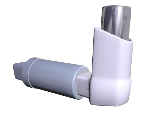
Acute management: oxygen to keep saturations at or above 92 per cent, then short-acting beta-2 agonist — nebulised albuterol 2.5 mg for a child under 20 kg and 5 mg for 20 kg and over, or weight-based at 0.15 mg/kg/dose with a 2.5 mg minimum, given every 20 minutes for three doses or continuously in severe disease. A metered-dose inhaler with a valved spacer, four to eight puffs, is at least as effective as a nebuliser for mild-to-moderate exacerbations and delivers less systemic side-effect. Add ipratropium for the first hour in moderate-to-severe attacks, and systemic corticosteroid early — prednisolone 1 to 2 mg/kg/day, maximum 60 mg/day, for 3 to 5 days. In severe exacerbation add magnesium sulfate 25 to 50 mg/kg intravenously over 20 minutes, maximum 2 g, monitoring for hypotension. Expect tremor, tachycardia and hypokalaemia from beta agonists and reassure families that these are pharmacological, not a sign of worsening disease. Chronic control rests on inhaled corticosteroids, and teaching must cover spacer use, rinsing the mouth after inhaled steroid to prevent oral candidiasis, distinguishing controller from reliever, trigger control, and a written asthma action plan shared with the school.
Pneumonia
In children under five, most community-acquired pneumonia is viral. The commonest bacterial cause across childhood is Streptococcus pneumoniae; in children over five, Mycoplasma pneumoniae causes a more insidious “walking pneumonia” with headache, malaise and a persistent dry cough. Bacterial pneumonia typically brings abrupt high fever, a toxic appearance, focal crackles or bronchial breathing and focal consolidation; viral pneumonia brings a more gradual course with diffuse findings and often coexisting wheeze. Tachypnoea is the single most sensitive sign of pneumonia in a child — more sensitive than auscultation. Note that lower-lobe pneumonia in a child frequently presents as abdominal pain, and upper-lobe pneumonia as neck stiffness.
First-line outpatient therapy for presumed bacterial pneumonia in an immunised child is high-dose amoxicillin 90 mg/kg/day divided every 12 hours, maximum 4 g/day; a macrolide is added or substituted where atypical organisms are suspected. Nursing care centres on oxygenation, hydration, antipyretics dosed by weight (acetaminophen 10 to 15 mg/kg every 4 to 6 hours, maximum 75 mg/kg/day and never exceeding the 4 g adult ceiling; ibuprofen 10 mg/kg every 6 to 8 hours in children over 6 months), positioning with the affected side down or upright as tolerated, and encouraging deep breathing through play — bubbles, pinwheels, incentive spirometry as a game. Watch for the complications: parapneumonic effusion or empyema, presenting as persistent fever beyond 48 to 72 hours of appropriate antibiotics with worsening pleuritic pain and diminished breath sounds.
Cystic fibrosis
Cystic fibrosis is an autosomal recessive disorder of the CFTR gene on chromosome 7, most commonly the F508del variant. The defective chloride channel produces dehydrated, viscous secretions in every exocrine organ. In the lung this means impaired mucociliary clearance, chronic infection with Staphylococcus aureus and later Pseudomonas aeruginosa, bronchiectasis and progressive obstruction. In the pancreas, plugged ducts cause exocrine insufficiency with steatorrhoea, fat-soluble vitamin deficiency and poor weight gain despite a voracious appetite. Sweat glands cannot reabsorb chloride, producing the salty-tasting skin families often report first. Ten to twenty per cent of affected newborns present with meconium ileus.
Diagnosis begins with the newborn screening immunoreactive trypsinogen assay and is confirmed by the sweat chloride test: 60 mmol/L or above is diagnostic, 30 to 59 mmol/L is intermediate and requires genetic testing, and below 30 mmol/L is normal. Daily management is demanding and family-centred: airway clearance one to four times daily by high-frequency chest wall oscillation vest, positive expiratory pressure device or manual physiotherapy, performed before meals or at least an hour afterwards to avoid vomiting; inhaled dornase alfa and hypertonic saline; inhaled antibiotics for chronic Pseudomonas; and CFTR modulator therapy for eligible genotypes. Nutrition is treatment, not a side issue — a high-calorie, high-protein diet with unrestricted fat, added salt, supplemental fat-soluble vitamins A, D, E and K, and pancreatic enzyme replacement taken with every meal and every snack, never crushed or chewed, and given within 30 minutes of eating. Adolescents need explicit conversation about fertility (most males have congenital absence of the vas deferens), CF-related diabetes, transition to adult services, and the isolation rules that keep people with CF apart from one another to prevent cross-infection.
Bronchiolitis is managed by suctioning, hydration and oxygen — not bronchodilators or steroids — while asthma demands early systemic corticosteroid, because only bronchospasm reverses in minutes and oedema and mucus plugging do not.
Section 4Congenital Heart Disease: Shunts, Obstruction and Cyanosis
The circulation changes at birth, and some lesions depend on that change not finishing
Congenital heart disease affects roughly 8 to 10 of every 1000 live births. In fetal life the lungs are fluid-filled and high-resistance, so blood bypasses them through the foramen ovale and the ductus arteriosus. With the first breaths, pulmonary vascular resistance falls, systemic resistance rises when the placenta is removed, the foramen ovale functionally closes, and the ductus arteriosus constricts in response to rising oxygen and falling prostaglandin, usually functionally closing within 24 to 72 hours. This transition is why many left-to-right shunts are silent at birth and only declare themselves at four to eight weeks, once pulmonary resistance has fallen enough to let blood cross the defect. It is also why a ductal-dependent lesion produces a catastrophic collapse on day two or three of life, precisely when the family have been discharged home.
Screening reflects this timing. Pulse oximetry screening for critical congenital heart disease is performed at 24 to 48 hours of age, measuring the right hand (preductal) and either foot (postductal). The screen passes when both readings are 95 per cent or higher and the difference between them is 3 per cent or less. It fails immediately at any reading below 90 per cent. Readings of 90 to 94 per cent in both limbs, or a difference greater than 3 per cent, prompt repeat measurement in one hour, up to three times, before failing. A failed screen requires echocardiography, not reassurance. The hyperoxia test helps distinguish cause: if the PaO2 stays below about 100 mmHg after 10 minutes on 100 per cent oxygen, the cyanosis is likely cardiac rather than pulmonary, because oxygen cannot fix a right-to-left shunt.
When a lesion is ductal-dependent — that is, when either pulmonary or systemic blood flow requires the duct to stay open — the treatment is prostaglandin E1 (alprostadil) as a continuous infusion, 0.01 to 0.1 mcg/kg/min, usually started around 0.05 mcg/kg/min. The nursing implications are specific and urgent: PGE1 causes apnoea (be prepared to intubate and have the equipment at the bedside), hypotension, fever, flushing, jitteriness and bradycardia. It is given through a secure dedicated line, never interrupted, and never bolused. Oxygen in these babies is used with caution — in a duct-dependent single-ventricle circulation, high oxygen lowers pulmonary resistance and can steal flow away from the body.
Lesions with increased pulmonary blood flow
When a defect connects the higher-pressure left side to the lower-pressure right side, blood recirculates through the lungs. These children are acyanotic but volume-overloaded: they show tachypnoea, diaphoresis with feeding, poor weight gain, recurrent respiratory infections and, over years, pulmonary vascular disease. Ventricular septal defect is the commonest congenital lesion and produces a harsh holosystolic murmur at the left lower sternal border; small muscular defects frequently close spontaneously in the first year. Atrial septal defect gives a softer systolic ejection murmur at the second left intercostal space with a characteristic fixed, widely split second heart sound. Patent ductus arteriosus produces a continuous machinery murmur, bounding pulses and a widened pulse pressure; in preterm infants it is closed pharmacologically with indomethacin, ibuprofen or acetaminophen, and surgically or by catheter device if that fails. Atrioventricular septal defect is strongly associated with trisomy 21 and causes early heart failure.
The complication to understand is Eisenmenger syndrome. Years of high-flow, high-pressure pulmonary circulation remodel the pulmonary arterioles until pulmonary resistance exceeds systemic resistance and the shunt reverses. A previously pink child becomes cyanotic, and the lesion becomes inoperable. This is the reason a “simple” shunt is repaired on schedule rather than watched indefinitely.
Obstructive lesions
Coarctation of the aorta is narrowing of the aorta, typically near the ductal insertion. The examination finding that makes the diagnosis costs nothing: bounding pulses and higher blood pressure in the arms, with weak or absent femoral pulses and lower pressure in the legs. Any infant with a murmur, and any child with hypertension, needs four-extremity blood pressures and femoral pulse palpation. Older children may report leg fatigue, cool feet and headaches or epistaxis from upper-body hypertension. A severe coarctation is ductal-dependent for systemic flow and presents with shock as the duct closes. Aortic stenosis causes exertional chest pain, syncope and a systolic ejection murmur radiating to the neck, and it is one of the causes of sudden death in young athletes. Pulmonary stenosis produces a systolic ejection murmur at the upper left sternal border and, when severe, right ventricular hypertrophy and cyanosis through a stretched foramen ovale.
Cyanotic lesions and the hypercyanotic (tet) spell
Cyanotic disease means deoxygenated blood reaches the systemic circulation. The classic group is remembered as the five Ts — Tetralogy of Fallot, Transposition of the great arteries, Truncus arteriosus, Tricuspid atresia and Total anomalous pulmonary venous return — to which hypoplastic left heart syndrome is added. Central cyanosis becomes visible when roughly 5 g/dL of haemoglobin is desaturated, so a polycythaemic child looks blue earlier and an anaemic child may be dangerously hypoxaemic while still looking pink. Chronic hypoxaemia drives erythropoiesis, raising haematocrit and blood viscosity, which in turn raises the risk of thrombosis and stroke — which is why maintaining hydration in a cyanotic child is a genuine neurological safeguard. Clubbing appears after months of desaturation.
| Lesion | Physiology | Hallmark finding | Priority management |
|---|---|---|---|
| Ventricular septal defect | Left-to-right shunt | Harsh holosystolic murmur, left lower sternal border | Calorie support, diuretics; surgical patch if large |
| Atrial septal defect | Left-to-right shunt | Fixed split S2 | Device or surgical closure |
| Patent ductus arteriosus | Left-to-right shunt | Machinery murmur, bounding pulses, wide pulse pressure | Indomethacin or ibuprofen in preterm; ligation or device |
| Coarctation of the aorta | Systemic obstruction | Arm-to-leg BP gradient, weak femoral pulses | PGE1 if ductal-dependent; balloon or surgical repair |
| Tetralogy of Fallot | Right-to-left shunt | Tet spells, boot-shaped heart, single S2 | Knee-chest first in a spell; complete repair in infancy |
| Transposition of the great arteries | Parallel circulations | Profound cyanosis within hours of birth | PGE1, balloon atrial septostomy, arterial switch |
| Hypoplastic left heart | Ductal-dependent systemic flow | Grey, shocked infant as the duct closes | PGE1; staged Norwood, Glenn, Fontan |
A hypercyanotic or “tet” spell is an acute increase in right ventricular outflow obstruction plus a fall in systemic vascular resistance, so more deoxygenated blood crosses the ventricular septal defect into the aorta. It is triggered by crying, feeding, defaecation, a warm bath or waking, and it looks like sudden deepening cyanosis, hyperpnoea, irritability and, if unbroken, limpness, syncope and seizure. The sequence is fixed and the first step requires no equipment: place the infant in the knee-chest position (an older child squats spontaneously), which kinks the femoral arteries and raises systemic vascular resistance so blood is pushed forward into the pulmonary artery instead of across the defect. Then calm the child and comfort rather than stimulate — agitation feeds the spell. Then give 100 per cent oxygen by blow-by, then morphine 0.1 mg/kg intravenously, intramuscularly or subcutaneously to reduce hyperpnoea and relax the infundibulum, then an intravenous fluid bolus to increase preload, and finally phenylephrine or a beta-blocker per protocol. Oxygen alone will not break a spell, because the problem is not gas exchange but where the blood is going.
Classify every congenital lesion by whether pulmonary blood flow is increased, decreased or obstructed — and in a hypercyanotic spell, the knee-chest position comes before oxygen, because the problem is shunt direction, not gas exchange.
Section 5Acquired Cardiac Disease, Heart Failure, Shock and Arrest
Kawasaki disease
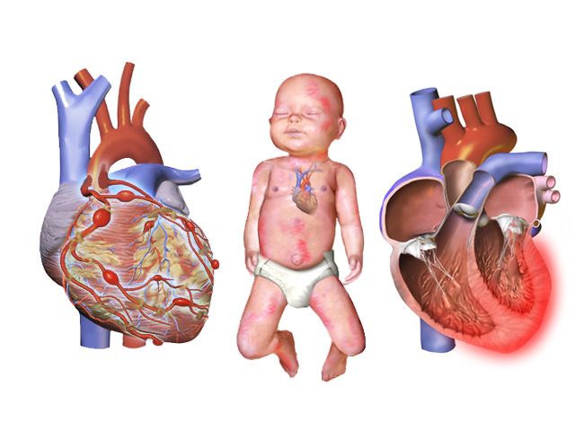
Kawasaki disease is an acute self-limited vasculitis of medium-sized arteries, most common under five years of age, and it is the leading cause of acquired heart disease in children in high-income countries. The danger is not the rash but the coronary arteries: without treatment, 20 to 25 per cent of children develop coronary artery aneurysms; with timely treatment, that falls to about 3 to 5 per cent. Diagnosis is clinical — fever lasting 5 days or more plus at least four of five features: bilateral non-exudative conjunctival injection; oral mucosal changes (cracked red lips, strawberry tongue); polymorphous rash; extremity changes (erythema and firm oedema of the hands and feet acutely, then periungual desquamation in the subacute phase); and cervical lymphadenopathy, usually unilateral and greater than 1.5 cm. The fever is high, prolonged and characteristically unresponsive to antipyretics and antibiotics, and the child is exceptionally irritable — irritability out of proportion to everything else is the sign experienced nurses trust.
Treatment is intravenous immunoglobulin 2 g/kg as a single infusion over 10 to 12 hours, given ideally within 10 days of fever onset and preferably by day 7, together with aspirin — high dose during the acute febrile phase, then reduced to a single antiplatelet dose of 3 to 5 mg/kg/day once the child has been afebrile for 48 to 72 hours, continued 6 to 8 weeks pending echocardiography. Echocardiograms are performed at diagnosis, at one to two weeks and at four to six weeks. Nursing priorities during IVIG are cardiac monitoring, strict intake and output because the infusion is a substantial volume load, and vigilance for infusion reaction. Two teaching points are frequently missed: live vaccines (measles-mumps-rubella and varicella) must be deferred for 11 months after IVIG because passive antibody blunts the response; and while the child is on aspirin, the family should report influenza or varicella exposure and the child should receive annual influenza vaccination, given the theoretical Reye syndrome risk. Warn families that peeling of the fingers and toes and joint pain are expected in weeks two and three and are not a relapse.
Acute rheumatic fever, infective endocarditis and myocarditis
Acute rheumatic fever is an immune-mediated sequel of untreated group A streptococcal pharyngitis, appearing one to five weeks after the sore throat, most often between 5 and 15 years. Antibodies raised against streptococcal M protein cross-react with cardiac, joint, skin and neural tissue. Diagnosis uses the Jones criteria: major manifestations are carditis, migratory polyarthritis, Sydenham chorea, erythema marginatum and subcutaneous nodules; minor manifestations include fever, arthralgia, prolonged PR interval and raised inflammatory markers — with evidence of preceding streptococcal infection required. Carditis is the manifestation that matters long-term, because valvulitis (most often mitral) scars into rheumatic heart disease. Chorea may appear months later and presents as clumsiness, deteriorating handwriting and emotional lability, which is easily mistaken for behavioural change. Acute treatment eradicates the streptococcus and manages inflammation; the enduring nursing message is secondary prophylaxis — intramuscular benzathine penicillin G every four weeks (600,000 units for children under 27 kg, 1.2 million units for 27 kg and over) for years, because a second attack causes far worse valve damage. Prevention is simpler still: complete the full course of antibiotics for streptococcal pharyngitis even after the throat feels better.
Infective endocarditis should be suspected in a child with structural heart disease, a prosthetic valve or a central line who develops unexplained prolonged fever, a new or changed murmur, splenomegaly, or embolic phenomena. Blood cultures are drawn from separate sites before antibiotics. Antibiotic prophylaxis before dental procedures is now reserved for the highest-risk groups — prosthetic valves or material, previous endocarditis, unrepaired cyanotic disease, and repairs with residual defects — but meticulous daily oral hygiene is advised for every child with a cardiac lesion. Myocarditis, usually viral, presents deceptively as a “viral illness” with disproportionate tachycardia, exercise intolerance, chest pain and an enlarged heart on chest radiograph, and it can progress to dilated cardiomyopathy and arrhythmia.
Paediatric heart failure and its medications
Heart failure in children arises from volume overload (left-to-right shunts), pressure overload (obstructive lesions), or myocardial failure (myocarditis, cardiomyopathy, arrhythmia, sepsis, severe anaemia). The clinical picture divides usefully into three groups: impaired myocardial function — tachycardia, gallop rhythm, weak pulses, cool extremities, sweating, fatigue; pulmonary congestion — tachypnoea, wheeze, cough, retractions, cyanosis, exercise intolerance; and systemic venous congestion — hepatomegaly, periorbital and facial oedema (dependent ankle oedema is an older-child sign, since infants lie flat), weight gain and neck vein distension in older children.
Nursing management is a set of concrete daily actions: daily weight on the same scale, same time, minimal clothing — a gain of more than 0.5 kg in 24 hours in a child is fluid, not growth; strict intake and output; clustering care to protect rest; upright or semi-Fowler positioning; and feeding support as described earlier. Diuresis with furosemide 1 mg/kg/dose intravenously requires monitoring of potassium, since hypokalaemia potentiates digoxin toxicity. Afterload reduction with an ACE inhibitor requires blood pressure and renal function monitoring and teaching about the dry cough. Digoxin demands specific nursing discipline: maintenance is roughly 5 to 10 mcg/kg/day divided twice daily, the therapeutic serum range is 0.8 to 2 ng/mL, and the apical pulse is counted for a full minute before every dose. Hold the dose and notify the prescriber if the apical rate is below 90 to 110 in an infant, below 70 in a child, or below 60 in an adolescent. Early toxicity in children shows as vomiting, anorexia, bradycardia and arrhythmia — the visual disturbances taught for adults are unreliable in a preverbal child. Teach families to give doses one hour before or two hours after feeds, never to mix digoxin into a bottle of formula, never to repeat a dose the child vomits, to give water afterwards to protect the teeth, and to store the medication out of reach, since the paediatric elixir is potent in very small volumes.
Shock, supraventricular tachycardia and cardiac arrest
Shock is inadequate delivery of oxygen and substrate to meet tissue demand. In children the commonest cause worldwide is hypovolaemia from gastroenteritis, followed by distributive shock from sepsis or anaphylaxis, then cardiogenic and obstructive causes. Because a child compensates with tachycardia and intense vasoconstriction, the early picture is a fast heart, cool mottled peripheries, delayed capillary refill, a narrow pulse pressure and irritability progressing to lethargy — with a normal blood pressure. Warm shock in early sepsis is the exception: bounding pulses, flash capillary refill and a widened pulse pressure. Treatment is oxygen, vascular access within five minutes — intraosseous access is the correct choice when two peripheral attempts fail — isotonic fluid boluses of 20 mL/kg (10 mL/kg for neonates, cardiac disease, malnutrition or DKA) with reassessment for hepatomegaly, crackles and worsening work of breathing after each, antibiotics within one hour in septic shock, glucose checked and corrected early because children deplete glycogen fast, and vasoactive infusions if fluid alone does not restore perfusion.
Supraventricular tachycardia is the commonest symptomatic arrhythmia of childhood. Suspect it when the rate exceeds roughly 220 bpm in an infant or 180 bpm in a child, is unvarying beat-to-beat, does not change with crying or comfort, and has no visible P waves — whereas sinus tachycardia varies with activity and has an identifiable cause. Infants may present after hours of undetected SVT with pallor, poor feeding and heart failure. If the child is stable, attempt vagal manoeuvres first: for infants, a plastic bag of iced water applied to the forehead and bridge of the nose for 15 to 30 seconds; for older children, blowing through an obstructed straw or bearing down. Never apply ocular pressure and never perform carotid massage in a child. If vagal manoeuvres fail, give adenosine 0.1 mg/kg by rapid intravenous push, maximum 6 mg, immediately followed by a rapid saline flush through a proximal large-bore line using a two-syringe or stopcock technique — the half-life is under 10 seconds, so a slow push simply does not work. A second dose of 0.2 mg/kg, maximum 12 mg, may follow. Warn the child and family that a brief pause and an unpleasant flushing sensation are expected, and run a continuous rhythm strip. If the child is unstable — hypotensive, altered, poorly perfused — the answer is synchronised cardioversion at 0.5 to 1 J/kg, escalating to 2 J/kg, with sedation if it does not delay treatment.
Paediatric cardiac arrest is usually the end of a respiratory or circulatory failure, not a sudden electrical event, so the rhythm you will most often meet is bradycardia progressing to asystole or pulseless electrical activity rather than ventricular fibrillation. This changes the priority: oxygenate and ventilate first. If the heart rate is below 60 with signs of poor perfusion despite adequate oxygenation and ventilation, begin chest compressions. Compress at 100 to 120 per minute to a depth of one third of the anteroposterior chest diameter — about 4 cm in an infant, 5 cm in a child — with full recoil, at 30 compressions to 2 breaths for a single rescuer and 15 to 2 for two rescuers. With an advanced airway in place, give continuous compressions with one breath every 2 to 3 seconds. Epinephrine is 0.01 mg/kg (0.1 mL/kg of the 0.1 mg/mL concentration) intravenously or intraosseously every 3 to 5 minutes, maximum single dose 1 mg. Defibrillation for a shockable rhythm is 2 J/kg first, then 4 J/kg, then up to 10 J/kg or the adult dose. Use a length-based resuscitation tape for every weight-based decision when the weight is unknown, and support the family’s presence at the resuscitation with a dedicated staff member assigned to them — family presence is associated with better bereavement outcomes and does not impede the team.
Paediatric arrest is usually respiratory in origin, so the resuscitation priority is oxygenation and ventilation — and compressions begin at a heart rate under 60 with poor perfusion, not at zero.
The chapter in one breath
Children compensate brilliantly and then fail suddenly, and almost everything in this chapter follows from that. A narrow airway makes small amounts of oedema catastrophic; a compliant chest wall and a fatigue-prone diaphragm mean sustained tachypnoea ends in exhaustion; a relatively fixed stroke volume means the heart rate carries the whole burden of compensation, so tachycardia is the earliest alarm and bradycardia is pre-arrest. Assess from the doorway — appearance, work of breathing, circulation to skin — then confirm with age-banded numbers rather than adult ranges, and remember that blood pressure is the last value to fall. Every dose is per kilogram, written out, and checked against the maximum adult dose; maintenance fluid follows 4-2-1, resuscitation follows 20 mL/kg (10 mL/kg in neonates, cardiac disease, malnutrition and DKA). Learn the sequences that save lives: dexamethasone and calm in croup, hands off the throat in epiglottitis, suction and hydration rather than bronchodilators in bronchiolitis, early steroid in asthma, knee-chest before oxygen in a tet spell, immunoglobulin within ten days in Kawasaki disease, vagal manoeuvre before adenosine, and compressions at a heart rate under 60 with poor perfusion. Escalate with SBAR, keep assessment, teaching, evaluation and the unstable child with the registered nurse, and keep the family beside the bed.
Words worth knowing
| Term | What it means |
|---|---|
| Compensated shock | Inadequate tissue perfusion with a still-normal blood pressure, revealed by tachycardia, delayed capillary refill, narrow pulse pressure and falling urine output. |
| 4-2-1 rule | Hourly maintenance fluid: 4 mL/kg/h for the first 10 kg, 2 mL/kg/h for the next 10 kg, 1 mL/kg/h for each kilogram beyond 20. |
| Stridor | Harsh, usually inspiratory noise from upper airway narrowing; at rest it signifies significant obstruction. |
| Grunting | Expiration against a partly closed glottis to generate end-expiratory pressure — an infant sign of alveolar disease and impending failure. |
| Westley croup score | Severity scale for croup combining stridor, retractions, air entry, cyanosis and level of consciousness. |
| Steeple sign / thumb sign | Radiographic subglottic tapering of croup; radiographic swollen epiglottis of epiglottitis. |
| Tripod position | Sitting forward on the hands with chin thrust out to maximise airway diameter; classic in epiglottitis. |
| Bronchiolitis | Viral obstruction of terminal bronchioles in infants, most often RSV, treated with suction, hydration and oxygen. |
| Silent chest | Absence of wheeze in a severe asthma exacerbation, indicating airflow too poor to generate sound — an emergency. |
| Peak flow zones | Green 80 per cent or more, yellow 50–79 per cent, red under 50 per cent of the child’s personal best. |
| Sweat chloride test | Diagnostic test for cystic fibrosis; 60 mmol/L or above is diagnostic, 30–59 intermediate, under 30 normal. |
| Ductal-dependent lesion | A congenital defect in which pulmonary or systemic blood flow requires a patent ductus arteriosus, maintained with prostaglandin E1. |
| Prostaglandin E1 (alprostadil) | Infusion at 0.01–0.1 mcg/kg/min that keeps the ductus open; causes apnoea, hypotension and fever — keep intubation equipment ready. |
| Critical CHD pulse oximetry screen | Right hand and foot saturations at 24–48 hours; passes at 95 per cent or above in both with a difference of 3 per cent or less. |
| Hypercyanotic (tet) spell | Acute right-to-left shunting in tetralogy of Fallot; treat with knee-chest position first, then calm, oxygen, morphine and fluid. |
| Eisenmenger syndrome | Reversal of a left-to-right shunt once pulmonary vascular resistance exceeds systemic resistance, rendering the lesion inoperable. |
| Kawasaki disease | Medium-vessel vasculitis with fever of five days or more; treated with IVIG 2 g/kg within 10 days to protect the coronary arteries. |
| Jones criteria | Diagnostic framework for acute rheumatic fever, requiring evidence of preceding group A streptococcal infection. |
| Digoxin hold parameters | Count the apical pulse for a full minute; hold and notify if under 90–110 in infants, under 70 in children, under 60 in adolescents. |
| Intraosseous access | Rapid vascular route into the marrow, indicated when peripheral access fails within about five minutes in a shocked child. |