Immunity, Inflammation & Hypersensitivity
Your immune system is a standing army. You never notice it until it wins too slowly, fights too hard, or turns on your own side. This chapter follows the whole arc. You will meet the invaders. You will see how the innate and adaptive defenses answer them. You will learn what inflammation costs. Then you will see what happens when immunity misfires as allergy, autoimmunity, or immunodeficiency.
By the end of this chapter you can
- Name the main groups of pathogens and how each one causes disease.
- Compare innate with adaptive immunity, and humoral with cell-mediated defense.
- Match each of the five antibody classes to the job it is known for.
- Explain the cardinal signs of inflammation by what causes each one.
- Tell the four hypersensitivity types apart, with a classic example of each.
- Describe how autoimmunity and immunodeficiency (including HIV/AIDS) start.
Section 1The invaders: types of pathogens
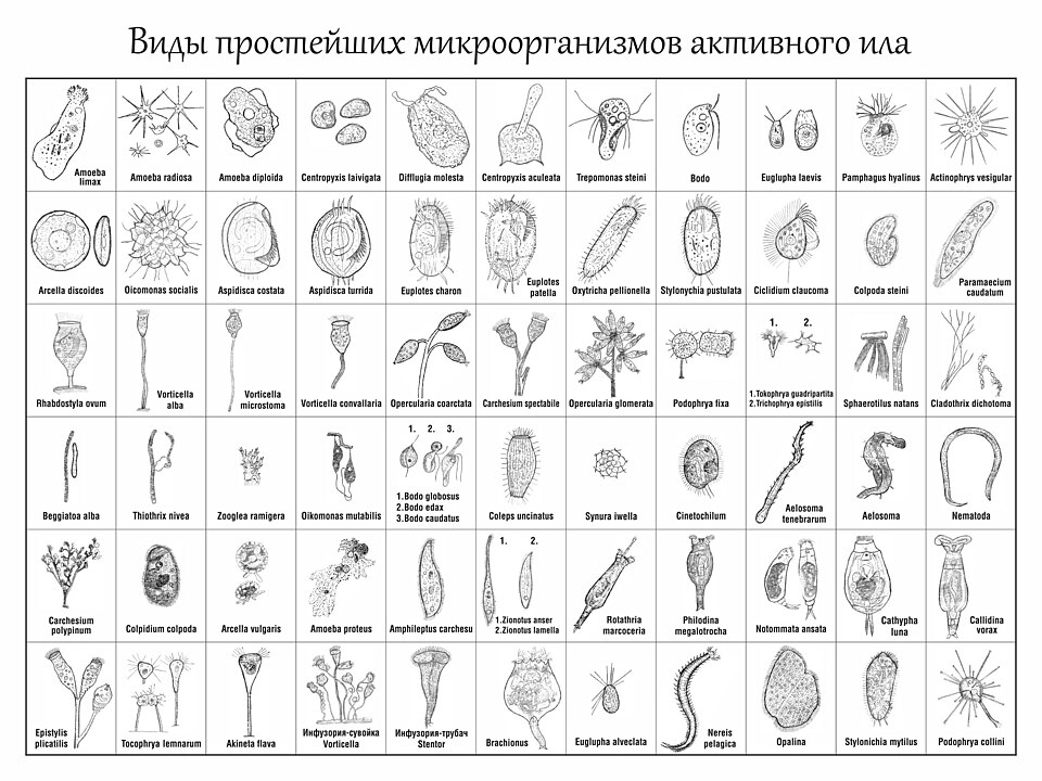
Right now, as you read this, trillions of microbes live on you and in you. That is completely normal. These resident microbes are your normal flora (also called your microbiota). They live on your skin, in your mouth, and most of all in your gut. They earn their keep. They crowd out troublemakers, help digest food, and even make some vitamins, like vitamin K. Most of the time, you and your flora help each other. A pathogen is different. It is any organism that can cause disease. It may invade tissue, release toxins, or set off an immune response that harms you. Some organisms are opportunistic. They are harmless in their usual spot. They turn dangerous when they reach a place they do not belong. Gut bacteria that slip into the blood after surgery are one example. So your first question is what kind of invader this is. The type of organism decides the treatment.
Bacteria: tiny, but they play dirty
Bacteria are single cells with their own tools for living and copying themselves. That is why many can grow on their own, outside our cells. We sort them two ways. First by shape, as round cocci or rod-shaped bacilli. Second, and more useful, by the Gram stain. Gram-positive bacteria have a thick peptidoglycan cell wall and no outer membrane. That thick wall traps and holds the purple stain. Gram-negative bacteria have a thin wall hidden under an extra outer membrane. They lose the purple and pick up a pink counterstain instead. That difference is more than a color. Gram-negative bacteria carry endotoxin (lipopolysaccharide, LPS) in that outer membrane. When the bacteria die and break apart, LPS pours out. It can drive high fever, dangerously low blood pressure, and septic shock. Bacteria harm us in two broad ways. One is invasion, where they spread into tissue and destroy it. The other is releasing toxins. Exotoxins are proteins that some bacteria send out to damage cells at a distance. The toxins behind tetanus and C. difficile colitis work this way. Endotoxin is different. It is part of the gram-negative wall itself.
Viruses: hijackers that cannot live alone
Viruses are not really cells. A virus is genetic material, either DNA or RNA, wrapped in a protein coat. Viruses are obligate intracellular parasites. That means a virus must get inside one of your living cells to do anything at all. It attaches to a host cell, enters, and releases its genes inside. Then it takes over the cell's own ribosomes and enzymes. The cell becomes a factory that turns out new virus copies, and it is often destroyed in the process. Viruses hide inside your cells and borrow your machinery. So antibiotics do nothing against them, and treatment is harder to aim without harming the host.
Fungi, parasites, and a word on prions
Fungi are more complex cells. They include yeasts, such as Candida, and molds. Fungi often turn harmful when the immune system is weak, or when antibiotics wipe out normal flora. Protozoa are single-celled parasites. One example is the Plasmodium that causes malaria. Helminths are the parasitic worms, and they are made of many cells. Prions are the strange outliers. They are misfolded proteins with no DNA or RNA at all. A prion makes other proteins misfold too, which leads to fatal brain disease.
Matching the treatment to the invader
Here is the payoff. Naming the class of organism guides the whole plan. Antibiotics attack features that only bacteria have, such as the cell wall. That is why they are useless against viruses. Giving them anyway breeds antibiotic resistance. It can also set off a C. difficile infection by killing the flora that protect you.
| Pathogen | Key trait | Treatment class |
|---|---|---|
| Bacteria | Own cells; toxins & invasion | Antibiotics |
| Viruses | Take over host cells | Antivirals |
| Fungi | Yeasts & molds | Antifungals |
| Parasites | Protozoa & worms | Antiparasitics |
Look at the main classes of pathogens in the figure below. It lines these families up side by side.
- Normal flora usually protect us. A pathogen is any organism that causes disease, and opportunists turn harmful in the wrong place or when defenses drop.
- Bacteria harm us by invasion and by toxins. Gram-negative endotoxin (LPS) can drive fever and septic shock, and the Gram stain guides the first drug choice.
- Viruses are obligate intracellular parasites that take over host cells to copy themselves. That is why antibiotics do not work on them.
- Fungi, protozoa, and helminths each need their own drug class. Prions are misfolded proteins with no nucleic acid.
- Match the drug to the organism: antibiotic, antiviral, or antifungal. Misusing antibiotics breeds resistance and can cause C. difficile.
Section 2The body’s defenses
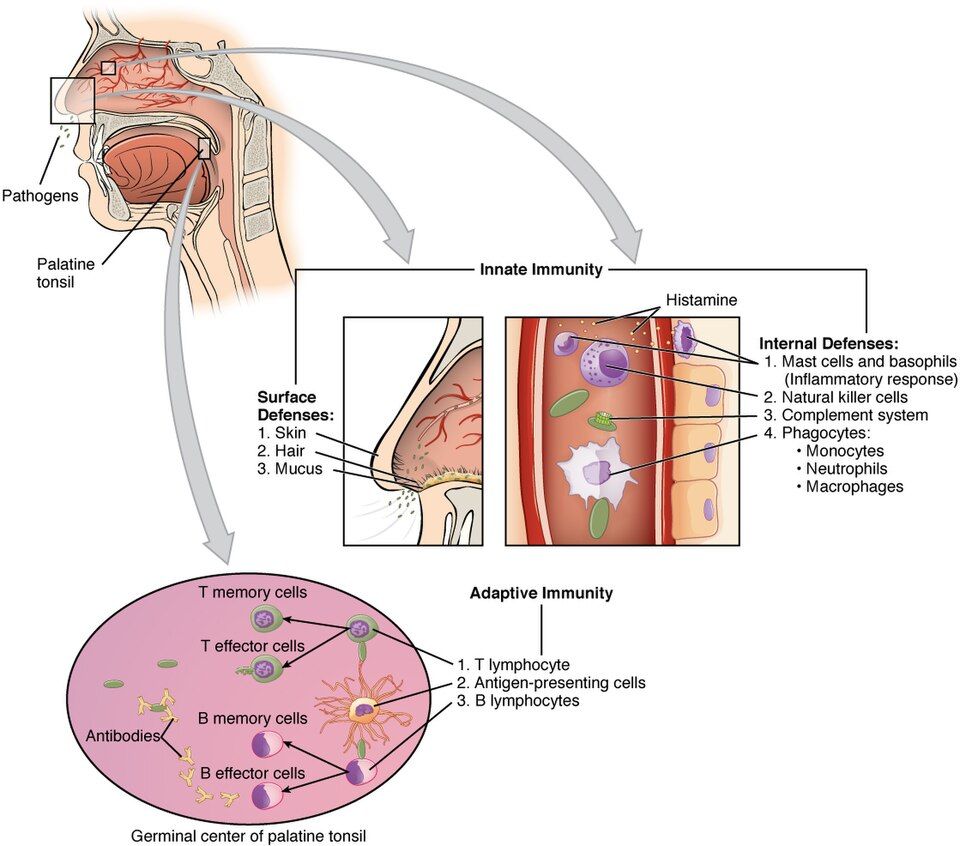
Every second of every day, your body fights off bacteria, viruses, and other invaders. You never notice. It does this with two teams that work together. One is a fast, general-purpose crew called innate immunity. The other is slower but far more exact, and it is called adaptive immunity. How these two teams work is the base for everything else in this chapter.
Innate immunity: fast, but nonspecific
Innate immunity is the defense you are born with. It acts within minutes to hours. But it does not care exactly who the invader is. It treats most threats the same way. Think of the locks, alarms, and guards in a building.
- Physical and chemical barriers come first. Unbroken skin is a wall that invaders cannot cross easily. Mucous membranes line the airways and gut and trap microbes, so cilia and secretions can sweep them out. Stomach acid and lysozyme in tears add a chemical defense. This is why any break in the skin, like an IV site or a pressure injury, is a real infection risk.
- Phagocytes are cells that eat invaders. Neutrophils get there first and fast. They lead the early response and are the main cell in pus. Macrophages arrive a bit later, swallow debris, and clean up. This eating process is called phagocytosis.
- Complement is a chain of plasma proteins. It punches holes in microbes, tags them for phagocytes (opsonization), and turns up inflammation.
- Natural killer (NK) cells destroy your own cells once they turn dangerous. They go after virus-infected cells and some tumor cells.
- Fever is a whole-body innate response. Chemical messengers called pyrogens reset the hypothalamus to a higher temperature. That slows microbial growth and speeds up immune cells. So a fever feels bad, but it is often a sign your defenses are working.
Adaptive immunity: specific, with memory
Adaptive immunity takes longer to start. A first exposure may take days. But it is specific, which means it aims at one invader. It also remembers. It has two branches.
- Humoral immunity is run by B cells. Once activated, B cells become plasma cells. Those cells pump antibodies (immunoglobulins) into the blood and body fluids. Antibodies shut down toxins, clump microbes together, and flag them to be destroyed. This branch is best against invaders outside your cells, such as bacteria in the bloodstream.
- Cell-mediated immunity is run by T cells. It handles threats inside cells, such as viruses, and it handles abnormal cells.
The T-cell coordinators and killers
Two kinds of T cells are worth knowing by name. Helper T cells (CD4) are the coordinators. They release cytokines that switch on B cells, cytotoxic T cells, and macrophages. HIV attacks CD4 cells. So a falling CD4 count leaves the whole system without a leader, and that is why HIV does so much damage. Cytotoxic T cells (CD8) are the killers. They destroy virus-infected and cancerous cells directly.
Active vs passive immunity
Immunity can be earned or borrowed. In active immunity your own body makes the response and builds memory. You can get there by getting sick, or by vaccination, which is a safe preview of the invader. Active immunity is slow to build but lasts a long time. In passive immunity you receive ready-made antibodies from someone else. Protection starts right away, but it is temporary, because you never made memory cells. Antibodies crossing the placenta to the fetus are one example. Antibodies in breast milk are another.
| Feature | Active immunity | Passive immunity |
|---|---|---|
| Source | Your own immune response | Borrowed antibodies |
| Onset | Slow (days to weeks) | Right away |
| Duration | Lasts a long time (memory) | Short (no memory) |
| Examples | Infection, vaccines | Placenta, breast milk, antibody shots |
Primary vs secondary response
The primary response is your body's first meeting with an invader. It is slow. Building up antibodies often takes a week or more, which is why you can feel sick that first time. But the immune system files away memory cells. On a repeat exposure, the secondary (memory) response is faster and stronger. It makes far more antibodies, far more quickly, so you may never feel sick at all. This is exactly why vaccines and booster doses work. They build memory safely, so the real invader meets a fast secondary response. Look at the figure below to see how the innate and adaptive teams fit together.
- Innate immunity is fast but nonspecific. It includes barriers, neutrophils and macrophages, complement, NK cells, and fever. Adaptive immunity is slower but specific, and it remembers.
- Adaptive immunity has two branches. Humoral means B cells make antibodies, best against threats outside cells. Cell-mediated means T cells, best against threats inside cells.
- Helper T cells (CD4) coordinate the response. Cytotoxic T cells (CD8) kill infected and abnormal cells.
- Active immunity comes from infection or a vaccine. You make it yourself and it lasts. Passive immunity comes from the placenta, breast milk, or antibody shots. It is borrowed, immediate, and temporary.
- The secondary (memory) response is faster and stronger than the primary response. That is the whole basis for vaccines and boosters.
Section 3Antibodies (immunoglobulins)
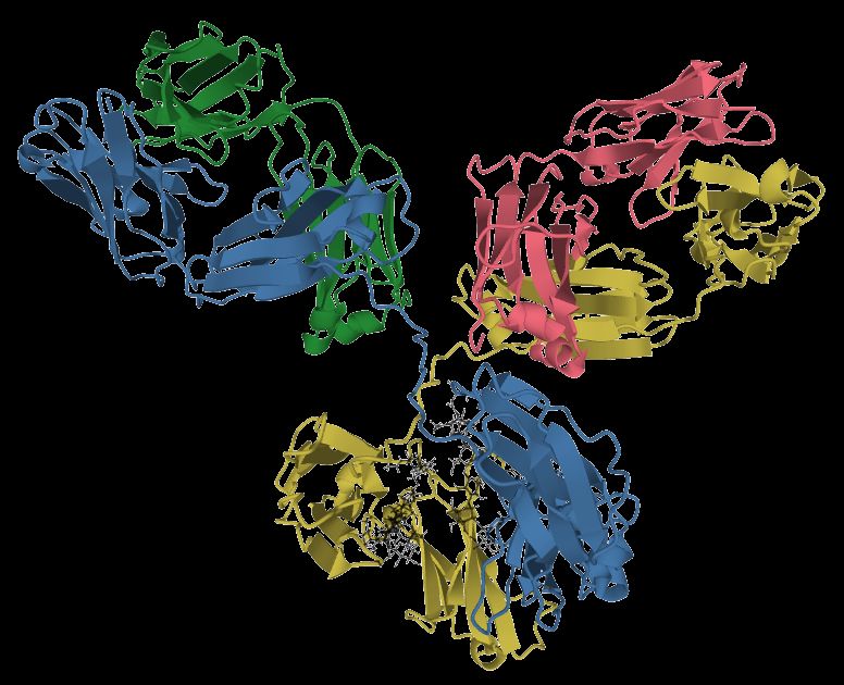
Your immune system faces a huge problem. How do you spot millions of different germs when each one looks a little different? The answer is antibodies, also called immunoglobulins (Ig). Picture a tiny Y-shaped protein tag that your body builds to match one target. The tips of the Y are the variable region. They are shaped to lock onto one antigen, which is a molecular fingerprint on a microbe. The stem of the Y is the constant region. It is the handle that the rest of the immune system grabs.
Antibodies are made by plasma cells. These are mature B lymphocytes that their antigen has switched on. Once an antibody is released, it does three main jobs. First, it tags the invader so phagocytes know to eat it. That tagging is called opsonization. Second, it can neutralize a threat. It coats a virus or a toxin so the threat can no longer attach to your cells. Third, it can activate complement. Complement is a chain of blood proteins that punches holes in bacterial membranes and turns up inflammation. In short: tag, neutralize, and activate.
The five classes and their signature jobs
All antibodies share that Y shape. But the constant-region handle comes in five flavors. Each flavor sends the antibody to a different job and a different place in the body.
- IgG is the most abundant antibody in blood. It is the workhorse of the secondary (memory) response. It is the only class that crosses the placenta. It is very good at opsonization, at neutralizing, and at activating complement.
- IgM is the first to appear in a new infection, and it is the largest antibody. It travels as a pentamer, which means five Y units joined together. Those ten binding arms make it a strong early complement activator.
- IgA guards the mucosal surfaces, the wet linings of the gut, lungs, and airways. It is concentrated in secretions like saliva, tears, and breast milk. That gives a nursing newborn ready-made protection.
- IgE drives allergy and type I hypersensitivity, and it defends against parasites. It binds to mast cells and basophils. When an antigen links those IgE molecules together, the cells release histamine.
- IgD mostly sits on the surface of naive B cells as part of the B-cell receptor. It helps those cells get switched on.
Reading antibody levels: recent versus past infection
IgM and IgG appear on different timelines. So a lab panel can tell you when an exposure happened. When you meet an antigen for the first time, IgM rises first, usually within days. It then fades over a few weeks. IgG rises later and stays high for months to years, which gives lasting immunity. Clinicians use this pattern all the time.
| Feature | IgM elevated | IgG elevated |
|---|---|---|
| What it suggests | Recent or current infection | Past infection or immunity |
| Timing | Shows up first, fades in weeks | Shows up later, stays for years |
| Response type | Primary response | Secondary / memory response |
| Classic example | Acute hepatitis or rubella | An old vaccine or an old exposure |
So IgM positive with IgG negative usually points to a fresh infection. IgG positive with IgM negative usually means the person was infected or vaccinated in the past and is now immune.
Why placenta-crossing IgG protects a newborn
A newborn's immune system is still learning. It cannot make strong antibody responses yet. Because IgG crosses the placenta during pregnancy, the mother loans her baby a whole library of antibodies. This passive immunity shields the infant for roughly the first few months of life, while the baby's own B cells grow up. IgA in breast milk adds a second layer right at the gut lining. The trade-off is that borrowed protection does not last. That is exactly why the infant vaccine schedule starts before it wears off.
The figure below lets you look at the five classes side by side.
- Antibodies (immunoglobulins) are Y-shaped proteins from plasma cells. They tag, neutralize, and activate complement against one specific antigen.
- There are five classes. The hook is GAMED: IgG (most abundant, crosses the placenta), IgA (mucosa and breast milk), IgM (first and largest, a pentamer), IgE (allergy and parasites), IgD (B-cell receptor).
- IgM rises first and fades, so it points to a recent infection. IgG rises later and stays, so it points to past infection or immunity.
- The mother's IgG crosses the placenta. It gives the newborn temporary passive immunity for the first months of life.
Section 4Inflammation & healing
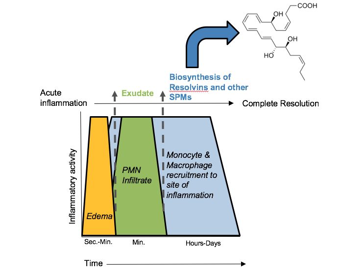
Think of inflammation as your body's first-responder team. It is not the same thing as infection. Infection means microbes have invaded. Inflammation is the response, and it fires for many triggers. Bacteria, a splinter, a burn, a sprain, or dying tissue will all set it off. In healthy tissue, capillaries run quietly and white blood cells patrol the blood. The moment cells are injured, that calm is broken on purpose. The goal is to contain the threat, clear the debris, and set up repair. To understand the response, you need a handful of chemical mediators and what they do to blood vessels.
The five cardinal signs and why each happens
Injured cells and nearby mast cells release histamine. They also release prostaglandins, bradykinin, and cytokines. These mediators cause two changes in blood vessels, and those two changes explain every classic sign.
- Redness (rubor) and heat (calor) come from vasodilation. The arterioles widen, so more warm blood floods the area.
- Swelling (tumor) comes from increased capillary permeability. The vessel walls loosen and let protein-rich fluid leak into the tissue. That fluid is edema.
- Pain (dolor) comes from bradykinin and prostaglandins firing nerve endings. Pressure from the swelling adds to it.
- Loss of function (functio laesa) is the sum of pain and swelling. A swollen, aching joint simply will not move well.
Meanwhile, neutrophils are the first white cells on the scene. They squeeze out of the vessels, a step called diapedesis. Then they begin phagocytosis of pathogens and debris. Monocytes follow them out and mature into macrophages, which take over the cleanup. So the order is neutrophils first, then monocytes and macrophages.
Acute versus chronic inflammation
Acute inflammation is fast and it stops on its own. It runs for hours to a few days, and ideally it ends in full healing. Chronic inflammation drags on for weeks, months, or years. Usually the trigger never clears, as in a lasting infection, an autoimmune disease, or ongoing irritation. The cast of cells changes too. Neutrophils give way to macrophages and lymphocytes. The cost to the tissue is real. Chronic inflammation drives fibrosis, which is scarring, plus tissue destruction and sometimes granulomas. The organ slowly loses working cells and gains stiff scar that does no work.
Exudates and the systemic response
The fluid that leaks out is called exudate. Serous exudate is thin and watery, like the fluid in a blister. Purulent exudate is thick pus, full of dead neutrophils and microbes, and it points to infection. Inflammation is not only local. Cytokines such as IL-1 and IL-6 travel to the brain and liver and cause systemic signs. These include fever, which is a raised set-point in the hypothalamus, and leukocytosis, which is a high white blood cell count. Acute-phase reactants rise as well. A climbing CRP is a true acute-phase protein. A rising ESR is an indirect marker driven by fibrinogen. Both are lab clues that inflammation is active somewhere in the body.
Healing: resolution versus repair
Two endings are possible. Resolution is the best case. The injury was mild and the cells can grow back, so the tissue returns to normal. Repair with scar (fibrosis) happens when the damage is wide, or when those cells cannot grow back. Fibroblasts lay down collagen and granulation tissue fills the gap. The scar holds, but it does not fully work.
| Feature | Primary intention | Secondary intention |
|---|---|---|
| Wound edges | Clean, lined up (sutured) | Wide and gaping, cannot be closed |
| Tissue loss | Very little | A lot |
| Healing time | Fast | Slower |
| Scar | Small | Larger, more granulation tissue |
| Infection risk | Lower | Higher |
Why anti-inflammatory drugs work
NSAIDs such as ibuprofen and aspirin block the COX enzymes that make prostaglandins. So they dull pain, fever, and swelling but leave much of the immune response in place. Corticosteroids such as prednisone act much further upstream. They shut down the genes for many mediators at once. They are far stronger, but they also suppress immunity more. That is why they raise infection risk and slow healing. Use the figure below to trigger the acute inflammatory response and watch the cardinal signs appear.
- All five cardinal signs (redness, heat, swelling, pain, loss of function) trace back to vasodilation and increased capillary permeability from histamine and prostaglandins.
- Acute inflammation is short and usually heals. Chronic inflammation lasts, and it costs tissue through fibrosis and destruction.
- Systemic signs of inflammation include fever, leukocytosis, and a high CRP/ESR. Purulent exudate suggests infection.
- Healing ends in resolution, which is back to normal, or repair with scar. Clean wounds heal by primary intention, gaping wounds by secondary.
- NSAIDs block prostaglandins through COX. Corticosteroids shut down many mediators at once and can mask the signs of infection.
Section 5When immunity overreacts: hypersensitivity
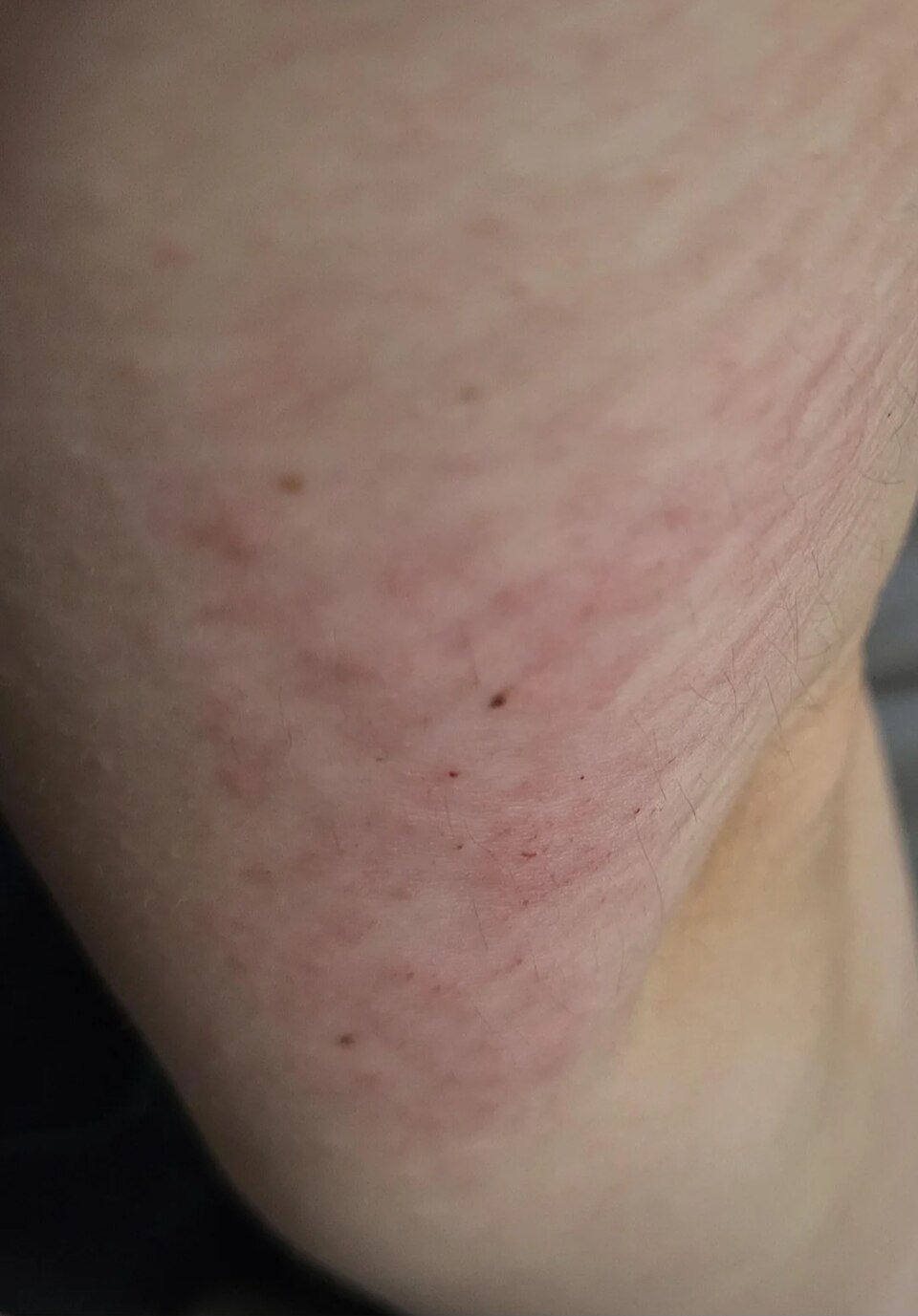
Your immune system is built to protect you. Sometimes it protects too hard. Hypersensitivity means the immune response is too big or aimed at the wrong target. The defense itself becomes the cause of tissue damage. To see how, recall how normal immunity works. Your body makes antibodies (immunoglobulins) that tag foreign invaders. It also makes special T cells that spot threats directly. Normally these tools attack germs and leave your own cells alone. In hypersensitivity, that aim breaks down. The immune system may react to something harmless, such as pollen, a food, or a medication. It may target your own cells. Or it may leave inflammatory debris behind that damages healthy tissue.
Clinicians sort these overreactions into four classic types. A simple memory device keeps them straight: ACID. That stands for Anaphylactic, Cytotoxic, Immune-complex, and Delayed. Antibodies drive the first three. Only the fourth one works without them.
Type I - Immediate (IgE-mediated): allergy and anaphylaxis
This is the classic allergy. On the first exposure, the body makes IgE antibodies against a harmless allergen. It parks that IgE on the surface of mast cells and basophils. This quiet step is called sensitization. On the next exposure, the allergen bridges those IgE molecules together. The mast cells then degranulate, dumping histamine and other mediators within minutes. Histamine causes vasodilation, which brings redness, warmth, and low blood pressure. It causes increased capillary permeability, which brings swelling and hives. It also causes bronchoconstriction, which brings wheezing. A local reaction is hay fever or hives. A whole-body reaction is anaphylaxis, where airway swelling and vasodilatory shock can kill quickly. This is why epinephrine is the first-line rescue drug. It reverses bronchoconstriction and vasodilation fast.
Type II - Cytotoxic (antibody against cells)
Here antibodies, usually IgG or IgM, bind right onto antigens on the surface of certain cells. That marks those cells for destruction by complement and phagocytes. The classic example is a mismatched blood transfusion. Give type A blood to a type B patient, and the patient's ready-made antibodies attack the donor red cells. The result is rapid hemolysis, with fever, flank pain, and dark urine. Another example is hemolytic disease of the newborn. An Rh-negative mother makes anti-Rh antibodies. Those antibodies cross the placenta and destroy the red cells of an Rh-positive baby.
Type III - Immune-complex mediated
In Type III, antibody and antigen clump into immune complexes. These clumps circulate, then settle in tissues such as joints, skin, kidney glomeruli, and blood vessel walls. Once lodged, they switch on complement and set off inflammation right where they landed. This is the mechanism behind systemic lupus erythematosus (SLE). It also causes serum sickness, a reaction to foreign proteins such as certain antivenoms. Here is the key difference from Type II. The antibody is not aimed at a fixed cell. It is floating debris that inflames wherever it lands.
Type IV - Delayed (T-cell mediated)
Type IV is the odd one out, because there is no antibody at all. Instead, sensitized T lymphocytes recognize the antigen and call in macrophages. This cell-driven process takes time, so the reaction peaks at 48 to 72 hours. That is exactly why you read a TB skin test (PPD) at 48 to 72 hours. The firm raised induration is delayed T-cell inflammation. Other examples are contact dermatitis, from poison ivy or nickel, and transplant (graft) rejection.
| Type | Mediator | Timing | Classic example |
|---|---|---|---|
| I - Anaphylactic | IgE, mast cells, histamine | Seconds to minutes | Anaphylaxis, hay fever |
| II - Cytotoxic | IgG/IgM against the cell surface | Minutes to hours | Transfusion reaction, HDN |
| III - Immune-complex | Antibody-antigen clumps in the blood | Hours | SLE, serum sickness |
| IV - Delayed | T cells (no antibody) | 48-72 hours | TB skin test, poison ivy |
Use the figure below to compare the four types side by side.
- ACID puts the four types in order: Anaphylactic (I), Cytotoxic (II), Immune-complex (III), Delayed (IV).
- Types I, II, and III are antibody-driven. Type IV is T-cell driven, with no antibody.
- Type I needs sensitization first. The severe reaction hits on re-exposure, and epinephrine is the rescue drug for anaphylaxis.
- Type II targets certain cells, as in a transfusion reaction or hemolytic disease of the newborn. Type III drops floating clumps that inflame tissue, as in SLE and serum sickness.
- Type IV is delayed 48 to 72 hours. That is the reason a TB skin test is read days later.
Section 6Autoimmunity & immunodeficiency
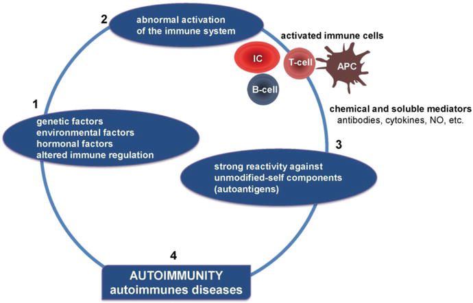
Your immune system has an almost impossible job. It must attack anything foreign, such as bacteria, viruses, and cancer cells. At the same time it must leave you completely alone. The skill of telling "self" from "non-self" is called self-tolerance, and it is built while the immune system develops. As lymphocytes mature, any that react strongly against your own tissue are normally deleted or switched off. That filtering is called central and peripheral tolerance. When it breaks down, the immune system turns its weapons inward. This section covers what happens when tolerance fails, which is autoimmunity. It also covers the flip side, when the immune system is too weak to defend at all, which is immunodeficiency.
When self-tolerance breaks down: autoimmunity
In autoimmune disease, the immune system loses self-tolerance. It starts making autoantibodies and self-reactive T cells that treat normal tissue like an invader. The result is chronic inflammation and tissue destruction wherever the immune system aims. The trigger is usually a mix of two things. One is genetic susceptibility, often certain HLA types. The other is an environmental push, such as an infection, hormones, or drugs. Here is a helpful way to learn these diseases. Ask one question: what tissue is being attacked? Use the figure below to watch tolerance break down step by step.
| Disease | Target attacked | Classic result |
|---|---|---|
| SLE (lupus) | Many tissues at once: skin, joints, kidneys, blood | Butterfly rash, joint pain, lupus nephritis; ANA usually positive |
| Rheumatoid arthritis | Synovial joints (the joint lining) | Matching joints on both sides are warm and swollen; morning stiffness |
| Multiple sclerosis | Myelin around CNS nerves | Nerve signals slow down: weakness, vision changes, numbness |
| Type 1 diabetes | Beta cells of the pancreas | No insulin is made, so blood glucose runs high |
| Hashimoto thyroiditis | Thyroid (destroys it) | Hypothyroidism: tired, cold, gaining weight |
| Graves disease | Thyroid (over-stimulates it) | Hyperthyroidism: heat intolerance, weight loss, exophthalmos |
Notice that Hashimoto and Graves hit the same organ with opposite results. In Hashimoto, autoantibodies and T cells slowly destroy thyroid tissue, so hormone output falls. In Graves, the autoantibody copies TSH and switches the gland on all the time. That floods the body with thyroid hormone. Same target, opposite mechanism. It is a favorite exam contrast.
The opposite problem: immunodeficiency
Sometimes the danger is not an overactive immune system. It is an underactive one. In immunodeficiency, part of the immune defense is missing or weak, so ordinary germs turn dangerous. There are two broad types.
- Primary (congenital) means the person is born with a genetic defect in immune development. These are usually rare. They often show up in infancy with repeated, severe, or unusual infections.
- Acquired (secondary) means the immune system grew normally but was damaged later. Causes include infection such as HIV, chemotherapy, long-term steroids or other immunosuppressants, poor nutrition, and aging. These are far more common.
HIV/AIDS and the CD4 count
The key example of acquired immunodeficiency is HIV (human immunodeficiency virus). HIV infects and destroys CD4+ T-helper cells. Those are the "quarterback" cells that coordinate almost the entire immune response. As the virus copies itself, the CD4 count drops steadily. A normal CD4 count is usually about 500 to 1,500 cells/mm³. The diagnosis becomes AIDS when the count falls below roughly 200 cells/mm³, or when an AIDS-defining opportunistic infection appears. At that point the body can no longer fight off opportunistic infections. These are organisms like Pneumocystis pneumonia that a healthy immune system would handle easily. So the CD4 count is basically a scoreboard of how much immune defense is left.
When inflammation goes body-wide: SIRS and sepsis
Inflammation is usually local and protective. But an infection can spill the inflammatory response into the whole bloodstream. Then the reaction loses its controls and turns dangerous. SIRS (systemic inflammatory response syndrome) is the older name for that body-wide inflammatory storm. It is scored on an abnormal temperature, a fast heart rate, fast breathing, and an abnormal white-cell count. That ladder is useful for spotting a sick patient, but it is not the current definition. The definition that counts is Sepsis-3. Under Sepsis-3, sepsis is infection plus new organ dysfunction, not infection alone. Septic shock is sepsis that still needs vasopressor drugs to hold a mean arterial pressure of 65 mmHg, with a blood lactate above 2 mmol/L. The middle term severe sepsis was retired in 2016, so you will see it only in older writing. In all of it, the same mediators meant to fight germs now cause widespread vasodilation and leaky vessels. Blood pressure drops and organs are starved of oxygen. It is a sobering reminder that too much immune activity can kill as surely as too little.
Why immunosuppressed patients are so fragile
The cause may be HIV, chemotherapy, anti-rejection drugs after a transplant, or steroids. Either way, an immunosuppressed patient has fewer or weaker defenders. Germs that a healthy person would shrug off can cause overwhelming infection. The usual warning signs may also be muted. A patient with a low white-cell count, called neutropenia, may not run a fever or form pus. So the infection hides until it is advanced. That is why care focuses on strict hand hygiene and neutropenic precautions. It is also why even a low-grade fever is treated as a possible emergency.
- Autoimmunity is a loss of self-tolerance, so the immune system attacks the body's own tissue. Learn each disease by asking what tissue is targeted: SLE hits many systems, RA hits joints, MS hits myelin, T1DM hits beta cells, and Hashimoto and Graves hit the thyroid.
- Hashimoto destroys the thyroid, so hormone levels fall. Graves over-stimulates it, so they rise. Same organ, opposite effect.
- Immunodeficiency is either primary, meaning born with it, or acquired. HIV is the key acquired example. It destroys CD4+ helper cells, and a CD4 count below about 200 cells/mm³ defines AIDS.
- Use Sepsis-3. Sepsis is infection plus new organ dysfunction. Septic shock is sepsis needing vasopressors to hold a MAP of 65 mmHg with a lactate above 2 mmol/L. SIRS is the older framing, and severe sepsis was retired in 2016.
- Immunosuppressed patients catch infections easily, and their signs may be blunted, with little fever or pus. So any fever is treated urgently.
The chapter in one breath
- Pathogens differ in structure and in treatment. Bacteria have a cell wall and answer to antibiotics. Viruses need a host cell and answer to antivirals. Fungi, protozoa and parasites, and misfolded-protein prions each call for a different defense and a different drug.
- Innate immunity is the fast, nonspecific first responder you are born with, built from skin, phagocytes, complement, and fever. Adaptive immunity is slower but specific, and it remembers the invader through B cells that make antibodies and T cells.
- The five immunoglobulin classes split the work. IgG is the most abundant and crosses the placenta. IgM is the first responder and the biggest. IgA guards mucous membranes and rides in breast milk. IgE drives allergy and fights parasites. IgD is the B-cell surface receptor.
- Inflammation is the alarm at the tissue level, with five cardinal signs: redness (rubor), heat (calor), swelling (tumor), pain (dolor), and loss of function. All of them come from vasodilation and increased capillary permeability driven by histamine and other mediators.
- The four hypersensitivity reactions run in order. I is immediate and IgE-mediated, as in anaphylaxis. II is cytotoxic, where antibody attacks cells, as in transfusion reactions. III is immune-complex deposition, as in lupus. IV is delayed and T-cell-mediated, as in the TB skin test and contact dermatitis.
- Autoimmunity is a loss of self-tolerance, where the immune system attacks the body's own tissue. It causes diseases such as rheumatoid arthritis, systemic lupus erythematosus, and type 1 diabetes.
- Immunodeficiency is a weak or missing defense. It can be primary, meaning congenital, or secondary, meaning acquired from chemo, steroids, or poor nutrition. Either way the patient is open to opportunistic infection.
- HIV is a retrovirus that destroys CD4+ helper T cells and cripples adaptive immunity. Once the CD4 count falls below 200 cells/mm³, or an opportunistic infection appears, the illness has become AIDS.
ReferenceWords worth knowing
Here is a quick reference for the terms in this chapter. Skim it now, or come back whenever a word trips you up.
- Anaphylaxis
- A severe, fast, whole-body type I hypersensitivity reaction. A flood of histamine causes vasodilation all over the body, bronchoconstriction, and dangerously low blood pressure. It can kill within minutes if it is not treated.
- Antibody
- A Y-shaped protein, also called an immunoglobulin, made by plasma cells. It binds one specific antigen to tag it for destruction, to neutralize it, or to call in other immune defenses.
- Antibody titer
- A lab measure of how much of one antibody is in the blood. It is used to check immunity from a vaccine or past infection, or to follow an active immune response.
- Antigen
- Any molecule, usually a protein or sugar on a germ or foreign cell, that the immune system reads as "not self." The body answers by making antibodies or by switching on T cells.
- Adaptive immunity
- The slower, learned arm of defense. It aims at one specific antigen and builds memory, so the next meeting with that same invader is met faster and harder.
- Autoimmunity
- A breakdown of self-tolerance. The immune system mistakes the body's own healthy tissue for a threat and attacks it, as in lupus or rheumatoid arthritis.
- Cell-mediated immunity
- The arm of adaptive immunity run by T lymphocytes rather than antibodies. It kills virus-infected and cancerous cells directly, and it coordinates other immune responses.
- Complement
- A chain of blood proteins. Once triggered, it punches holes in microbe membranes, coats germs so they will be eaten, and turns up inflammation.
- Cytokine
- A small signaling protein, such as an interleukin or an interferon. Immune cells release cytokines to talk to each other, call in help, and set the strength and type of the response.
- Exudate
- The protein-rich fluid, cells, and debris that leak from blood vessels into inflamed tissue. It ranges from watery (serous) to full of pus (purulent), depending on the cause.
- Humoral immunity
- The arm of adaptive immunity run by B lymphocytes and the antibodies they release. Those antibodies travel in body fluids to neutralize pathogens and toxins.
- Immunodeficiency
- A weak or missing part of the immune system. It may be inherited or acquired, as in HIV or chemotherapy, and it leaves a person open to frequent or unusual infections.
- Inflammation
- The body's protective response to injury or infection. Blood vessels widen and immune cells rush in, which produces the classic redness, heat, swelling, and pain.
- Innate immunity
- The fast, nonspecific first line of defense you have from birth. It includes skin barriers, phagocytes, and inflammation, and it attacks any invader with no prior exposure needed.
- Pathogen
- Any disease-causing microbe, such as a bacterium, virus, fungus, or parasite, that can invade the body and set off an immune response.
- Phagocyte
- A cell-eating white blood cell, such as a neutrophil or a macrophage. It swallows and digests germs, dead cells, and debris to clear an infection.
DrillFlashcards for this unit
These are the Week 2 cards, the same deck as the hub, pulled in here so you can review as soon as you finish reading. Tap a card to flip it. Hit Shuffle to mix them up. To grade yourself and track mastery across the whole deck, open the full flashcards page.