Foundations: The Cell, ABGs & Genetics
No organ fails on its own. Something goes wrong first in the cell, in the fluids around it, or in the genes that built it. This chapter is the floor the rest of the book stands on. You will see how cells cope and how they die. You will see how water and electrolytes stay in balance. You will see how the body guards its pH. And you will see how traits and disorders run in families.
By the end of this chapter you can
- Tell reversible cell injury from irreversible, and name the five cellular adaptations.
- Compare apoptosis with necrosis, and spot the main necrosis types.
- Explain how fluid moves between compartments, plus the four ways edema forms.
- Predict what sodium, potassium, and calcium imbalances do.
- Read a basic ABG. Is it respiratory or metabolic? Is it compensating?
- Use inheritance patterns and a Punnett square to predict risk. Know trisomy 21 and the sex-chromosome disorders.
Section 1Cells: injury, adaptation & death
Every cell in your body is a tiny, hard-working machine. Like any machine, it needs fuel and it needs to dump waste. Two supplies matter most: oxygen and glucose. Cells burn glucose with oxygen inside the mitochondria. That makes ATP, the energy cash that runs every pump, every repair, and every heartbeat. Take the oxygen away and the whole factory starts to fail. That is why the most common cause of cell injury is a lack of oxygen.
When the fuel runs out: hypoxia and ischemia
Hypoxia means too little oxygen reaches the tissue. Ischemia is a worse version of the same problem. Blood flow itself is blocked. So the cell loses oxygen, loses glucose, and cannot wash its waste away. Without ATP the sodium-potassium pump quits. Sodium and water rush in, and the cell swells. That swelling is cellular edema, the first change you can see. Bring oxygen back fast and the cell recovers. That stage is reversible injury. If the insult drags on, mitochondria rupture. Calcium floods in. Enzymes start to digest the cell from the inside. Now the damage is irreversible.
Free radicals and oxidative damage
Injury also comes from unstable molecules called free radicals (reactive oxygen species). Each one carries an unpaired electron. It grabs electrons from whatever is nearby. That damages membranes, proteins, and DNA. This harm is called oxidative stress. It lies under radiation injury, reperfusion damage, and aging. The body fights back with antioxidants and enzymes. When radicals outrun those defenses, the cell is harmed.
Five ways cells adapt
Cells try to adjust to long-term stress before they give up. There are five classic adaptations:
- Atrophy - shrinking. A leg in a cast for weeks loses muscle bulk from disuse.
- Hypertrophy - cells get bigger. The heart wall thickens against high blood pressure. A weightlifter's muscles enlarge.
- Hyperplasia - more cells. The uterus grows a thicker lining each cycle under hormones. The liver regrows after damage.
- Metaplasia - one mature cell type is swapped for a tougher one. In a smoker's airway, delicate ciliated cells turn into flat squamous cells. In the lower esophagus, reflux pushes the change the other way. There the squamous lining is replaced by columnar cells (Barrett esophagus).
- Dysplasia - cells look disordered and abnormal. This is not a normal adaptation. It signals trouble, and it can be a precancerous change, as on an abnormal Pap smear.
Two ways cells die
When adaptation fails, cells die in one of two very different ways. Apoptosis is programmed, tidy cell death. The cell shrinks, packs itself into neat bundles, and is cleared quietly. Nothing spills. It is normal and useful. It carves out fingers in the womb and clears worn-out cells. Necrosis is messy, unplanned death from injury. The cell bursts, spills its enzymes, and provokes inflammation in the tissue around it.
| Feature | Apoptosis | Necrosis |
|---|---|---|
| Trigger | Programmed, a normal body process | Injury, usually ischemia or a toxin |
| Energy | Needs ATP | ATP is used up |
| Membrane | Stays whole | Bursts and spills its contents |
| Inflammation | None | Yes, often strong |
Necrosis comes in patterns worth knowing:
- Coagulative - firm tissue that keeps its outline. This is the classic look after an ischemic infarct in the heart or kidney.
- Liquefactive - tissue melts into liquid. Think of the brain after a stroke, and of bacterial abscesses.
- Caseous - a crumbly center that looks like soft cheese. It is the hallmark of tuberculosis.
Signs of chronic stress inside the cell
Cells under long strain often show accumulations. These are substances that pile up because the cell cannot use them or ship them out. Fat builds up in liver cells, which is called steatosis. So does brown lipofuscin, the wear-and-tear pigment of aging. Cells and dead tissue may also show calcification, which means calcium salts get laid down. When that happens in damaged or dead tissue while the blood calcium is normal, it is dystrophic calcification. When a high blood calcium drives deposits into otherwise normal tissue, it is metastatic. Both are quiet fingerprints of an injury the body has fought for a long time.
The five adaptations are hard to keep straight in words alone. Use the interactive figure below. It puts atrophy, hypertrophy, hyperplasia, metaplasia, and dysplasia side by side.
- Cells need a steady supply of oxygen and glucose to make ATP. Hypoxia and ischemia are the top causes of injury. Ischemia is worse, since it also cuts off glucose and waste removal.
- Injury is reversible at first, with cell swelling from failed pumps. Once mitochondria rupture and calcium floods in, it turns irreversible. Free radicals add oxidative damage.
- The five adaptations are atrophy (shrink), hypertrophy (bigger cells), hyperplasia (more cells), metaplasia (swapped cell type), and dysplasia (disordered, often precancerous).
- Apoptosis is tidy, programmed, and causes no inflammation. Necrosis is messy and sparks inflammation. Its patterns are coagulative (infarct), liquefactive (brain or abscess), and caseous (TB).
- Long cell stress leaves footprints: odd accumulations such as fat and lipofuscin, plus calcification.
Section 2Fluid balance & edema
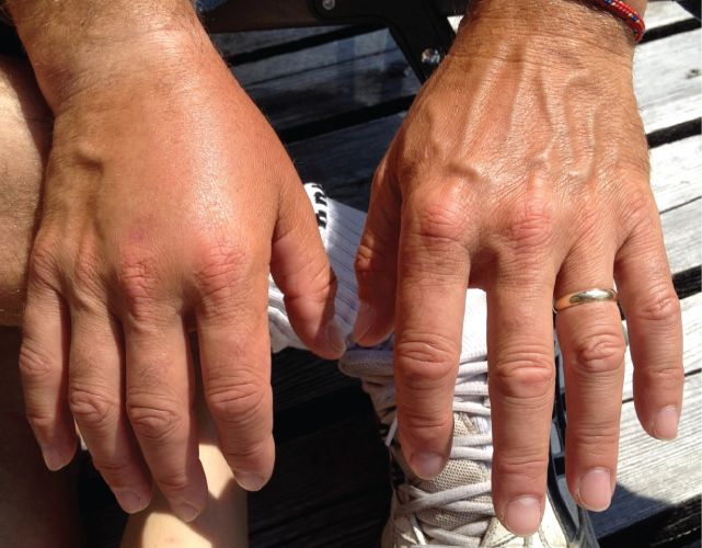
Your body is mostly water. Where that water sits matters as much as how much you have. In a healthy adult, water makes up about 60% of body weight. A handy memory trick is the 60-40-20 rule. About 60% of body weight is total water. About 40% sits inside cells. About 20% sits outside them.
The fluid compartments
Picture two big rooms. The intracellular fluid (ICF) compartment is all the water inside your cells. It holds the most, roughly two-thirds of body water. The extracellular fluid (ECF) compartment is everything outside the cells, the other one-third. The ECF splits again into two parts. Interstitial fluid bathes the spaces between cells. Intravascular fluid is the plasma inside your blood vessels. Fluid shifts between these spaces all day long. Keeping that balance is what keeps cells alive and blood pressure steady.
Osmosis and tonicity
Water follows solute. Osmosis is water moving across a semipermeable membrane. It moves from where solute is low toward where solute is high. It chases the particles it cannot easily follow. Tonicity tells you how a solution changes cell volume. It is one of the most important ideas behind choosing IV fluids.
| Solution | What it does to the cell | Why |
|---|---|---|
| Isotonic (e.g., 0.9% normal saline) | No net change. The cell stays the same. | The solute matches the cell, so water does not shift. |
| Hypotonic (e.g., 0.45% saline) | Cell swells and may burst (lyse). | The outside is more dilute, so water rushes into the cell. |
| Hypertonic (e.g., 3% saline) | Cell shrinks (crenates). | The outside is more concentrated, so water is pulled out. |
Starling forces: the tug-of-war at the capillary
At every capillary, two pressures fight. One pushes fluid out and one pulls it back. We call them the Starling forces. Hydrostatic pressure is the outward push of fluid on the vessel wall. It is basically the pressure of pumped blood, and it drives fluid out into the tissue. Oncotic pressure is the inward pull. It is also called colloid osmotic pressure, and albumin creates most of it. Albumin is a large protein that stays in the blood and draws water back in. Normally fluid leaves at the arterial end and is reabsorbed at the venous end. The lymph system mops up the small amount left over. When that balance tips, fluid piles up in the tissue. That is edema.
The four mechanisms of edema
Edema is not one problem. It is any break in that Starling balance or in the drainage system. Four mechanisms cause it:
- Increased hydrostatic pressure. Too much outward push forces fluid into tissue. This is classic in heart failure, where blood backs up. It also comes from venous obstruction such as a DVT. Holding on to sodium and water belongs here too, since the extra volume is how the pressure rises. Aldosterone or kidney failure often drives that. You see dependent swelling in the ankles and feet.
- Decreased plasma oncotic pressure. Low albumin means a weak inward pull, so fluid stays out. Look for it in cirrhosis, nephrotic syndrome, and severe malnutrition.
- Lymphatic obstruction. Lymph channels normally drain the leftover fluid. If they are blocked, protein-rich fluid backs up as lymphedema. It often follows lymph node removal or radiation in cancer care.
- Leaky capillaries. Inflammation, burns, or sepsis widen the gaps in the capillary wall. Fluid and even protein escape straight into the tissue, so the swelling is local and warm in inflammation, and body-wide in sepsis.
Third spacing
Third spacing is fluid that leaves the vessels and the tissue spaces and gets trapped. It sits where it does no useful work. That can be the belly (ascites), the pleural cavity, or the bowel wall after surgery or in sepsis. This matters because the fluid is lost to the circulation. The patient can look puffy and swollen and still be intravascularly dry. Look for a low blood pressure, a rising heart rate, and poor urine output. You may be giving fluids to someone who appears fluid-overloaded. So judge perfusion, not just how the patient looks. Try the interactive figure below to move fluid between the compartments.
- Total body water is about 60% of weight. Roughly 40% is inside cells and 20% is outside them, as interstitial plus intravascular fluid. That is the 60-40-20 rule.
- Tonicity predicts cell volume. Hypotonic fluid swells cells, hypertonic fluid shrinks them, and isotonic fluid leaves them unchanged.
- Starling forces weigh the outward hydrostatic push against the inward oncotic pull from albumin. Tip that balance and you get edema.
- Edema has four mechanisms: high hydrostatic pressure, low oncotic pressure from low albumin, leaky capillaries, or blocked lymph drainage. Holding on to sodium and water is not a fifth mechanism. It is how hydrostatic pressure rises.
- Third spacing traps fluid where it cannot circulate. So a patient can look swollen yet be short on blood volume. Judge by perfusion and vital signs.
Section 3The electrolytes that matter most
Electrolytes are the tiny charged particles dissolved in your body's water. They make nerves fire, muscles squeeze, and the heart beat on rhythm. Because they carry a charge, small shifts change how excitable your cells are. The body works hard to hold them in a narrow range. When it cannot, the nervous system and the heart usually complain first. Let's walk through the big three, plus a quiet helper.
Sodium: the master of water balance
Sodium is the main electrolyte outside your cells. Normal is roughly 135 to 145 mEq/L. Where sodium goes, water follows. That single rule explains almost every symptom. Sodium sets where water sits, so sodium problems are really water problems. The brain feels them the most, because it is packed inside a rigid skull.
In hyponatremia the sodium is low, so the blood outside the cells is dilute. Water moves into cells by osmosis and they swell. In brain cells that means cerebral edema. So patients get headache, confusion, and lethargy. If the drop is severe or fast, they can seize. In hypernatremia the outside is too salty. Water is pulled out of cells and they shrink. The brain literally dries out. You see thirst, restlessness, irritability, and altered mental status. Notice the pattern. Sodium imbalances are neurologic, because they move water in and out of neurons.
Potassium: the heart's electrolyte
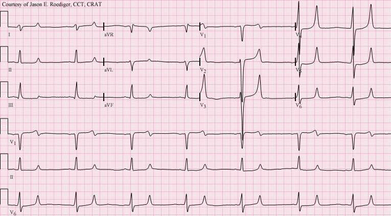
Potassium is the major electrolyte inside cells. Normal is about 3.5 to 5.0 mEq/L. It sets the electrical resting membrane potential. That is what lets heart muscle depolarize and repolarize on cue. This is why potassium is often called the most dangerous electrolyte. Too little and too much can both stop the heart. The danger shows up on the ECG.
In hypokalemia, heart cells turn sluggish to repolarize. That gives flattened T waves, ST depression, and U waves. Patients also get muscle weakness and risk lethal arrhythmias. In hyperkalemia, cells depolarize too easily at first. That gives the classic peaked (tall, tented) T waves. Then the QRS widens. Last comes a sine-wave pattern that can slide into ventricular fibrillation or asystole. Both extremes cause arrhythmias, so potassium sits at the top of the emergency list.
| Feature | Hypokalemia | Hyperkalemia |
|---|---|---|
| Level | < 3.5 mEq/L | > 5.0 mEq/L |
| ECG | Flat T waves, U waves, ST depression | Peaked T waves, wide QRS |
| Muscle | Weakness, cramps, ileus | Weakness, then paralysis |
| Main danger | Fatal arrhythmia | Fatal arrhythmia or arrest |
Calcium: nerves, muscles, and PTH
Calcium steadies nerve and muscle membranes. Total normal is roughly 8.5 to 10.5 mg/dL, so a total below 8.5 mg/dL is hypocalcemia. Parathyroid hormone (PTH) guards it. PTH raises calcium in three ways. It pulls calcium out of bone, saves it in the kidney, and turns on vitamin D. Here the rule flips. Low calcium makes cells more excitable.
In hypocalcemia, membranes turn twitchy and fire too easily. That causes tetany, muscle cramps, and tingling. Two signs help you catch it. Trousseau sign is carpal spasm when a blood-pressure cuff is inflated. Chvostek sign is a facial twitch when you tap the cheek over the facial nerve. In hypercalcemia, membranes are over-stabilized, so things slow down. The old line is "stones, bones, groans, and psychiatric moans". That means kidney stones, bone pain, constipation with belly pain, and lethargy or confusion. Too much PTH or a cancer is often the cause.
Magnesium: the quiet partner
Magnesium tends to travel with potassium and calcium. Normal is about 1.5 to 2.5 mEq/L. Low magnesium makes potassium hard to replace. It also worsens a low calcium. So you often cannot fix a stubborn hypokalemia until you correct the magnesium first. Think of it as the electrolyte that keeps the other two honest.
Which imbalances are true emergencies? The ones that threaten the heart or brain fastest. Severe hyperkalemia and hypokalemia can cause lethal arrhythmias within minutes. Symptomatic hyponatremia with seizures means the brain is swelling. Severe hypocalcemia can cause laryngospasm. Use the interactive figure below to see how each imbalance shifts.
- Sodium controls water, so its imbalances are neurologic. Low sodium swells brain cells, with edema and seizures. High sodium shrinks them, with thirst and confusion. Correct both slowly.
- Potassium is the most dangerous electrolyte, since both hypo- and hyperkalemia cause lethal arrhythmias. Watch the ECG for flat T waves and U waves when low, peaked T waves and a wide QRS when high.
- Calcium reverses the rule. Low calcium raises excitability and causes tetany, with positive Trousseau and Chvostek signs. PTH is the main regulator.
- Magnesium tracks with potassium and calcium. A low magnesium often blocks a successful potassium replacement.
- True emergencies are the ones hitting the heart or brain fastest: severe hyperkalemia, severe hypokalemia, and symptomatic hyponatremia.
Section 4Acid-base balance & the ABG
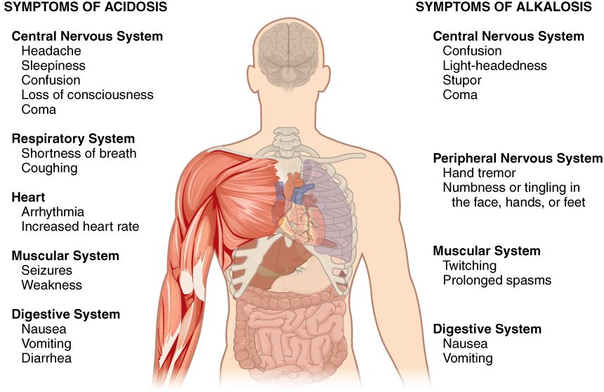
Your blood is fussy about one thing above almost all others. That thing is its pH. Every enzyme, every heartbeat, and every nerve signal depends on a narrow window of 7.35 to 7.45. That sits just barely on the alkaline (basic) side of neutral. Your body guards it closely. Slip below 7.35 and we call it acidosis. Drift above 7.45 and it is alkalosis. Go much past 6.8 or 7.8 in either direction and cells simply stop working. So the body runs two teams whose whole job is defending that pH.
Two defenders: the lungs (fast) and the kidneys (slow)
Picture pH as a seesaw between an acid and a base. The acid side is carbon dioxide (CO2), a waste gas that dissolves into carbonic acid. The base side is bicarbonate (HCO3), the buffer that soaks up extra acid. The lungs control the CO2 side, and they are fast. Breathe harder and you blow off CO2 within minutes, so there is less acid. Breathe slower and CO2 builds up. The kidneys control the bicarbonate side, and they are slow. They take hours to days to hold onto or dump HCO3. Fast lungs, slow kidneys. Remember that and half of ABGs make sense.
The four primary disorders
Every simple acid-base problem is a story about one of those two chemicals going wrong. If the trouble starts with CO2, it is a breathing problem, so it is respiratory. If it starts with HCO3, it is a metabolic or kidney problem, so it is metabolic.
| Disorder | Primary change | Classic cause |
|---|---|---|
| Respiratory acidosis | CO2 high, so acid is kept in | COPD, hypoventilation (slow breathing), opioid overdose |
| Respiratory alkalosis | CO2 low, blown off | Hyperventilation (fast breathing), anxiety, early sepsis |
| Metabolic acidosis | HCO3 low, too much acid | DKA, shock with lactic acid, diarrhea |
| Metabolic alkalosis | HCO3 high | Vomiting, NG suction, diuretics |
Reading an ABG in three steps
You do not need to memorize a chart. Just walk down three numbers in order. The standard normals are pH 7.35-7.45, PaCO2 35-45 mmHg, and HCO3 22-26 mEq/L.
- Look at the pH first. Below 7.35 is acidosis. Above 7.45 is alkalosis. This tells you which way the body has actually gone.
- Look at the CO2 (normal 35-45 mmHg). Above 45 is acidic. Below 35 is alkalotic, because CO2 is an acid. Now compare its direction with the pH. If the two moved opposite ways, the lungs are the culprit. It is respiratory.
- Look at the HCO3 (normal 22-26 mEq/L). Below 22 is acidic. Above 26 is alkalotic. Compare its direction with the pH. If the two moved the same way, the kidneys and metabolism are the culprit. It is metabolic.
Those two checks have a name. It is ROME, for Respiratory Opposite, Metabolic Equal. Use one rule only, so the answer never flips on you. Here is an example. The pH is 7.28, which is low, and the CO2 is 60 mmHg, which is high. The two numbers went opposite ways, so the lungs are to blame. This is respiratory acidosis. You often see that in a COPD patient who is retaining CO2. Now flip it. If the pH is 7.28 and the HCO3 is 14 mEq/L, both numbers fell together. Equal directions point to the metabolic side, as in DKA.
Compensation: the other team pitches in
Here is the elegant part. When one system fails, the other one tries to drag the pH back toward normal. If the lungs cause an acidosis, the kidneys slowly retain bicarbonate to buffer it. If the kidneys or metabolism cause an acidosis, the lungs speed up to blow off CO2. Those deep, sighing Kussmaul respirations in DKA are exactly that rescue in action. The compensating system pushes its own number the same way as the primary problem. So a compensated respiratory acidosis shows a high CO2, which is the problem, plus a high HCO3, which is the kidney rescue.
Watch the pH to judge how far compensation has gone. If the pH is back inside 7.35-7.45, it is fully compensated. If the pH is still abnormal but the second value has clearly shifted to help, it is partially compensated. If only the primary system is off and the other has not budged, it is uncompensated. Remember, compensation nudges pH toward normal but rarely overshoots it. The body does not overcorrect past neutral.
Tie it to real patients and it sticks. The vomiting patient loses stomach acid and swings alkalotic, which is metabolic alkalosis. The panicking patient hyperventilates and blows off CO2, which is respiratory alkalosis. The septic or bleeding patient in shock pours out lactic acid, which is metabolic acidosis. The sedated patient after surgery barely breathes and retains CO2, which is respiratory acidosis. Use the step-by-step ABG interpreter below to practice walking those three numbers yourself.
- Normal blood pH is 7.35-7.45. Below that is acidosis and above it is alkalosis. The lungs defend pH fast with CO2, the kidneys slowly with bicarbonate.
- CO2 problems are respiratory. HCO3 problems are metabolic. High CO2 or low HCO3 means acidosis. Low CO2 or high HCO3 means alkalosis.
- Read an ABG in order: pH, then CO2, then HCO3, and use ROME: Respiratory Opposite, Metabolic Equal. A CO2 moving opposite the pH is respiratory. An HCO3 moving with the pH is metabolic.
- Compensation is the opposite system shifting to pull pH back toward normal. The kidneys retain HCO3 in a respiratory acidosis. The lungs blow off CO2 in DKA. It rarely overshoots normal.
- Classic causes: DKA and shock give metabolic acidosis. COPD and hypoventilation give respiratory acidosis. Vomiting gives metabolic alkalosis. Hyperventilation gives respiratory alkalosis.
Section 5Genes & inheritance
Every cell in your body carries a full instruction manual written in DNA. Think of DNA as a long string of just four chemical letters: A, T, C, and G. A gene is one meaningful sentence in that manual. Each gene tells the cell how to build one protein. Proteins do almost everything. They form structures, carry oxygen, speed up reactions as enzymes, and open and close channels. The flow is simple to remember. DNA is copied (transcribed) into mRNA. Then the mRNA is read (translated) into a protein. When the recipe is spelled correctly, the protein works.
What a mutation actually does
A mutation is a spelling change in a gene. Sometimes it is harmless. Other times it swaps one letter and changes one amino acid. Then the protein folds wrong or stops working. In sickle cell disease, a single letter change turns normal hemoglobin into a sticky version. That warps red cells into a sickle shape. So the whole chain matters. A tiny DNA typo can leave a missing or broken protein, and a broken protein shows up as disease. This is why genetic conditions look so much alike inside one family. Everyone inherits the same misspelled recipe.
Autosomal dominant vs autosomal recessive
You inherit two copies of most genes, one from each parent. These genes live on the autosomes, chromosomes 1 through 22. Whether a condition appears depends on how many broken copies it takes.
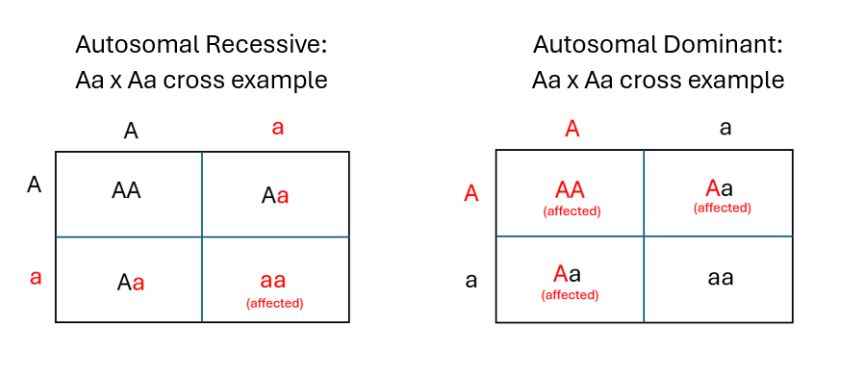
In autosomal dominant inheritance, one faulty copy is enough to show the trait. So it usually appears in every generation, and it affects males and females equally. Huntington disease is the classic example. In autosomal recessive inheritance, you need two faulty copies to be affected. People with just one copy are healthy carriers. They carry the gene silently and can pass it on. Cystic fibrosis and sickle cell disease follow this pattern. That is why they often seem to skip generations, then appear when two carriers have a child.
| Feature | Autosomal dominant | Autosomal recessive |
|---|---|---|
| Copies needed to show | One | Two |
| Family pattern | Usually every generation | Often skips generations |
| Carriers | Rare idea here, since one copy shows | Yes. One copy is a silent carrier. |
| Examples | Huntington disease | Cystic fibrosis, sickle cell |
X-linked recessive: why males are hit harder
Some genes sit on the X chromosome. Females are XX and males are XY. In X-linked recessive conditions like hemophilia, the faulty gene is on the X. A female has a backup X. One healthy copy usually covers for the broken one, so she is typically a carrier. A male has only one X and no second copy to cover for it. So a single faulty gene shows. That is why X-linked recessive disorders affect males far more often. A carrier mother has a 50 percent chance of passing the affected X to each son.
Predicting the odds with a Punnett square
A Punnett square is a simple grid. It lays out each parent's possible gene copies and shows the chance of each combination in their children. Use capital letters for dominant and lowercase for recessive. Take two carriers of a recessive condition, both Aa. The grid gives four boxes: AA, Aa, Aa, and aa. So there is about a 25 percent chance of an affected child (aa). There is a 50 percent chance of a carrier and a 25 percent chance of neither. Each pregnancy is its own independent roll. The odds reset every time. You can explore the patterns and squares in the interactive figure below.
Penetrance and expressivity
Penetrance is whether a gene shows at all. It is the share of people with the gene who actually develop the trait. Expressivity is how strongly it shows. The same gene can look mild in one person and severe in another.
- DNA holds the recipe. A gene builds a protein. A mutation is a spelling change that can break that protein and cause disease.
- Autosomal dominant needs one copy and appears every generation, as in Huntington disease. Autosomal recessive needs two copies and has healthy carriers, as in cystic fibrosis and sickle cell.
- X-linked recessive conditions such as hemophilia mostly affect males, because they have only one X and no backup copy.
- A Punnett square predicts the odds for children. Two recessive carriers have about a 25 percent chance of an affected child in each pregnancy.
- Penetrance is whether the gene shows. Expressivity is how strongly it shows.
Section 6Chromosomal & congenital disorders
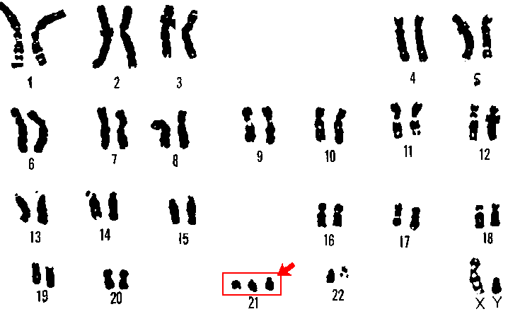
Every cell in your body normally carries 46 chromosomes in 23 pairs. That is 22 pairs of autosomes plus one pair of sex chromosomes, XX in females and XY in males. You inherit exactly half from each parent. This tidy sorting happens during meiosis, the special cell division that makes eggs and sperm. When meiosis works, each gamete ends up with exactly 23 chromosomes. Fertilization then restores the full set of 46. Chromosomal disorders happen when that sorting goes wrong. They also happen when an outside exposure damages a developing embryo. Use the interactive figure below to see how one extra chromosome reshapes the picture.
Nondisjunction: chromosomes that refuse to separate
The most common mistake is nondisjunction. A pair of chromosomes fails to pull apart during meiosis. One gamete ends up with an extra copy, and the other is left short. If that gamete joins in fertilization, the embryo carries the wrong count. We call that state aneuploidy. Three copies of a chromosome instead of two is a trisomy. Having only one copy is a monosomy. Chromosomes are packed with genes. So even one extra or missing chromosome throws off the dose of hundreds of proteins. That is why these disorders affect so many body systems at once. Nondisjunction becomes more likely with advancing maternal age. That is why the risk of trisomies climbs after about age 35.
Trisomy 21 and its cousins
Trisomy 21 (Down syndrome) is the most common surviving autosomal trisomy. It means three copies of chromosome 21. Classic features usually include intellectual disability and a flat facial profile with upslanting palpebral fissures, the slant of the eyelid openings. A single palmar (simian) crease is common, along with hypotonia, which means low muscle tone. That extra genetic material affects many systems. Congenital heart defects are common, often an atrioventricular septal defect. There is also a higher risk of leukemia and of early Alzheimer-type changes. Two rarer and far more severe trisomies are worth knowing. They are trisomy 18 (Edwards syndrome) and trisomy 13 (Patau syndrome). Both usually cause profound impairment and many organ malformations. Most affected infants do not survive past infancy.
When the sex chromosomes are off
Nondisjunction can also hit the sex chromosomes. Turner syndrome (45,X) is a monosomy. It is a female with only one X. It usually shows up as short stature, a webbed neck, and ovarian dysgenesis. That means the ovaries do not work, so there is infertility and no puberty without hormone therapy. Coarctation of the aorta, a narrowed aorta, is also common. Klinefelter syndrome (47,XXY) is a male with an extra X. It often appears around puberty with tall stature, small firm testes, and gynecomastia, which is breast tissue growth. Testosterone is reduced, which leads to infertility. Notice the pattern. An extra sex chromosome tends to be tolerated better than an extra autosome. That is largely because the body naturally silences extra X chromosomes.
| Feature | Turner (45,X) | Klinefelter (47,XXY) |
|---|---|---|
| Karyotype | One X, with a sex chromosome missing | Two X plus one Y |
| Phenotype | Female | Male |
| Stature | Short | Tall |
| Classic clue | Webbed neck, coarctation | Small testes, gynecomastia |
| Fertility | Usually infertile, from ovarian dysgenesis | Usually infertile, from low testosterone |
Teratogens: when the environment is the culprit
Not every congenital disorder is genetic. A teratogen is any agent that disrupts normal fetal development. It can be a drug, an infection, a chemical, or radiation. Timing matters enormously. The embryonic period runs roughly from week 3 to week 8. Organs are first forming then, so it is the window of greatest risk. An exposure during it can cause major structural defects. Later exposure tends to affect growth and function instead. One key cluster to memorize is the TORCH infections. TORCH stands for Toxoplasmosis, Other (including syphilis), Rubella, Cytomegalovirus, and Herpes simplex. These infections in the mother can cross the placenta. They cause malformations, growth restriction, hearing loss, and neurologic damage. Alcohol is one of the most preventable teratogens. Exposure before birth can produce fetal alcohol spectrum disorder, with characteristic facial features, growth deficits, and lifelong problems with thinking and learning. There is no known safe amount.
Multifactorial disorders
Finally, many common conditions are multifactorial. Neural tube defects, cleft lip and palate, and congenital heart disease are examples. They arise from several genes plus environmental influences, not from one single cause.
- Nondisjunction means chromosomes fail to separate in meiosis. It causes trisomies, with an extra copy, and monosomies, with a copy missing.
- Trisomy 21 (Down syndrome) is the most common autosomal trisomy. Trisomy 18 (Edwards) and trisomy 13 (Patau) are rarer and usually fatal in infancy.
- Sex-chromosome disorders include Turner (45,X), a short and infertile female, and Klinefelter (47,XXY), a tall and infertile male.
- Teratogens include the TORCH infections and alcohol. They do the most structural harm during the embryonic period, weeks 3 to 8.
- Multifactorial disorders arise from genetic and environmental causes together.
The chapter in one breath
- Cellular adaptation is the cell's survival plan under stress. It runs in five modes: atrophy (shrink), hypertrophy (enlarge), hyperplasia (multiply), metaplasia (swap to a tougher cell type), and dysplasia (disordered growth that can come before cancer).
- When stress outpaces adaptation you get reversible injury, which is cell swelling from failing Na+/K+ pumps. If hypoxia and ATP loss keep going, it tips into irreversible injury and cell death.
- Apoptosis is tidy, programmed, energy-using single-cell suicide with no inflammation. Necrosis is messy, accidental death. Cells rupture, spill their contents, and trigger inflammation.
- Body water splits into intracellular fluid, about two-thirds, and extracellular fluid. Edema happens when fluid leaks into tissue from high hydrostatic pressure, low albumin and so a weak oncotic pull, leaky capillaries, or blocked lymphatics.
- The must-know electrolytes are sodium (water balance and nerve firing), potassium (the cardiac rhythm ion, dangerous high or low), calcium (bones, clotting, muscle contraction), and magnesium (the quiet cofactor that steadies nerves and heart).
- Acid-base balance is a tug-of-war between the lungs, which are fast and blow off CO2, and the kidneys, which are slow and adjust HCO3-. Together they hold arterial pH in the tight 7.35-7.45 window.
- Read an ABG by checking pH first, acid or alkaline. Then use ROME, for Respiratory Opposite and Metabolic Equal: a CO2 that moved opposite the pH is respiratory, and an HCO3 that moved with the pH is metabolic. Then decide whether the other system is compensating to nudge pH back toward normal.
- Inheritance patterns follow the pedigree: autosomal dominant (one copy shows, every generation), autosomal recessive (needs two copies, carriers hidden), and X-linked recessive (hits males hardest, as in hemophilia).
- Chromosomal disorders come from wrong chromosome numbers, classically trisomy 21 (Down syndrome) from nondisjunction. Many congenital defects such as neural tube defects arise from gene-environment hits during development, for example low folate.
ReferenceWords worth knowing
A quick reference for the terms in this chapter. Skim it now, or circle back whenever a word trips you up.
- Apoptosis
- Apoptosis is orderly, programmed cell death. The cell shrinks and packages itself neatly for removal without spilling its contents, so no inflammation follows.
- Atrophy
- Atrophy is a drop in the size of cells, and so of the whole tissue or organ. It follows disuse, loss of nerve supply, aging, or reduced blood supply, as when a casted limb shrinks.
- Autosomal recessive
- Autosomal recessive describes an inheritance pattern that needs two copies of a mutated gene, one from each parent, on a non-sex chromosome. Unaffected carriers with one copy can pass it on silently.
- Compensation (acid-base)
- Compensation is the body's attempt to restore normal blood pH using the system that is not primarily broken. The lungs adjust CO₂ for a metabolic problem, and the kidneys adjust bicarbonate for a respiratory problem.
- Dysplasia
- Dysplasia is disordered growth in which cells vary in size, shape, and arrangement. It is not cancer, but it is often a warning change that can come before it.
- Edema
- Edema is a buildup of extra fluid in the interstitial space, the tissue between cells. It shows as visible swelling, and it sometimes pits when you press on it.
- Hyperplasia
- Hyperplasia is a rise in the number of cells in a tissue, driven by more cell division. An example is the endometrium thickening under hormonal stimulation.
- Hypertrophy
- Hypertrophy is a rise in the size of single cells, with no new cells added, which enlarges the organ. An example is heart muscle thickening against high blood pressure.
- Hyponatremia
- Hyponatremia is a blood sodium level below normal, usually under about 135 mEq/L. It often means too much water for the amount of sodium, and it can cause confusion or seizures.
- Hypoxia
- Hypoxia is too little oxygen reaching the tissues. It starves cells of the fuel they need to make energy.
- Ischemia
- Ischemia is reduced blood flow to a tissue. It limits delivery of both oxygen and nutrients while letting wastes build up, and it is a common cause of hypoxia.
- Metaplasia
- Metaplasia is the reversible swap of one mature cell type for another, tougher one under stress. Airway cells changing under long-term cigarette smoke is one example. The swap withstands the stress but loses the original tissue's special job, and it can come before dysplasia.
- Necrosis
- Necrosis is disorderly, unplanned cell death from injury. Cells swell and burst, spill their contents, and trigger inflammation.
- Nondisjunction
- Nondisjunction is the failure of chromosomes to separate properly during cell division. It leaves a cell with an extra or missing chromosome, as in trisomy 21 (Down syndrome).
- Oncotic pressure
- Oncotic pressure is the inward pull that plasma proteins, mainly albumin, exert to keep fluid inside blood vessels. When it drops, fluid leaks out and edema forms.
- Tonicity
- Tonicity is how a solution's solute concentration moves water in or out of a cell. The cell swells in a hypotonic solution, shrinks in a hypertonic one, and stays unchanged in an isotonic one.
DrillFlashcards for this unit
These are the Week 1 cards. It is the same deck as the hub, pulled in here so you can review the moment you finish reading. Tap the card to flip it. Hit Shuffle to mix them up. To grade yourself and track mastery across the whole deck, open the full flashcards page.