Pulmonary
Every cell in the body runs on oxygen, and the lungs are where it gets in and carbon dioxide gets out. This chapter follows a breath from the air to the bloodstream and back — how ventilation and perfusion must match, what happens when airways narrow or a clot lodges, and how to read the story in a patient’s lung sounds.
By the end of this chapter you can
- Explain ventilation, perfusion, and V/Q mismatch (dead space vs shunt).
- Contrast chronic bronchitis with emphysema in COPD.
- Describe the three problems in asthma and rescue vs controller inhalers.
- Recognize a pulmonary embolism and its setup (Virchow’s triad).
- Match lung sounds (crackles, wheezes, stridor) to their causes.
Section 1How breathing works: ventilation, perfusion & V/Q
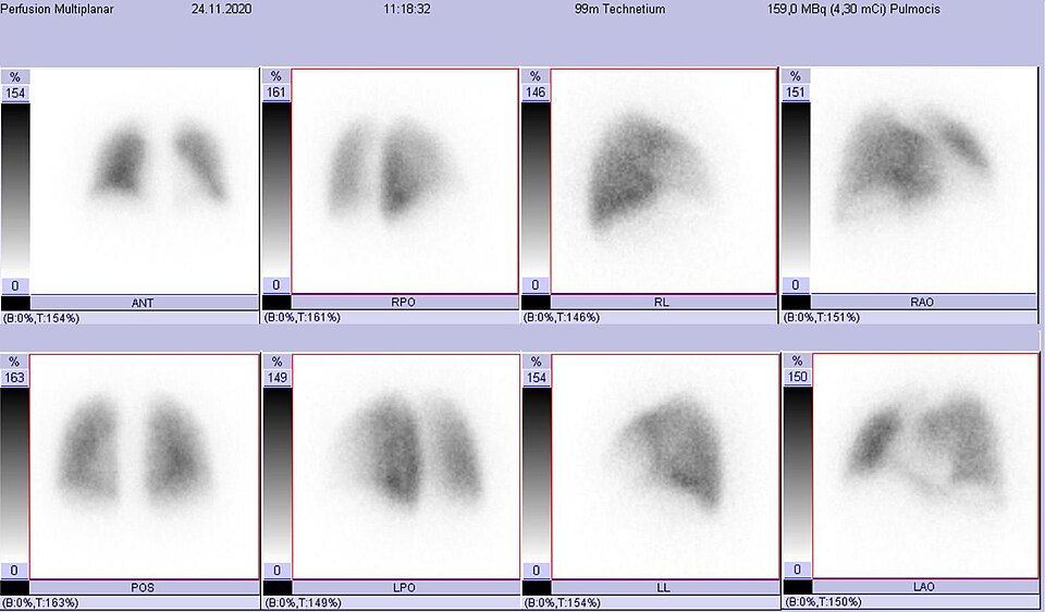
Every breath you take is really two jobs happening at once, and they have to be perfectly coordinated. Ventilation is the movement of air in and out of the lungs, delivering fresh oxygen to the tiny air sacs called alveoli and carrying carbon dioxide away. Perfusion is the flow of blood through the pulmonary capillaries that wrap around those alveoli. Think of it like a delivery system: ventilation brings the packages (oxygen) to the loading dock, and perfusion is the truck fleet that picks them up. If the packages arrive but no trucks show up, or the trucks show up but there are no packages, the whole system stalls. For gas exchange to work, air and blood must meet at the same place, at the same time.
Matching air to blood: the V/Q ratio
Nurses describe how well ventilation and perfusion line up using the V/Q ratio, where V is ventilation and Q is perfusion (blood flow). In an ideal, healthy lung the overall ratio is about 0.8 usually, because we perfuse a touch more than we ventilate. That number is not the same everywhere in the lung, though. Because of gravity, the bases (bottoms) of the lungs get more blood flow, while the apices (tops) get relatively more air, so the ratio naturally varies from top to bottom. What matters clinically is that when air and blood are reasonably balanced, oxygen crosses into the blood and carbon dioxide is blown off efficiently. When they fall out of balance, we call it a V/Q mismatch, and it comes in two flavors.
Two ways it goes wrong: dead space and shunt
A high V/Q means ventilation without matching perfusion. The air arrives, but no blood is there to pick up the oxygen. This wasted ventilation is called dead space. The classic cause is a pulmonary embolism, where a clot blocks a pulmonary artery. The alveoli fill with air, but the blood truck never arrives.
A low V/Q means perfusion without matching ventilation. Blood keeps flowing past alveoli that are not getting fresh air, so it returns to the heart still low on oxygen. This is called a shunt. Think pneumonia filling alveoli with pus, pulmonary edema flooding them with fluid, or atelectasis (collapsed alveoli). One clinical clue: pure shunt often responds poorly to supplemental oxygen, because the extra oxygen cannot reach blood flowing past blocked alveoli.
| Feature | Dead space (high V/Q) | Shunt (low V/Q) |
|---|---|---|
| What is missing | Perfusion (blood flow) | Ventilation (air) |
| Plain-English picture | Air arrives, no blood to load it | Blood arrives, no fresh air to load |
| Classic cause | Pulmonary embolism | Pneumonia, pulmonary edema, atelectasis |
| Response to oxygen | Good (the still-perfused lung takes up the extra oxygen) | Poor and refractory (blood bypasses any ventilated alveolus) |
Gas exchange across the alveolar membrane
Where air and blood actually meet is the alveolar-capillary membrane, an incredibly thin barrier just a few cells thick. Gases cross it by diffusion, always moving from high pressure to low pressure. Oxygen is more concentrated in the alveolus than in the incoming blood, so it diffuses into the blood; carbon dioxide is more concentrated in the blood, so it diffuses out into the alveolus to be exhaled. Anything that thickens this membrane (fluid, scar tissue, inflammation) slows diffusion and worsens gas exchange, even when ventilation and perfusion are otherwise present.
Hypoxemia vs hypoxia, and how oxygen travels
These two words sound alike but mean different things, and nursing exams love to test the difference. Hypoxemia is low oxygen level in the blood (measured as a low PaO2 on an arterial blood gas or a low SpO2 on the pulse oximeter). Hypoxia is low oxygen reaching the tissues themselves. They often travel together, but not always. Severe anemia, for example, can cause tissue hypoxia even when the blood oxygen level (PaO2) looks normal, because there simply are not enough carriers to deliver it.
And those carriers are the key to the whole system: about 97 percent of oxygen in the blood rides bound to hemoglobin inside red blood cells, with only a tiny fraction dissolved in plasma. Explore the figure below to see ventilation, perfusion, and V/Q matching working together.
- Ventilation (air in and out) and perfusion (blood flow past alveoli) must match at the same place and time for gas exchange to work; the normal V/Q ratio is about 0.8.
- Dead space is ventilation without perfusion (high V/Q, classic cause pulmonary embolism); shunt is perfusion without ventilation (low V/Q, classic causes pneumonia, edema, atelectasis).
- Gases cross the thin alveolar-capillary membrane by diffusion from high to low pressure; a thickened membrane slows oxygen uptake.
- Hypoxemia is low oxygen in the blood; hypoxia is low oxygen in the tissues, and they do not always occur together.
- About 97 percent of oxygen is carried bound to hemoglobin, so oxygen delivery depends on carriers, not just lung function.
Section 2Obstructive disease: COPD
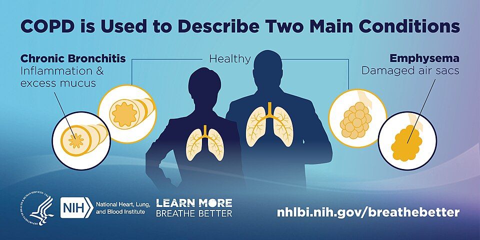
Every time you breathe, air travels down your windpipe, splits into smaller and smaller tubes (the bronchi and bronchioles), and finally reaches millions of tiny balloon-like sacs called alveoli. This is where oxygen slips into the blood and carbon dioxide slips out. Two things make this work: the airways must stay open, and the lung tissue must be springy. That springiness, called elastic recoil, is what quietly squeezes air back out when you exhale. Normal exhalation is passive and effortless.
In chronic obstructive pulmonary disease (COPD), that easy exhale breaks down. The airways narrow and the springiness is lost, so air flows in but gets stuck on the way out. Think of COPD as a disease of getting air OUT, not in. Stale air piles up behind the blockage, a problem called air trapping, and the lungs stay over-inflated. This is why patients often push air out through pursed lips and why their exhale takes so long.
Why air gets trapped
COPD is really two overlapping problems, and most patients have some of each.
Chronic bronchitis is an airway problem. Long-term irritation inflames the bronchial lining, so it swells and the mucus glands grow larger and pump out thick secretions. The airway is now narrowed by both swelling and mucus, and the tiny sweeping hairs (cilia) that clear that mucus are damaged. Mucus sits and grows bacteria, so infections keep coming. It is defined clinically by a a chronic productive cough for at least 3 months in each of 2 consecutive years.
Emphysema is a tissue problem. The walls between alveoli are destroyed and merge into fewer, larger, floppy sacs. That destroys surface area for gas exchange and destroys the elastic recoil that normally props the small airways open and drives exhalation. Without that support, airways collapse on exhale and trap air even more.
| Feature | Chronic bronchitis (blue bloater) | Emphysema (pink puffer) |
|---|---|---|
| Core problem | Inflamed, mucus-clogged airways | Destroyed alveolar walls, lost recoil |
| Classic look | Cyanotic, often overweight, edematous | Thin, barrel chest, leaning forward |
| Cough & mucus | Heavy productive cough | Minimal cough, little sputum |
| Oxygen level | Lower early (hypoxemia, blue tint) | Kept closer to normal by fast breathing |
| Breathing style | Wet, congested | Pursed-lip, working hard to exhale |
The nicknames are memory hooks, not real patient types. The blue bloater is cyanotic and puffy from fluid backup; the pink puffer stays pinker by breathing fast and hard. The over-inflated, air-trapped chest that becomes rounded and rigid is called a barrel chest (an increased anteroposterior diameter).
The leading cause
Cigarette smoking is by far the number-one cause. Smoke keeps the airways inflamed and floods the lung with enzymes that digest alveolar walls, while overwhelming the body's natural defense against those enzymes. Over years, this drives both the bronchitis and the emphysema. (A smaller group inherits alpha-1 antitrypsin deficiency, which removes that natural defense and causes emphysema even in nonsmokers.) Quitting smoking is the single most powerful thing that slows the disease.
When the strain reaches the heart
Chronically low oxygen in the lungs makes lung blood vessels clamp down (hypoxic pulmonary vasoconstriction). That raises pressure in the pulmonary arteries (pulmonary hypertension), and the right side of the heart must pump against it. Over time the right ventricle enlarges and fails, a condition called cor pulmonale. Nurses see this as jugular vein distension, liver enlargement, and peripheral edema, which is part of why the blue bloater looks swollen.
You can explore how the clogged airway and the destroyed alveoli differ in the interactive figure below.
- COPD is trouble getting air OUT: narrowed airways and lost elastic recoil cause air trapping and a prolonged, labored exhale.
- Chronic bronchitis is inflamed, mucus-filled airways (productive cough, blue bloater); emphysema is destroyed alveolar walls with lost recoil (barrel chest, pink puffer).
- Smoking is the leading cause and quitting is the most effective way to slow progression.
- Chronic hypoxia and pulmonary hypertension can strain the right heart into cor pulmonale (edema, JVD).
- Give oxygen carefully, usually targeting SpO2 88 to 92 percent, since very high-flow O2 can worsen CO2 retention.
Section 3Asthma
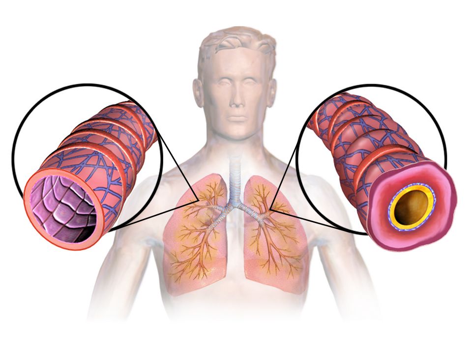
Take a slow breath in. Air rushes down your windpipe, splits into two bronchi, then branches again and again into ever-smaller bronchioles before reaching the tiny air sacs where gas exchange happens. Those small airways are wrapped in a thin layer of smooth muscle, lined with a moist membrane, and are supposed to stay open and springy so air moves in and out with almost no effort. In a healthy lung, breathing out is passive and quiet: the airways stay wide, and the air leaves easily.
In asthma, those small airways become hyperreactive. They overreact to things that should be harmless, and the airway narrows. The key word for asthma is reversible obstruction: unlike the fixed damage of emphysema, an asthma attack can open back up with treatment or even on its own. But while it is happening, air gets trapped, and the person has to work hard to move it.
Three problems at once
An asthma attack is not one thing going wrong; it is three processes hitting the airway together, which is why it can escalate fast.
- Bronchospasm. The ring of smooth muscle around the bronchioles squeezes and tightens, physically narrowing the tube. This is the fast part, and it is what rescue inhalers target.
- Airway inflammation. The lining swells (edema) as immune cells release histamine, leukotrienes, and other mediators. A swollen wall makes an already-narrow tube even tighter.
- Mucus. Irritated airways pump out thick mucus that clogs the passage from the inside.
Muscle squeeze plus swollen walls plus mucus plugs equals a much smaller opening for the same amount of air. Explore the interactive figure below to see how the bronchospasm and inflammation stack up.
What sets it off
Asthma airways are twitchy, and different people have different triggers. Common ones include allergens (pollen, dust mites, pet dander, mold), exercise (especially in dry air), cold air, respiratory infections like the common cold, cigarette smoke, and strong odors or air pollution. Part of good asthma care is helping the patient identify and avoid their personal triggers.
Why it sounds the way it does
When air is forced through those narrowed airways, it makes a high-pitched musical whistle called a wheeze, classically heard on expiration. Here is the mechanism that trips up new nurses: getting air in is easier than getting it out. Breathing in naturally widens the airways; breathing out lets them collapse a little. So air gets trapped, and you see prolonged expiration (a long, labored breath out) as the patient struggles to empty their lungs.
The two inhalers
Patients often carry two inhalers that do very different jobs, and confusing them is dangerous.
| Feature | Rescue (reliever) | Controller (preventer) |
|---|---|---|
| Drug class | Short-acting beta-agonist (SABA), e.g. albuterol | Inhaled corticosteroid (ICS) |
| What it does | Relaxes bronchial smooth muscle fast (bronchodilation) | Reduces underlying airway inflammation over time |
| When to use | During an attack, for quick relief | Every day, even when feeling well |
| Onset | Minutes | Days to weeks of regular use |
The rescue inhaler opens the muscle spasm in minutes but does nothing for the inflammation. The controller quiets the inflammation but is useless in a sudden attack. A patient reaching for their rescue inhaler more and more often is a red flag that their disease is poorly controlled.
When it will not break: status asthmaticus
Status asthmaticus is a severe, prolonged attack that does not respond to the usual rescue treatment. It is a medical emergency. The patient tires out, gas exchange fails, and carbon dioxide climbs. Watch for the silent chest, use of accessory muscles, a rising respiratory rate that then slows as they fatigue, cyanosis, and a rising PaCO2 on blood gas (a dangerous sign, because a tiring asthmatic should normally be blowing off CO2, not retaining it). These patients need aggressive care and often intensive monitoring.
- Asthma is reversible airway obstruction driven by three things at once: bronchospasm, inflammation/edema, and mucus.
- Common triggers include allergens, exercise, cold air, and respiratory infections.
- Expect wheezing and prolonged expiration; a silent chest means almost no air is moving and is an ominous emergency sign.
- Rescue SABA inhalers (albuterol) relieve spasm fast; daily controller inhaled corticosteroids treat the inflammation but do not help an acute attack.
- Status asthmaticus is a severe attack unresponsive to standard treatment; a rising PaCO2 signals impending respiratory failure.
Section 4Pulmonary embolism
Every breath you take is only half the job. Your lungs pull in oxygen, but that oxygen is useless unless blood flows past the air sacs to pick it up and carry it to the rest of your body. Normally, the right side of your heart pumps blood through the pulmonary arteries into a lacy network of capillaries wrapped around millions of tiny air sacs called alveoli. There, oxygen loads onto red blood cells and carbon dioxide unloads. Air (ventilation) and blood (perfusion) meet in perfect partnership. A pulmonary embolism (PE) is what happens when that partnership is suddenly broken by a plug in the pipe.
What actually goes wrong
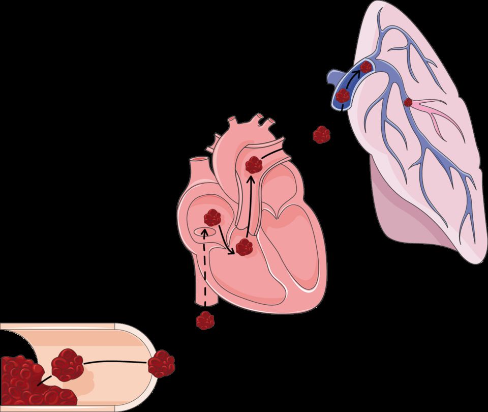
A PE is a clot that lodges in a pulmonary artery and blocks blood flow to part of the lung. Most of the time the clot did not form in the lung at all. It usually starts as a deep vein thrombosis (DVT) in a leg or pelvic vein, then a piece breaks loose. Once free, that traveling clot is called an embolus. It rides the venous blood up to the right heart, gets pumped into the pulmonary circulation, and wedges wherever the vessel becomes too narrow to pass. The lung tissue downstream still gets air, but no blood arrives to collect the oxygen.
Why the clot forms: Virchow's triad
Clots do not appear at random. Back in the 1800s, Rudolf Virchow described three conditions that set the stage, and nurses still use his triad today to spot at-risk patients:
- Venous stasis (blood pooling and sitting still). Long flights, bed rest, surgery, immobility, and heart failure all slow the flow so clotting factors accumulate instead of washing away.
- Hypercoagulability (blood that clots too easily). Pregnancy, estrogen-containing contraceptives, cancer, dehydration, and inherited clotting disorders tip the balance toward clotting.
- Endothelial injury (damage to the vessel's inner lining). Trauma, surgery, central lines, and inflammation expose the surfaces that trigger clot formation.
When two or three of these overlap, a DVT becomes far more likely, and a DVT is the seed of most PEs.
Dead space: ventilation without perfusion
Here is the core problem in one phrase: ventilation without perfusion. The blocked region of lung is still being filled with air, but no blood flows past it, so that air is wasted. Physiologists call this wasted, air-but-no-blood region alveolar dead space. Gas exchange simply cannot happen there. Meanwhile, blood that should have gone to that region is shoved into the remaining open vessels, and the mismatch drops the oxygen level in the blood. The result is hypoxemia (low blood oxygen), which is why a PE is a true medical emergency: a large clot can crash oxygen levels and strain the right heart within minutes.
Why the signs happen
PE symptoms come on suddenly, and each one traces straight back to the mechanism:
- Sudden dyspnea (breathlessness) and hypoxia because a chunk of lung can no longer oxygenate blood, so the body is starved of oxygen and screams for more air.
- Pleuritic chest pain (sharp pain that worsens with a deep breath) because the oxygen-starved lung tissue and its lining, the pleura, become irritated and inflamed.
- Tachycardia (fast heart rate) because the heart speeds up to push blood past the blockage and compensate for falling oxygen. You may also see tachypnea, anxiety, and a cough.
| Feature | Normal lung unit | Pulmonary embolism |
|---|---|---|
| Air in alveoli | Yes | Yes (still ventilated) |
| Blood flow past alveoli | Yes | No (clot blocks perfusion) |
| Gas exchange | Efficient | Fails in the blocked zone |
| Result | Normal oxygen | Dead space, hypoxemia |
Explore the figure below to see a clot lodged in a pulmonary artery and how it cuts off blood flow to the lung beyond it.
- A PE is usually a leg DVT that broke loose, traveled as an embolus, and lodged in a pulmonary artery, blocking blood flow to part of the lung.
- Virchow's triad (stasis, hypercoagulability, endothelial injury) sets up the clot in the first place, so prevention targets these three.
- The blocked lung is ventilated but not perfused, creating alveolar dead space (ventilation without perfusion) and dropping blood oxygen.
- Classic signs are sudden dyspnea, pleuritic chest pain, tachycardia, and hypoxia, each explained by lost gas exchange and the body's compensation.
- PE is a medical emergency; a swollen, tender calf is an early clue that a DVT could embolize.
Section 5Pneumonia & respiratory infections
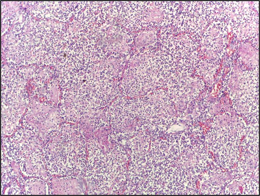
Every time you take a breath, air travels down to about 300 million tiny air sacs called alveoli. These sacs are wrapped in a fine mesh of capillaries, and their walls are astonishingly thin, roughly one cell thick, so oxygen can slip across into the blood while carbon dioxide slips out. This trade is called gas exchange, and it depends on those air sacs staying open, dry, and full of air. Pneumonia is what happens when that clean, air-filled space becomes a battleground.
What goes wrong: the alveoli flood
Pneumonia is an infection of the lung tissue itself, most often caused by bacteria (classically Streptococcus pneumoniae), but also viruses and fungi. When a pathogen reaches the alveoli, the immune system rushes in. White blood cells, fluid, and cellular debris pour into the air sacs, creating pus and inflammatory exudate. The problem is that a sac full of fluid and pus can no longer hold air. Oxygen cannot cross a wall of liquid nearly as well as it crosses air, so gas exchange breaks down. This is why the hallmark of pneumonia is hypoxemia (low blood oxygen) and shortness of breath, or dyspnea. When enough neighboring alveoli fill and clump into a solid, airless region, we call it consolidation, and it shows up as a white patch on a chest X-ray.
The classic signs, and the mechanism behind each
Pneumonia tends to announce itself with a recognizable cluster:
- Fever and chills are the body's response to infection. Chemical signals called pyrogens reset the brain's thermostat upward to make the environment hostile to microbes.
- Productive cough brings up thick, often yellow, green, or rust-colored sputum, which is the pus and debris being cleared from flooded airways.
- Tachypnea and tachycardia (fast breathing and heart rate) are compensations. The body speeds up to move more air and pump oxygen-poor blood faster.
- Pleuritic chest pain, a sharp pain that worsens with a deep breath, appears when inflammation reaches the lining of the lung.
On imaging, that patch of consolidation confirms the flooded region. In older or immunocompromised patients, though, the picture is often quieter, and confusion or simply "not acting right" may be the only early sign.
Where you caught it matters
Nursing care changes depending on where the infection was acquired, because the likely organisms and their drug resistance differ.
| Feature | Community-acquired (CAP) | Hospital-acquired (HAP / VAP) |
|---|---|---|
| Where acquired | Out in daily life; onset before or within 48 hrs of admission | 48+ hrs after admission; VAP is a subtype tied to a ventilator |
| Common organisms | S. pneumoniae, respiratory viruses, atypicals | Often Pseudomonas, Staph aureus including MRSA |
| Drug resistance | Usually lower | Often higher, harder to treat |
| Typical severity | Ranges mild to severe | Frequently sicker, higher mortality |
Ventilator-associated pneumonia (VAP) deserves special note: a breathing tube bypasses the natural filtering and cough defenses of the upper airway, giving bacteria a direct highway into the lungs. This is why nurses follow a "VAP bundle," including keeping the head of the bed elevated and providing regular oral care.
Who is most at risk
Anyone whose defenses are weakened is more vulnerable. This includes the very young and adults over 65, smokers, and people with chronic lung disease such as COPD. It also includes patients who are immunocompromised, those with difficulty swallowing (who may aspirate food or secretions into the lungs), and anyone whose cough reflex is blunted by sedation or a decreased level of consciousness.
A word on tuberculosis
Tuberculosis (TB) is a distinct, airborne infection from Mycobacterium tuberculosis in which the immune system walls the bacteria inside a granuloma; it can stay dormant for years as latent TB (no symptoms, not contagious) before reactivating into active TB with cough, weight loss, and night sweats. Explore the interactive figure below to see how infection fills the alveoli.
- Pneumonia fills the alveoli with fluid and pus, so oxygen cannot cross into the blood, producing hypoxemia, dyspnea, and consolidation on imaging.
- Classic signs are fever, productive cough, crackles from fluid-filled sacs re-opening, and pleuritic chest pain; older adults may only show confusion.
- Community-acquired pneumonia (CAP) is usually less resistant; hospital-acquired (HAP) and ventilator-associated (VAP) pneumonia involve tougher organisms like Pseudomonas and MRSA.
- Highest risk: the very young, adults over 65, smokers, COPD, immunocompromised, and those who aspirate; vaccines and deep breathing help prevent it.
- TB is a separate airborne infection walled into granulomas, existing as latent (dormant, non-contagious) or active disease.
Section 6Lung sounds & assessment
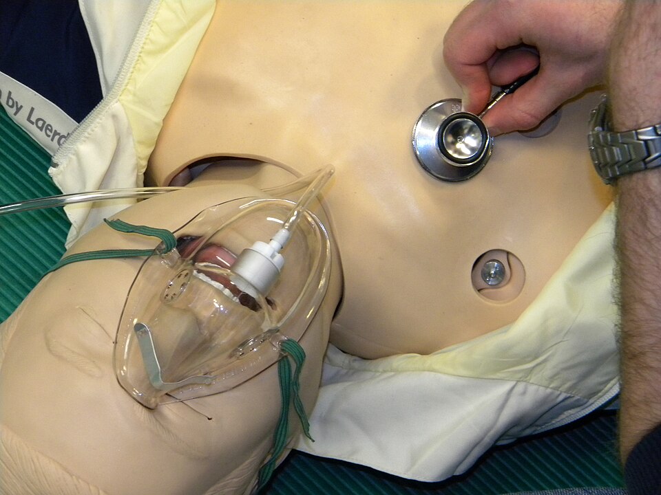
Every time you breathe, air rushes down your windpipe, splits into smaller and smaller tubes, and finally reaches millions of tiny air sacs called alveoli. When those airways are open and the air sacs are dry and elastic, air moves smoothly and quietly. That smooth flow is exactly what a stethoscope is listening for. When you place your stethoscope on someone's chest, you are eavesdropping on how easily air is getting in and out, and whether anything, fluid, mucus, swelling, or scarring, is getting in the way.
Normal breath sounds: the quiet baseline
Over most of the lung fields you should hear vesicular breath sounds, a soft, low, rustling whoosh that is louder on inspiration and fades quickly on expiration. Over the large central airways (the trachea and around the sternum) sounds are naturally louder and more tubular (bronchial and bronchovesicular). The key idea for students: normal air movement is clear and quiet. Anything extra you hear layered on top is called an adventitious (added) sound, and each type tells a different mechanical story about what is happening inside the airways.
Crackles: air popping open through fluid
Crackles (older term: rales) are brief, discontinuous popping or bubbling sounds, often compared to Velcro pulling apart or hair rubbed between your fingers. Mechanistically, they happen when air suddenly pushes open small airways and alveoli that were stuck shut or filled with fluid. You hear them most on inspiration because that is when the collapsed spaces snap open. Crackles usually point toward fluid or collapse in the air sacs, classically pneumonia (inflammatory exudate) or heart failure (fluid backing up into the lungs, called pulmonary edema). In heart failure they often start at the lung bases, where gravity pools fluid.
Wheezes: air squeezing through narrowed tubes
Wheezes are high-pitched, musical, whistling sounds, usually loudest on expiration. Picture blowing through a nearly pinched straw. They happen when airways are narrowed by bronchospasm, swelling (edema), or mucus, so air is forced through a tight channel and the walls vibrate. Wheezes are the signature of asthma and COPD. One important safety note: a wheeze that suddenly goes silent in someone struggling to breathe is ominous, it can mean airflow has dropped so low that no sound is being made (a "silent chest"), which is an emergency, not an improvement.
Stridor: the upper-airway alarm bell
Stridor is a harsh, high-pitched crowing sound heard without a stethoscope, typically on inspiration. It comes from obstruction high up in the upper airway, the larynx or trachea, from swelling (croup, epiglottitis, anaphylaxis) or a foreign body. Treat stridor as an airway emergency: the tube that keeps a person alive is narrowing, and it can close.
Pleural friction rub: surfaces that should glide, grating instead
The lungs are wrapped in two thin membranes (the pleura) that normally slide silently past each other. When they become inflamed (pleuritis, often from infection), they grate together, producing a pleural friction rub, a low-pitched, grating, leathery sound heard on both inspiration and expiration, often with pain on breathing.
| Feature | Crackles | Wheezes |
|---|---|---|
| Sound | Popping / bubbling | Musical / whistling |
| Cause | Fluid or collapsed air sacs | Narrowed airways |
| Timing | Mainly inspiration | Mainly expiration |
| Points toward | Pneumonia, heart failure | Asthma, COPD |
Pulse oximetry: seeing the result of all this airflow
Listening tells you how air is moving; the pulse oximeter (SpO2) tells you the result, how well oxygen is actually reaching the blood. It clips on a finger and estimates the percent of hemoglobin carrying oxygen, usually 95 to 100% in healthy adults. When crackles, wheezes, or obstruction block oxygen from reaching the alveoli, the SpO2 often falls (hypoxemia).
Tie this back to the arterial blood gas (ABG) from Week 1. The pulse ox is a quick, noninvasive estimate; the ABG is the precise lab measurement, giving true PaO2, PaCO2, and pH. Remember the oxyhemoglobin dissociation curve: because of its S-shape, an SpO2 that drops below roughly 90% often signals a much larger fall in PaO2 than the number suggests. Trust the ABG for the full picture, especially for carbon dioxide, which the pulse ox cannot see at all.
Explore the interactive figure below to hear how normal breath sounds differ from crackles, wheezes, stridor, and a friction rub.
- Normal breath sounds are clear and quiet; any added (adventitious) sound signals something interfering with airflow.
- Crackles = fluid or reopening air sacs (pneumonia, heart failure), mostly on inspiration; wheezes = narrowed airways (asthma, COPD), mostly on expiration.
- Stridor is a harsh inspiratory sound from upper-airway obstruction, treat it as an emergency; a pleural friction rub is grating and painful from inflamed pleura.
- Pulse oximetry (SpO2, usually 95 to 100%) estimates oxygen saturation noninvasively but cannot measure CO2, reads unreliably or falsely low in a cold or poorly perfused finger, and reads falsely high in carbon monoxide poisoning.
- The ABG confirms true oxygenation, ventilation, and pH; always weigh the number against how the patient actually looks.
The chapter in one breath
- Ventilation is air moving in and out of alveoli; when it drops (hypoventilation), CO2 backs up and the pH falls, giving respiratory acidosis.
- Perfusion is blood flow through the pulmonary capillaries, and gas exchange only works where ventilated alveoli meet perfused capillaries.
- A normal V/Q ratio is about 0.8; a blocked airway (pneumonia, mucus) creates a low-V/Q shunt, while a blocked vessel (PE) creates a high-V/Q dead space.
- COPD is chronic airflow limitation from emphysema plus chronic bronchitis, where air-trapping and a barrel chest cause a prolonged expiratory phase and chronically retained CO2.
- Asthma is reversible bronchospasm with airway inflammation and mucus, producing expiratory wheezing; a silent chest with a rising CO2 signals dangerous fatigue, not improvement.
- Pulmonary embolism is a clot (often from a DVT) lodging in the pulmonary artery, causing sudden dyspnea, pleuritic chest pain, tachycardia, and hypoxemia with a low CO2 from hyperventilating.
- Pneumonia fills alveoli with inflammatory exudate, so you see fever, productive cough, crackles, and consolidation that impairs oxygenation.
- Adventitious lung sounds localize the problem: crackles mean fluid or reopening alveoli, wheezes mean narrowed airways, and stridor means a dangerous upper-airway obstruction.
ReferenceWords worth knowing
A quick reference for the terms in this chapter — skim it now, or circle back whenever a word trips you up.
- Bronchospasm
- Sudden tightening of the smooth muscle wrapping the bronchi and bronchioles that narrows the airways, making it harder to move air (especially out) and often causing wheezing.
- Chronic bronchitis
- A form of COPD defined clinically as a productive cough lasting at least 3 months in each of 2 consecutive years, caused by chronic airway inflammation, mucus-gland hypertrophy, and excess sputum.
- Consolidation
- Lung tissue that has filled with fluid, pus, blood, or inflammatory cells instead of air (as in pneumonia), making that region solid and impairing gas exchange.
- COPD (chronic obstructive pulmonary disease)
- A progressive, largely irreversible airflow-limiting disease—usually from smoking—that blends emphysema and chronic bronchitis and traps air on exhalation.
- Crackles
- Brief, discontinuous popping or bubbling sounds heard (usually on inspiration) when collapsed or fluid-filled alveoli and small airways suddenly snap open, often signaling fluid or infection.
- Dead space
- Air that is inhaled but does not take part in gas exchange—the conducting airways (anatomic) plus alveoli that are ventilated but not perfused (alveolar); added together the two make up physiologic, or total, dead space.
- Emphysema
- A form of COPD in which the alveolar walls are destroyed and enlarged, reducing surface area for gas exchange and the elastic recoil that normally pushes air out.
- Hypoxemia
- A lower-than-normal level of oxygen in the arterial blood (usually a PaO2 below about 80 mmHg or SaO2 under about 95%), which starves tissues of oxygen if uncorrected.
- Perfusion
- The flow of blood through the pulmonary capillaries that surround the alveoli, releasing carbon dioxide to be exhaled and picking up oxygen from the alveolar air.
- Pulmonary embolism
- A blockage of a pulmonary artery—most often by a clot that traveled from a deep vein—that suddenly cuts off blood flow to part of the lung, creating dead space and often causing sharp dyspnea and hypoxemia.
- Shunt
- Blood that passes through the lungs without being oxygenated because it flows past alveoli that are perfused but not ventilated (as in consolidation or collapse), a cause of hypoxemia that oxygen alone often cannot fix.
- Status asthmaticus
- A severe, prolonged asthma attack that does not respond to usual bronchodilators, a medical emergency that can progress to respiratory failure.
- Stridor
- A harsh, high-pitched sound heard mainly on inspiration that signals narrowing or obstruction of the upper airway (larynx or trachea) and warrants urgent attention.
- Ventilation
- The mechanical movement of air into and out of the lungs, bringing oxygen to the alveoli and carrying carbon dioxide away.
- V/Q ratio
- The balance between ventilation (V, airflow) and perfusion (Q, blood flow) in a lung region, normally about 0.8; mismatches in either direction impair gas exchange.
- Wheeze
- A continuous, musical, usually high-pitched whistling sound—often loudest on expiration—produced when air is forced through narrowed lower airways, as in asthma or COPD.
DrillFlashcards for this unit
These are the Week 10 cards — the same deck as the hub, pulled in here so you can review the moment you finish reading. Tap the card to flip it; hit Shuffle to mix them up. To grade yourself and track mastery across the whole deck, open the full flashcards page.