Oncology: How Cancer Works
Cancer is not one disease but a process — a single cell that stops obeying the body’s rules and starts dividing on its own. This chapter follows that process from the first mutation to a spreading tumor: how a normal cell turns malignant, how we tell benign from dangerous, how cancer travels, what sets it off, and how it is graded, staged, and treated.
By the end of this chapter you can
- Explain how a normal cell becomes cancer through mutated oncogenes and tumor-suppressor genes.
- Contrast benign and malignant tumors, and use the -oma / carcinoma / sarcoma naming.
- Describe how cancer invades, and the routes and common sites of metastasis.
- Name major carcinogens and the roles of p53 and BRCA.
- Tell grading from staging (TNM), and recognize tumor lysis syndrome.
Section 1From a normal cell to cancer
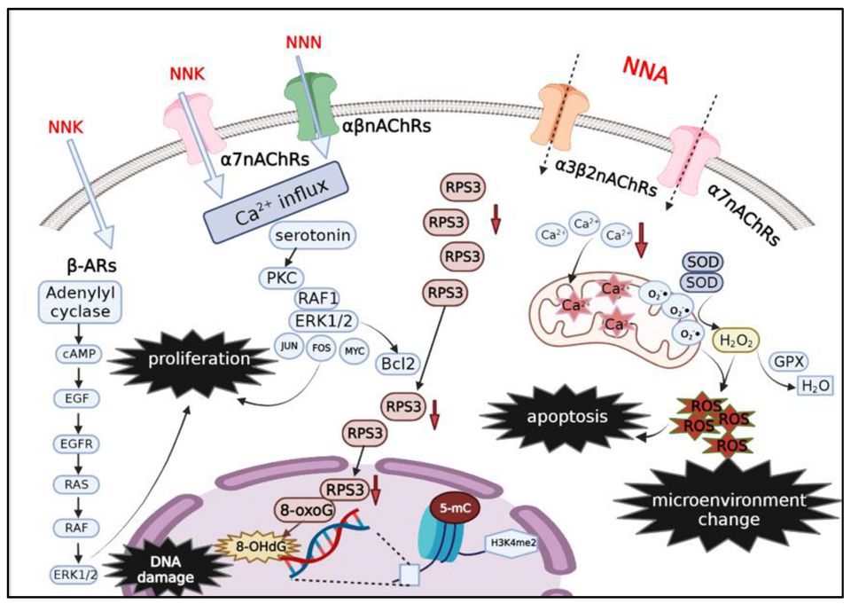
Every cell in your body lives by a simple rule: divide only when you are told to, and stop when the job is done. A healthy cell reads chemical signals from its neighbors, copies its DNA carefully, checks that copy for errors, and either keeps going or quietly retires. That balance between growing and stopping is controlled by genes. When those control genes get damaged, the balance tips, and a normal cell can slowly turn into a cancer cell. Cancer is not an invader from outside. It is one of your own cells that has lost its instructions.
The accelerator and the brakes
Think of cell division like driving a car. Two sets of genes keep it under control. Proto-oncogenes are the accelerator: they normally tell the cell when it is time to grow and divide. Tumor-suppressor genes are the brakes: they slow division, fix damaged DNA, or order a broken cell to self-destruct. In a normal cell these work together, so growth happens only when it should.
Mutations break this system in two different ways. When a proto-oncogene mutates, it becomes an oncogene a stuck accelerator that screams "grow!" even with no signal. This is a gain of function, and usually only one copy of the gene needs to change. When a tumor-suppressor gene mutates, the brakes fail. This is a loss of function, and typically both copies must be knocked out before protection is lost. The famous tumor suppressor p53 (often called the "guardian of the genome") normally halts a damaged cell and triggers apoptosis; when p53 fails, damaged cells survive and keep dividing.
The multistep journey: how tissue changes over time
Cancer does not appear overnight. Tissue drifts through recognizable stages as mutations accumulate:
- Hyperplasia too many cells, but they still look normal and stay organized.
- Dysplasia cells start looking abnormal disordered shapes, sizes, and arrangement. This is pre-cancerous but still reversible.
- Carcinoma in situ cells look fully cancerous but stay contained above the basement membrane. Nothing has spread yet, so cure rates are high.
- Invasive cancer cells break through the basement membrane into surrounding tissue and can travel to distant sites (metastasis).
What makes a cancer cell dangerous: the hallmarks
Researchers describe cancer through shared "hallmark" behaviors. In plain terms, cancer cells learn to:
- Divide without permission the stuck-accelerator, failed-brakes problem, so they multiply even when the body signals stop.
- Evade apoptosis normal cells with broken DNA are told to die (programmed cell death). Cancer cells silence that signal often by disabling p53 so damaged cells refuse to die.
- Grow their own blood supply a process called angiogenesis. Once a tumor grows past roughly 1 to 2 mm, it starves for oxygen, so it releases signals that recruit new blood vessels to feed itself and, later, to give cancer cells a highway to spread.
Tumor is not the same as cancer
Students mix these up constantly. A tumor (or neoplasm) is simply a lump an abnormal mass of cells. Cancer means the tumor is malignant: it invades and can spread. A benign tumor, like a lipoma, is a lump that stays put.
| Feature | Benign tumor | Malignant tumor (cancer) |
|---|---|---|
| Growth | Usually slow | Often rapid |
| Border | Well-defined, often encapsulated | Irregular, invades nearby tissue |
| Cell appearance | Looks like normal tissue (well-differentiated) | Abnormal, disorganized (poorly differentiated) |
| Spread (metastasis) | No | Yes via blood or lymph |
| Danger | Usually harmless unless pressing on something | Life-threatening if untreated |
Explore the figure below to watch a cell move from normal, through adaptation, into dysplasia the same stepwise drift described here.
- Proto-oncogenes are the accelerator and tumor-suppressor genes are the brakes; cancer needs a stuck accelerator (oncogene, one hit) and failed brakes (tumor suppressor, usually two hits).
- Cancer develops in steps: hyperplasia to dysplasia to carcinoma in situ to invasive cancer, as mutations pile up over time.
- Hallmark behaviors: uncontrolled division, evading apoptosis (often by disabling p53), and angiogenesis growing its own blood supply.
- A tumor is any lump; only a malignant tumor is cancer, defined by its ability to invade and metastasize.
- Invasion is marked by breaking through the basement membrane the line between contained and spreading disease.
Section 2Benign vs malignant
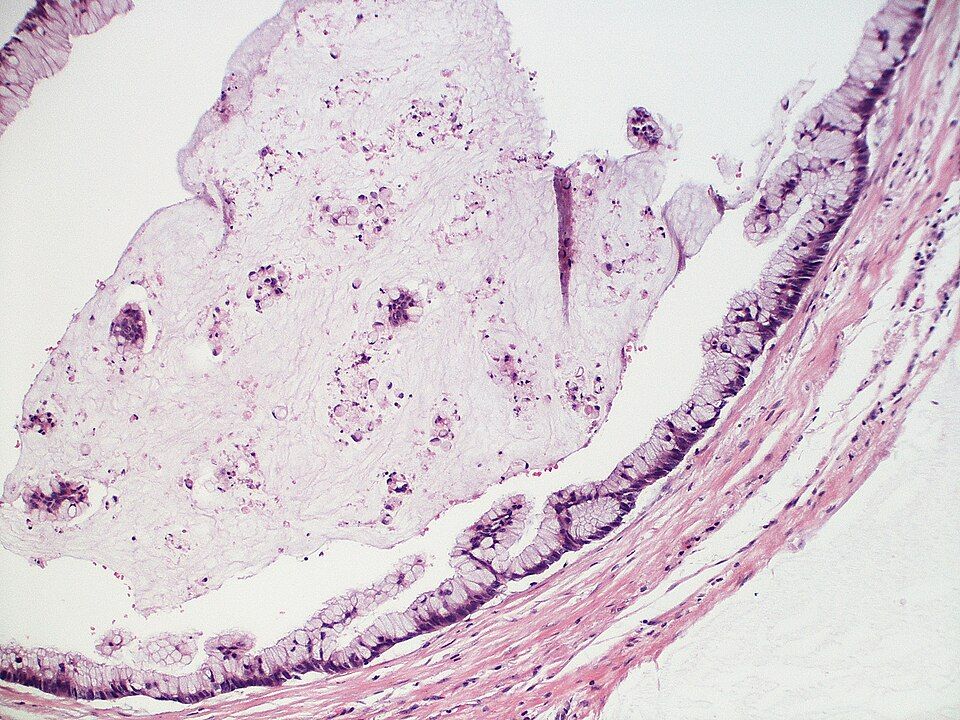
Your body makes new cells constantly. Skin, gut lining, blood cells, and more are always being replaced, and normally this happens under tight control: cells divide only when signaled, mature into a specific job, and stop dividing when the tissue is repaired. This maturing process is called differentiation. A well-differentiated cell looks and acts like the tissue it came from. A neoplasm (literally "new growth") is a lump of cells that has escaped normal growth controls and keeps dividing when it should not. The big clinical question is always the same: is this growth benign or malignant?
Benign growths: slow, walled-off, and well-behaved
A benign tumor grows slowly and stays put. Because it grows gradually, the surrounding tissue often has time to form a fibrous capsule around it, so the mass is encapsulated and can usually be shelled out cleanly by a surgeon. Its cells are well-differentiated, meaning they still closely resemble the normal tissue of origin and often still do that tissue's job. Benign tumors do not invade nearby structures and do not metastasize (spread to distant sites). Under the microscope the cells look orderly, with normal-sized nuclei and few dividing cells (low mitotic rate).
Malignant growths: fast, invasive, and mobile
Malignant tumors (true cancer) behave the opposite way. They grow quickly, so they rarely form a tidy capsule; instead they send finger-like projections that invade and destroy surrounding tissue, which is why the cut surface can look crab-like (the origin of the word "cancer"). Their cells are poorly differentiated or undifferentiated, meaning they no longer look like or act like the parent tissue. Most importantly, malignant cells can break away, travel through blood or lymph, and seed new tumors elsewhere. That spread is metastasis, and it is the feature that makes cancer so dangerous and so hard to cure.
| Feature | Benign | Malignant |
|---|---|---|
| Growth rate | Slow | Rapid, often erratic |
| Border | Encapsulated, well-defined | Invasive, poorly defined |
| Differentiation | Well-differentiated | Poorly differentiated to anaplastic |
| Metastasis | Never | Common |
| Recurrence after removal | Rare | Common |
How tumors are named
Naming follows a pattern worth memorizing. The suffix -oma usually signals a benign tumor named for its tissue of origin: a lipoma is a benign fat tumor, an adenoma a benign glandular tumor, a leiomyoma a benign smooth-muscle tumor (a uterine "fibroid"). Malignant tumors get one of two labels based on their tissue of origin: carcinomas from epithelial tissue, sarcomas from connective (mesenchymal) tissue. A carcinoma arises from epithelial tissue (skin, gland, and organ linings) and is by far the most common category, including adenocarcinoma of the colon or breast. A sarcoma arises from connective tissue such as bone (osteosarcoma), cartilage, muscle, or fat. A few dangerous "-oma" names break the rule, notably melanoma and lymphoma, which are malignant despite the friendly suffix.
Differentiation and anaplasia
Pathologists describe cancers by how far the cells have drifted from normal. Grading measures differentiation. A well-differentiated (low-grade) cancer still resembles its origin and usually behaves less aggressively. Anaplasia is the extreme opposite: cells so primitive and disorganized that they barely resemble any tissue. Anaplastic cells show pleomorphism (variable size and shape), large dark nuclei with a high nucleus-to-cytoplasm ratio, and abundant, sometimes abnormal, mitoses. In general, the less differentiated the cell, the more aggressive the tumor and the worse the prognosis.
When "benign" still hurts you
Benign does not always mean harmless. A tumor that would be trivial elsewhere can be life-threatening because of where it sits. A benign meningioma in the skull has nowhere to expand; as it grows it raises intracranial pressure and compresses brain tissue, causing headaches, seizures, or neurologic deficits. A benign adenoma can secrete hormones (for example, insulin from a pancreatic tumor, causing dangerous hypoglycemia), and a benign mass can obstruct a hollow organ or press on a vital vessel. Location, secretion, and compression, not just the label, drive the risk.
- Benign tumors are slow, encapsulated, well-differentiated, and stay put; malignant tumors are fast, invasive, poorly differentiated, and metastasize.
- Metastasis is the defining danger of malignancy and the main reason cancer is hard to cure.
- Naming: -oma usually means benign, carcinoma means malignant epithelial, sarcoma means malignant connective tissue; watch the exceptions melanoma and lymphoma.
- Differentiation drives grade; anaplasia (primitive, pleomorphic, high mitotic cells) signals the most aggressive tumors.
- A benign tumor can still be deadly because of its location, hormone secretion, or compression, classically a meningioma raising intracranial pressure.
Section 3How cancer grows and spreads
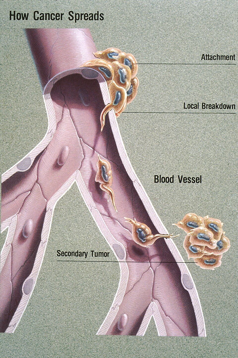
Normally, your tissues are beautifully organized. Cells stay in their own neighborhood, held in place by a thin, tough sheet called the basement membrane that separates the surface layer (epithelium) from the deeper connective tissue below. Cells also respect their neighbors: they stop dividing when they touch (contact inhibition), they anchor down where they belong, and they call for new blood vessels only when the body genuinely needs them. Cancer is what happens when a cell breaks every one of these rules at once, and understanding how it grows and travels is really a story about a cell that has forgotten how to stay put. Explore it below as we walk through each step.
Local invasion: breaking through the basement membrane
A tumor usually starts as a well-behaved lump that stays on its own side of the basement membrane. As long as it has not crossed that barrier, we call it carcinoma in situ ("in place") - and this is often curable, because it has not gone anywhere yet. The dangerous turning point is local invasion, when cancer cells secrete enzymes called matrix metalloproteinases (MMPs) that literally digest the basement membrane and the surrounding extracellular matrix. Once that fence is dissolved, the cells lose their normal stickiness (they down-regulate E-cadherin, the "glue" protein) and crawl into the deeper tissue. This is the moment a cancer becomes invasive, and it is why pathologists care so much about whether the basement membrane is intact.
Metastasis: how cancer travels to distant sites
Metastasis is the spread of cancer from the original (primary) tumor to a distant site. It is the single biggest reason cancer becomes life-threatening. There are three main routes.
- Lymphatic spread - cells enter lymphatic vessels and travel to nearby lymph nodes first. This is the classic route for carcinomas (epithelial cancers like breast and colon). It is why nurses palpate lymph nodes and why the sentinel lymph node - the first node draining a tumor - is biopsied to check whether spread has begun.
- Hematogenous spread - cells invade blood vessels and ride the bloodstream to far-off organs. This is the favored route for sarcomas (connective-tissue cancers) and explains distant organ metastases.
- Seeding of body cavities - cells shed off the tumor surface and scatter across a cavity lining, like peritoneal seeding in ovarian cancer. This can cause malignant ascites (fluid buildup) or pleural effusions.
| Feature | Lymphatic spread | Hematogenous spread |
|---|---|---|
| Typical cancer type | Carcinomas (epithelial) | Sarcomas (connective tissue) |
| First stop | Regional lymph nodes | Distant organs |
| Clinical clue | Enlarged, firm nodes | Masses in lung, liver, bone, brain |
| Assessed by | Sentinel node biopsy | CT/PET imaging, bone scan |
Where cancer tends to land
Metastases are not random. Cells often lodge in the first capillary bed they reach, and certain organs also provide fertile "soil" for tumor cells to settle - a concept called the seed-and-soil hypothesis. The four most common landing sites are lung, liver, bone, and brain. The lung and liver catch a large share of circulating cells because they act as biological filters with dense capillary networks (the liver receives portal blood; the lungs filter all venous return). Bone metastases are common in breast and prostate cancer and often cause deep pain, pathologic fractures, and high calcium levels (hypercalcemia). Brain metastases can raise intracranial pressure and cause headaches, seizures, or new neurologic deficits.
Angiogenesis: the tumor builds its own supply lines
A clump of cancer cells can only grow to about 1-2 mm before the cells in the center start starving - they are too far from any blood vessel to get oxygen and nutrients by diffusion. To keep growing, the tumor pulls off a striking trick called angiogenesis: it releases chemical signals, especially vascular endothelial growth factor (VEGF), that coax nearby blood vessels to sprout new branches straight into the tumor. This "angiogenic switch" gives the tumor its own supply line for continued growth and, unfortunately, an on-ramp into the bloodstream for metastasis. These new vessels are usually leaky and disorganized, which contributes to swelling and makes drug delivery unpredictable. Because VEGF is so central, several cancer drugs (anti-angiogenic agents like bevacizumab) work by blocking it to try to starve the tumor.
- Cancer becomes invasive when it uses enzymes (MMPs) to break through the basement membrane; before that, carcinoma in situ is often curable.
- Metastasis spreads through three routes: lymphatic (typical of carcinomas), hematogenous/bloodstream (typical of sarcomas), and seeding of body cavities.
- The most common landing sites are lung, liver, bone, and brain; new bone pain in a cancer patient is a red flag for metastasis.
- Angiogenesis, driven by VEGF, lets a tumor recruit its own blood supply to grow past 1-2 mm and also opens a route for cells to escape and spread.
Section 4What causes cancer
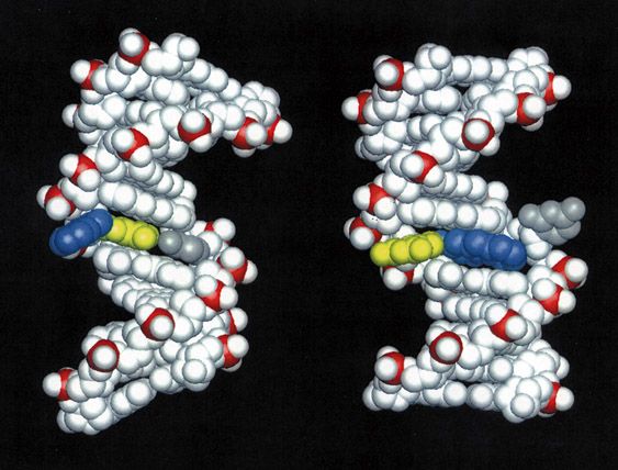
Every day, your body makes billions of new cells. Old cells wear out, get injured, or reach the end of their lifespan, so a healthy cell divides to make a fresh copy of itself. This is normally a tightly controlled process. Before a cell divides, it copies its entire DNA, then pauses at built-in checkpoints to proofread that copy. If the DNA looks damaged, the cell either fixes it or, if the damage is too severe, deliberately kills itself through a tidy self-destruct program called apoptosis. Cancer begins when this careful system breaks down and a cell starts dividing on its own schedule, ignoring the stop signals, and refusing to die.
So the real question is: what damages the DNA and the control genes badly enough to let a cell go rogue? The answer is usually a slow buildup of mutations (permanent changes in the DNA sequence) hitting the specific genes that run the cell cycle.
Carcinogens: the things that damage DNA
A carcinogen is any agent that causes cancer, usually by damaging DNA directly or by forcing cells to divide so fast that copy errors pile up. They fall into a few broad groups:
- Chemical — the classic example is tobacco smoke, which carries dozens of DNA-damaging compounds. Others include asbestos, benzene, and certain dyes. Many act at the point of contact (asbestos in the lungs, dyes in the bladder), but some, like benzene, are absorbed and cause blood cancers such as leukemia.
- Radiation — ultraviolet (UV) light from the sun damages skin-cell DNA and drives melanoma and other skin cancers. Ionizing radiation (X-rays, radon) does the same internally.
- Viral and microbial — some infections insert their own genes or cause chronic irritation. HPV (cervical and oropharyngeal cancers), EBV (some lymphomas), hepatitis B (liver cancer), and the bacterium H. pylori (stomach cancer) are the ones to know.
Oncogenes and tumor-suppressor genes
Two families of genes normally keep cell division in check, and cancer usually involves damage to both. Think of driving a car: you need a working gas pedal and working brakes.
Proto-oncogenes are the gas pedal. They tell a cell when it is time to grow and divide. When a proto-oncogene mutates and gets stuck in the "on" position, it becomes an oncogene that floors the accelerator, driving nonstop division.
Tumor-suppressor genes are the brakes. They slow division, run the DNA proofreading checkpoints, and trigger apoptosis when a cell is too damaged. The most famous is p53, nicknamed the guardian of the genome because it halts the cell cycle to allow repair, and orders apoptosis when repair fails. When p53 is knocked out, damaged cells sail through checkpoints and accumulate even more mutations. The BRCA1 and BRCA2 genes are also tumor suppressors; they help repair broken DNA, so when they fail, breast and ovarian tissue lose a key safety net.
| Feature | Oncogene | Tumor-suppressor gene |
|---|---|---|
| Normal job | Promotes cell growth (gas pedal) | Restrains growth, repairs DNA, triggers apoptosis (brakes) |
| Effect of mutation | Gain of function - stuck ON | Loss of function - brakes fail |
| Copies needed to cause trouble | Usually one mutated copy is enough | Usually both copies must be lost |
| Examples | Mutated RAS, HER2 | p53, BRCA1/2, RB |
Inherited vs acquired mutations, and why some cancers run in families
Most cancers come from acquired (somatic) mutations - damage that builds up in one person's cells over a lifetime from carcinogens, random copying errors, and aging. These are not passed to children, which is why the majority of cancers are sporadic.
A smaller group involves inherited (germline) mutations, present in every cell from birth because they came from a parent's egg or sperm. Someone who inherits one broken copy of a tumor-suppressor gene like BRCA1 only needs to lose the second copy for that safety net to fail - they are already halfway there. That head start is why these cancers appear earlier in life, often in both breasts or in multiple family members. This is the mechanism behind familial patterns and the basis for genetic counseling.
Chronic inflammation and failed immune surveillance
Two background factors quietly raise the odds. First, chronic inflammation (as in long-standing hepatitis, H. pylori gastritis, or inflammatory bowel disease) keeps tissue in a constant cycle of injury and repair. Every extra round of division is another chance for a mutation to slip in. Second, your immune system normally patrols for abnormal cells and destroys them - a process called immune surveillance. Cancer cells that learn to hide from or exhaust these immune patrols escape destruction, which is why immunosuppressed patients often have higher cancer rates.
- Cancer starts when DNA damage disables the normal controls on cell division, letting a cell divide without limit and dodge apoptosis.
- Carcinogens - chemical (tobacco), radiation (UV), and viral/microbial (HPV, EBV, hepatitis B, H. pylori) - cause cancer largely by damaging DNA; many are preventable through vaccines and lifestyle.
- Oncogenes are a stuck gas pedal (gain of function); tumor suppressors like p53 (guardian of the genome) and BRCA1/2 are failed brakes (loss of function).
- Most mutations are acquired over a lifetime; inherited germline mutations give a head start toward cancer, explaining earlier onset and familial clustering.
- Chronic inflammation and failed immune surveillance are contributors that let mutated cells accumulate and escape destruction.
Section 5Grading, staging & living with cancer
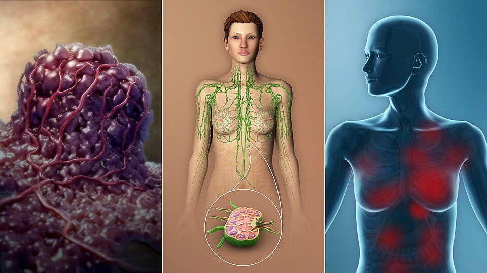
Your cells normally follow strict rules. They divide only when told to, they mature into a specific job (a skin cell, a colon cell), and they stay in their own neighborhood. Cancer breaks all three rules: the cells divide without permission, they fail to mature, and they invade and travel. Once a tumor exists, two of the first questions a care team asks are how ugly do these cells look under the microscope? and how far has this spread? Those are two different questions, and mixing them up is one of the most common student errors.
Grading vs staging
Grading is about appearance. A pathologist looks at the tumor cells and asks how much they still resemble the normal tissue they came from. Well-differentiated (low-grade) cells still look and behave a lot like the parent tissue and tend to grow slowly. Poorly differentiated or anaplastic (high-grade) cells look wild and disorganized and usually behave more aggressively. Grading is usually reported as G1 through G4.
Staging is about spread - how big the tumor is and how far it has traveled. The most widely used framework is the TNM system: T for the size and extent of the primary tumor, N for how many regional lymph nodes are involved, and M for distant metastasis (spread to far organs). Higher numbers mean more advanced disease, and staging drives treatment decisions and prognosis more powerfully than grade does.
| Feature | Grading | Staging |
|---|---|---|
| Question it answers | How abnormal do the cells look? | How far has it spread? |
| How it is measured | Differentiation under the microscope | TNM: Tumor, Nodes, Metastasis |
| Typical scale | G1 (well-differentiated) to G4 (anaplastic) | Stage I to Stage IV |
| Main use | Predicts aggressiveness | Guides treatment and overall prognosis |
The CAUTION warning signs
The American Cancer Society mnemonic CAUTION lists classic early clues that should prompt evaluation. They matter because early detection usually means a lower stage and better outcomes:
- C - Change in bowel or bladder habits
- A - A sore that does not heal
- U - Unusual bleeding or discharge
- T - Thickening or a lump (breast or elsewhere)
- I - Indigestion or difficulty swallowing
- O - Obvious change in a wart or mole
- N - Nagging cough or hoarseness
When the tumor causes trouble from a distance
Paraneoplastic syndromes happen when a tumor secretes hormones or hormone-like substances, causing symptoms far from the tumor itself - classic examples are SIADH (small cell lung cancer making ADH, driving down sodium) and hypercalcemia (from PTH-related peptide or bone breakdown).
Tumor lysis syndrome (TLS) is an oncologic emergency. When large numbers of cancer cells are destroyed quickly - often right after chemotherapy starts - they dump their contents into the blood. The result is high potassium (hyperkalemia), high phosphate, high uric acid, and low calcium, which can trigger fatal arrhythmias and acute kidney injury. Nurses watch labs closely and push hydration.
The four treatment pillars
Most cancer care combines four approaches. Surgery removes the tumor. Radiation damages the DNA of cells in a targeted area. Chemotherapy uses systemic drugs to kill dividing cells throughout the body. Immunotherapy retrains or unleashes the patient's own immune system to recognize and attack cancer.
Here is the key mechanism behind chemo's side effects: most chemotherapy targets rapidly dividing cells, because that is what cancer does. But some healthy tissues also divide fast, so they take collateral damage. That is exactly why chemo predictably causes hair loss (alopecia) from fast-dividing hair follicles, nausea, vomiting, and mouth sores (mucositis) from the fast-turnover gut lining, and bone marrow suppression (myelosuppression) - dropping white cells, red cells, and platelets. The low white count (neutropenia) is the scary one, because it leaves the patient wide open to infection.
- Grade describes how abnormal cells look (differentiation, G1-G4); stage describes how far cancer has spread using TNM (Tumor, Nodes, Metastasis).
- The CAUTION mnemonic flags early warning signs that warrant evaluation and can catch cancer at a lower, more treatable stage.
- Paraneoplastic syndromes (like SIADH and hypercalcemia) cause distant hormone-driven symptoms; tumor lysis syndrome is an emergency with hyperkalemia, hyperphosphatemia, hyperuricemia, and hypocalcemia.
- The four pillars are surgery, radiation, chemotherapy, and immunotherapy.
- Chemo harms fast-dividing normal cells too, causing alopecia, mucositis/GI upset, and myelosuppression - watch for neutropenic fever.
The chapter in one breath
- Carcinogenesis is a stepwise loss of control: a normal cell mutates until it ignores stop signals, dodges apoptosis, and divides on its own, becoming a clone of rogue cells.
- Cancer arises when proto-oncogenes mutate into oncogenes stuck in the on position (accelerator) and tumor suppressor genes like p53 fail (brakes), so growth and DNA-repair checkpoints stop working.
- Benign tumors are usually slow, encapsulated, well-differentiated, and stay put; malignant tumors grow fast, lack a capsule, look abnormal, and spread.
- Invasion is local spread as cancer cells release enzymes to chew through basement membrane and infiltrate nearby tissue, which is why margins matter in surgery.
- Metastasis is distant spread through blood or lymph to form secondary tumors; common landing sites are lung, liver, bone, and brain, and it signals advanced disease.
- Causes are multifactorial carcinogens and risk factors: tobacco, radiation, UV light, certain chemicals, oncogenic viruses (HPV, hepatitis, EBV), chronic inflammation, and inherited mutations (like BRCA).
- Grading describes how abnormal the cells look under the microscope (differentiation), while staging (often TNM: Tumor size, Nodes, Metastasis) describes how far it has spread and drives prognosis.
- Treatment follows a few core principles: surgery removes local disease, radiation targets a region, and chemotherapy works systemically by hitting rapidly dividing cells, often combined for better cure rates.
- Chemotherapy is not selective, so it also harms fast-turnover normal cells, explaining the classic toxicities: bone marrow suppression, hair loss, nausea, and mucositis.
ReferenceWords worth knowing
A quick reference for the terms in this chapter — skim it now, or circle back whenever a word trips you up.
- Anaplasia
- Anaplasia is the extreme loss of a cell's normal appearance and organization, where cancer cells look so wild and disorganized (with big, irregular nuclei and chaotic mitoses) that you often can't tell what tissue they came from, marking a highly malignant tumor.
- Angiogenesis
- Angiogenesis is the growing of new blood vessels, which a tumor hijacks by releasing signals (like VEGF) to build its own blood supply so it can keep feeding and expanding beyond a couple of millimeters.
- Benign
- A benign tumor is a growth that stays put, grows slowly, is usually well-differentiated and encapsulated, and does not invade nearby tissue or spread to distant sites, though it can still cause harm by pressing on nearby structures.
- Carcinogen
- A carcinogen is any agent that can cause cancer by damaging DNA or promoting abnormal growth, such as tobacco smoke, UV and ionizing radiation, certain chemicals, or oncogenic viruses like HPV.
- Carcinoma
- A carcinoma is a cancer that arises from epithelial cells, meaning the cells that line and cover body surfaces and glands (skin, breast, lung, colon, prostate), and it is the most common category of cancer.
- Carcinoma in situ
- Carcinoma in situ is an early cancer whose malignant cells are still confined to their original layer and have not yet broken through the basement membrane to invade, so it is often curable if caught and removed at this stage.
- Differentiation
- Differentiation is how closely a cell resembles the mature, specialized tissue it came from; well-differentiated cells look and behave almost normally, while poorly differentiated ones look primitive and tend to be more aggressive.
- Dysplasia
- Dysplasia is disordered but still reversible cell growth where cells vary in size, shape, and organization, a pre-cancerous change that can progress to cancer if the trigger continues.
- Malignant
- A malignant tumor is a cancerous growth that invades surrounding tissue, tends to be poorly differentiated, and can spread (metastasize) to distant parts of the body, making it life-threatening if untreated.
- Metastasis
- Metastasis is the spread of cancer cells from the original (primary) tumor to distant sites through the blood or lymphatic system, where they seed new (secondary) tumors, and it is the main reason cancer becomes deadly.
- Neoplasm
- A neoplasm, literally "new growth," is a mass of cells that keep dividing beyond normal limits and serve no useful purpose, and it may be either benign or malignant.
- Oncogene
- An oncogene is a mutated or overactive version of a normal growth-promoting gene (a proto-oncogene) that, once switched on, pushes a cell to divide relentlessly, acting like a gas pedal stuck to the floor.
- Paraneoplastic syndrome
- A paraneoplastic syndrome is a set of symptoms caused by substances (like hormones or cytokines) a tumor secretes rather than by the tumor's physical bulk, so a patient might show effects (such as low sodium or high calcium) far from the actual cancer site.
- Sarcoma
- A sarcoma is a cancer that arises from connective and supportive tissues, such as bone, cartilage, muscle, fat, or blood vessels, and it is far less common than carcinoma.
- TNM staging
- TNM staging is the standard system for describing how far a cancer has progressed by rating three things: T for the size and extent of the primary tumor, N for spread to regional lymph nodes, and M for distant metastasis.
- Tumor-suppressor gene
- A tumor-suppressor gene (such as p53 or BRCA) normally slows cell division, repairs DNA, or triggers a damaged cell to self-destruct, so when it is lost or inactivated the cell's brakes fail and cancer can develop.
DrillFlashcards for this unit
These are the Week 3 cards — the same deck as the hub, pulled in here so you can review the moment you finish reading. Tap the card to flip it; hit Shuffle to mix them up. To grade yourself and track mastery across the whole deck, open the full flashcards page.