Immune Disorders
The immune system is your patient's greatest protector — and, when it misfires, one of their most dangerous threats. Sometimes it overreacts to something harmless, like pollen or a dose of penicillin, and the response itself does the damage. Sometimes it turns against the body's own tissues. Sometimes it is too weak to defend at all. And sometimes, when we deliberately introduce blood or an organ from another person, it attacks the very help we are trying to give. This chapter walks through these problems — hypersensitivity, allergy and anaphylaxis, autoimmunity, immunodeficiency, and transfusion and transplant reactions — and shows you what each looks like at the bedside and what nursing actions keep patients safe.
By the end of this chapter you can
- Describe what hypersensitivity is and distinguish the four types (I–IV) by mediator, timing, and mechanism.
- Explain type I allergy from sensitization through IgE, mast-cell degranulation, and anaphylaxis.
- State why epinephrine is the first-line treatment for anaphylaxis and recognize the emergency.
- Explain how self-tolerance breaks down and name common autoimmune diseases and their targets.
- Distinguish primary (congenital) from secondary (acquired) immunodeficiency.
- Summarize the course of HIV/AIDS in terms of CD4 counts and opportunistic infections.
- Explain ABO and Rh blood typing and the cause of a hemolytic transfusion reaction.
- Distinguish the major kinds of graft and describe rejection and graft-versus-host disease.
Section 1Hypersensitivity: when defense becomes damage
An immune response that harms the host
A healthy immune response is protective — it clears an invader and then quiets down. A hypersensitivity reaction is an immune response that is exaggerated, misdirected, or inappropriate, so that the response itself injures the body's own tissues. The trigger may be genuinely dangerous (a microbe), harmless (pollen, a food, a drug), or even part of the patient (their own cells). What all hypersensitivities share is that the damage comes from the immune system's activity, not directly from the trigger.
Almost every hypersensitivity requires a first, silent exposure called sensitization. On that first encounter the immune system does not cause symptoms; it simply learns the antigen and builds a stockpile of antibodies or primed T cells against it. The dramatic reaction happens on a later exposure, when that pre-built machinery fires. This is why a patient can take a drug uneventfully once and react violently the second time — and why "no reaction last time" never guarantees safety.
The four types (Gell and Coombs)
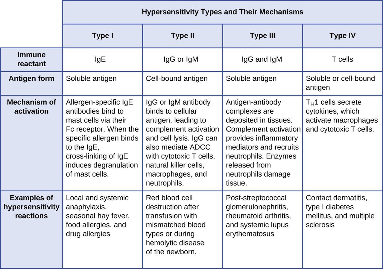
In the 1960s Gell and Coombs sorted hypersensitivities into four types by the immune mechanism behind them. The first three are driven by antibodies; the fourth is driven by T cells. A classic memory aid is ACID: type I Anaphylactic/allergic, type II Cytotoxic, type III Immune-complex, type IV Delayed. Types I–III happen within minutes to hours because antibodies are already present; type IV takes a day or more because T cells must be recruited to the site.
| Type | Also called | Mediator | Timing | Mechanism & example |
|---|---|---|---|---|
| I | Immediate / allergic / anaphylactic | IgE on mast cells | Seconds to minutes | Allergen cross-links IgE → mast-cell degranulation. Hay fever, food allergy, anaphylaxis |
| II | Cytotoxic | IgG / IgM vs. cell-surface antigen | Minutes to hours | Antibody binds a cell, marking it for destruction (complement, phagocytes). Hemolytic transfusion reaction, HDN, autoimmune hemolytic anemia |
| III | Immune-complex | IgG in antigen–antibody complexes | Hours | Soluble complexes deposit in vessels, joints, kidneys and inflame them. Serum sickness, SLE, some glomerulonephritis |
| IV | Delayed-type / cell-mediated | T cells (no antibody) | 1–3 days | Sensitized T cells release cytokines, recruit macrophages. Contact dermatitis (poison ivy, nickel), TB skin test |
Keep the antibody-versus-T-cell divide front of mind, because it explains the tempo you see clinically. If a patient breaks out in hives within minutes of a bee sting, you are watching type I. If a rash appears two days after a new topical cream, that slow onset points to type IV. Timing is a genuine diagnostic clue, not a trivia detail.
Why nurses care about all four
Each type maps to real bedside events. Type I is the drug allergy and the anaphylaxis you must be ready to treat in seconds. Type II is the mismatched blood transfusion you exist to prevent. Type III underlies flares of lupus and the joint and kidney damage of immune-complex disease. Type IV is the positive tuberculin skin test you read at 48–72 hours and the contact dermatitis a patient blames on their gloves or tape.
- Hypersensitivity is an immune response that damages the host's own tissues.
- Most reactions need a silent sensitization exposure first; the reaction fires on a later exposure.
- Types I–III are antibody-mediated (fast); type IV is T-cell-mediated (delayed 1–3 days).
- Remember ACID: Anaphylactic, Cytotoxic, Immune-complex, Delayed.
Section 2Allergy and anaphylaxis
Sensitization first, reaction later
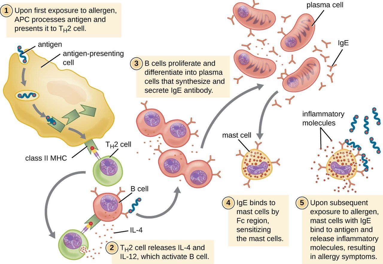
A type I reaction — what most people mean by "an allergy" — unfolds in two acts. During sensitization, a first exposure to an otherwise harmless allergen (pollen, peanut, penicillin, bee venom) is taken up and presented, and the immune system responds by making IgE antibody specific to it. That IgE then coats the surface of mast cells in the tissues and basophils in the blood, arming them. Nothing dramatic happens yet; the patient feels fine.
On the next exposure, the allergen bridges two adjacent IgE molecules on a mast cell — cross-linking them — and that is the trigger. Within seconds the mast cell undergoes degranulation, dumping preformed histamine and other mediators, and begins synthesizing leukotrienes and prostaglandins. Those mediators cause the familiar package: vasodilation (redness, warmth, dropping blood pressure), increased vascular permeability (swelling, hives), smooth-muscle contraction (bronchospasm, cramping), and increased secretions (runny nose, watery eyes).
From a local nuisance to a systemic emergency
Where the reaction happens decides how serious it is. A localized reaction stays near the point of contact: allergic rhinitis (hay fever), hives at a skin site, or asthma in the airways. A systemic reaction — anaphylaxis — occurs when mediators are released body-wide, usually because the allergen entered the bloodstream (an injected drug, an IV contrast agent, a sting, an ingested food). Anaphylaxis is a life-threatening emergency: widespread vasodilation causes blood pressure to crash (distributive shock), and airway swelling plus bronchospasm can choke off breathing within minutes.
| Feature | Localized allergy | Anaphylaxis (systemic) |
|---|---|---|
| Where mediators act | Near contact site | Whole body |
| Typical signs | Hives, itching, sneezing, wheeze | Hives plus hypotension, throat/airway swelling, stridor, wheeze, GI cramps, sense of doom |
| Main danger | Discomfort | Airway closure and shock — can kill in minutes |
| First-line treatment | Antihistamine; avoid trigger | Epinephrine, IM, immediately |
Recognizing and treating the emergency
The drug that reverses anaphylaxis is epinephrine, given intramuscularly into the outer thigh, and it must be given at the first sign — not after antihistamines, not after a wait-and-see. Epinephrine constricts blood vessels to restore blood pressure, relaxes airway smooth muscle to open breathing, and stabilizes mast cells. Antihistamines and corticosteroids are helpful adjuncts but are far too slow and too weak to be relied on alone. Alongside the drug, protect the airway, give oxygen, lay the patient flat with legs raised (unless breathing is easier sitting up), run IV fluids for the shock, and monitor closely — a biphasic reaction can return hours later even after the patient looks recovered.
Prevention is nursing work too. A documented allergy must be verified, banded, and honored on every medication pass; the single most common cause of a preventable drug reaction is a missed or ignored allergy history. For patients with severe allergies, teaching correct use of an epinephrine auto-injector — and to call emergency services and go to the hospital even after using it — is part of discharge.
- Type I allergy = sensitization (make IgE, arm mast cells) then a later reaction when allergen cross-links IgE.
- Mast-cell degranulation releases histamine, causing vasodilation, swelling, bronchospasm, and secretions.
- Anaphylaxis is systemic type I: airway swelling and distributive shock, potentially fatal in minutes.
- Treat anaphylaxis with IM epinephrine immediately; watch for a biphasic return.
Section 3Autoimmunity: the body against itself
How self-tolerance breaks down
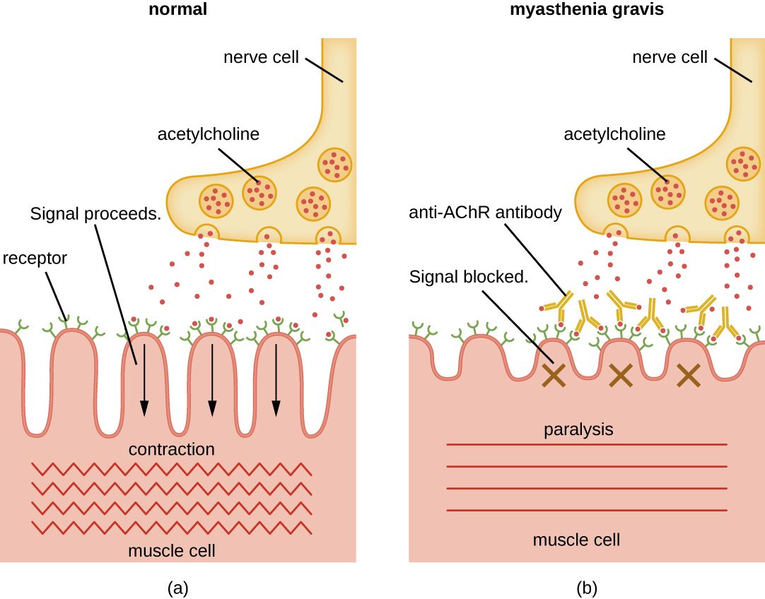
The immune system normally learns, early in life, to ignore the body's own molecules — a state called self-tolerance. Developing lymphocytes that react too strongly against self are deleted or disabled before they mature. Autoimmunity is the failure of that tolerance: self-reactive T cells or antibodies (autoantibodies) escape control and attack the body's own tissues, producing an autoimmune disease. The attack uses the same weapons as any hypersensitivity — often type II (autoantibodies against cell-surface targets), type III (immune complexes), or type IV (self-reactive T cells).
Why tolerance fails is usually a mix of factors. A genetic predisposition (many autoimmune diseases run in families and link to particular HLA/MHC types) sets the stage, and something in the environment — often an infection — pulls the trigger. One well-studied mechanism is molecular mimicry: a microbial antigen resembles a human protein closely enough that antibodies raised against the microbe also attack the body. The classic example is rheumatic fever, where antibodies against Streptococcus pyogenes cross-react with heart tissue. Autoimmune diseases also fall more often on women, and many wax and wane in flares and remissions.
Organ-specific versus systemic disease
It helps to sort autoimmune diseases by how much of the body they target. Organ-specific diseases aim autoantibodies or T cells at a single tissue — the thyroid, the pancreatic islets, the neuromuscular junction — so the damage and the symptoms are focused. Systemic diseases, like systemic lupus erythematosus, generate autoantibodies against widespread components (such as the cell nucleus), so they injure many organs at once: skin, joints, kidneys, blood, and heart.
| Disease | Target | Effect | Scope |
|---|---|---|---|
| Graves’ disease | TSH receptor (stimulating antibody) | Overactive thyroid (hyperthyroidism) | Organ-specific |
| Hashimoto thyroiditis | Thyroid tissue | Underactive thyroid (hypothyroidism) | Organ-specific |
| Type 1 diabetes mellitus | Pancreatic islet β-cells | No insulin production | Organ-specific |
| Myasthenia gravis | Acetylcholine receptor | Muscle weakness, ptosis | Organ-specific |
| Multiple sclerosis | CNS myelin sheath | Nerve conduction failure | Organ-specific (CNS) |
| Rheumatoid arthritis | Joint synovium | Symmetric joint inflammation, deformity | Systemic |
| Systemic lupus erythematosus | Nuclear antigens (anti-nuclear antibodies) | Skin, joints, kidney, blood, heart | Systemic |
Living with an autoimmune disease
There is no cure for most autoimmune diseases, so care aims to control the immune attack and manage what it has damaged. Treatment leans on immunosuppression — corticosteroids and other drugs that quiet the immune response — which brings its own hazard: a patient who is deliberately immunosuppressed is also more vulnerable to infection, blurring the line between an autoimmune problem and an immunodeficiency one. Some organ-specific diseases are managed by replacing what was lost rather than suppressing anything, as with insulin in type 1 diabetes or thyroid hormone in Hashimoto disease.
- Autoimmunity is a loss of self-tolerance: autoantibodies or self-reactive T cells attack the body.
- Causes combine genetic risk and an environmental trigger; molecular mimicry (e.g., rheumatic fever) is one route.
- Diseases are organ-specific (Graves, Hashimoto, type 1 diabetes, myasthenia gravis, MS) or systemic (RA, SLE).
- Care centers on immunosuppression or hormone replacement; immunosuppression raises infection risk.
Section 4Immunodeficiency and HIV/AIDS
When defenses are too weak
If hypersensitivity and autoimmunity are the immune system doing too much, immunodeficiency is it doing too little. A part of the immune system is missing or not working, so the patient cannot clear infections that a healthy person would shrug off — and they become prey to opportunistic organisms, microbes that rarely trouble the immunocompetent. Immunodeficiencies split into two groups by origin.
Primary immunodeficiencies are congenital — the person is born with a genetic defect in some component of immunity. They range from mild (selective IgA deficiency, often silent) to catastrophic. The most severe is severe combined immunodeficiency (SCID), in which both B cells and T cells are absent or nonfunctional; affected infants have almost no defense and, without treatment such as a bone-marrow transplant, do not survive infancy — the origin of the "bubble" isolation once used to protect them.
Secondary (acquired) immunodeficiencies are far more common and develop after birth from some external cause: malnutrition (the leading cause worldwide), certain cancers (especially leukemias and lymphomas), chemotherapy and other immunosuppressive drugs, radiation, aging, and viral infection — most importantly HIV. Many of your patients are secondarily immunodeficient by treatment: anyone on chemotherapy, high-dose steroids, or anti-rejection drugs.
| Feature | Primary immunodeficiency | Secondary immunodeficiency |
|---|---|---|
| Origin | Congenital (genetic) | Acquired after birth |
| Onset | Usually infancy/childhood | Any age |
| Examples | SCID, selective IgA deficiency, DiGeorge syndrome | HIV/AIDS, malnutrition, chemotherapy, leukemia, steroids |
| Frequency | Rare | Common — many hospitalized patients |
HIV and AIDS
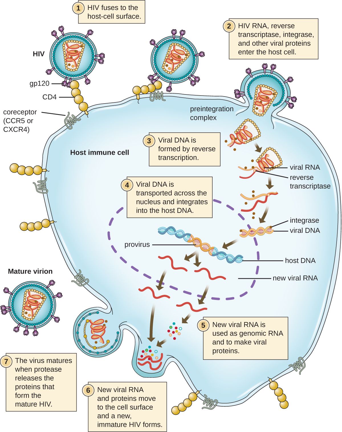
The human immunodeficiency virus (HIV) is a retrovirus that targets the very cell that coordinates the whole immune response: the CD4 helper T cell. Over years, HIV steadily infects and destroys these cells. As the CD4 count falls, the immune system loses its conductor, and the patient becomes progressively unable to fight off infection. HIV is transmitted through blood, sexual contact, and from mother to child in pregnancy, birth, or breastfeeding — not through casual contact.
AIDS (acquired immunodeficiency syndrome) is the advanced stage of HIV infection, defined by a CD4 count that falls below 200 cells/µL (normal is roughly 500–1,500) or by the appearance of an AIDS-defining opportunistic illness — infections and cancers that a healthy immune system holds at bay, such as Pneumocystis jirovecii pneumonia, Candida of the esophagus, disseminated tuberculosis, cytomegalovirus, and Kaposi sarcoma. The lower the CD4 count, the wider the range of opportunists that can take hold.
The story is far more hopeful than it once was. Antiretroviral therapy (ART) — combinations of drugs that block different steps of the viral life cycle — can suppress HIV to undetectable levels, preserve the CD4 count, prevent progression to AIDS, and render the virus untransmissible sexually. HIV has become, for many, a chronic manageable condition. Nursing priorities include supporting adherence to a lifelong regimen, watching for opportunistic infections, and applying standard precautions consistently — the same barrier practices that protect against all bloodborne pathogens.
- Immunodeficiency = too little immune function, opening the door to opportunistic infections.
- Primary = congenital (e.g., SCID); secondary = acquired (HIV, malnutrition, chemotherapy, steroids) and far more common.
- HIV destroys CD4 helper T cells; AIDS = CD4 < 200 cells/µL or an AIDS-defining opportunistic illness.
- ART suppresses HIV and preserves immunity; nurses support adherence and use standard precautions.
Section 5Transfusion and transplant reactions
ABO and Rh: the blood you can safely give
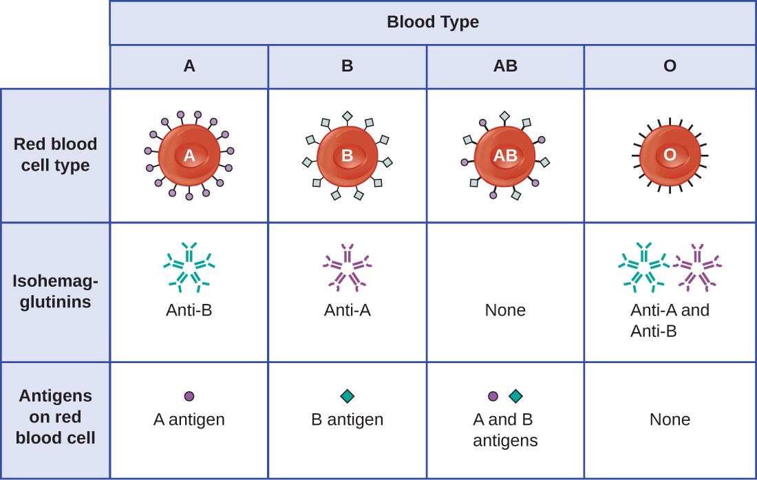
Red blood cells carry surface molecules that the immune system reads as antigens. The most important set is the ABO system: type A cells carry the A antigen, type B carry B, type AB carry both, and type O carry neither. Crucially, each person naturally makes antibodies against the ABO antigen they lack — a type A person has anti-B antibodies, a type O person has both anti-A and anti-B. This is why blood must be matched: transfusing the wrong ABO type sends antibody against donor red cells, and they are destroyed.
A second system, Rh, adds "positive" or "negative": Rh-positive cells carry the D antigen, Rh-negative cells do not. Unlike ABO, anti-Rh antibodies are not present from birth — an Rh-negative person makes them only after being exposed to Rh-positive blood. Because type O-negative blood carries none of these antigens, it is the emergency universal donor for red cells; type AB-positive, carrying no anti-A/anti-B antibodies, is the universal recipient.
| Blood type | Antigens on red cells | Antibodies in plasma | Can receive red cells from |
|---|---|---|---|
| A | A | anti-B | A, O |
| B | B | anti-A | B, O |
| AB | A and B | none | A, B, AB, O (universal recipient) |
| O | none | anti-A and anti-B | O only (universal donor) |
When a transfusion goes wrong
An acute hemolytic transfusion reaction is a type II hypersensitivity and a medical emergency, almost always caused by an ABO mismatch — usually a clerical or identification error at the bedside. The recipient's antibodies bind the transfused donor cells and destroy (hemolyze) them, triggering fever, chills, flank or back pain, dark urine, hypotension, and potentially kidney failure and death. The nurse's response is immediate: stop the transfusion, keep the IV line open with saline, and notify the provider and blood bank. Because these reactions come from misidentification, the two-person bedside verification of patient and unit is not paperwork — it is the actual safeguard against a fatal error.
A related problem crosses the placenta. Hemolytic disease of the newborn (HDN) occurs when an Rh-negative mother, sensitized to Rh-positive blood in an earlier pregnancy, makes anti-Rh antibodies that cross into a subsequent Rh-positive fetus and destroy its red cells. It is largely preventable: giving Rh immune globulin (RhoGAM) to Rh-negative mothers stops them from becoming sensitized in the first place.
Transplants and rejection
Transplanting tissue raises the same problem in a different form: the recipient's immune system recognizes donor cells as foreign (chiefly by their MHC/HLA molecules) and attacks the graft. How much it attacks depends on how closely donor and recipient are matched. An autograft (self to self, like a skin graft) and an isograft (between identical twins) are not rejected. An allograft (between two genetically different people — the usual organ transplant) is rejected unless the immune response is suppressed, and a xenograft (between species) faces the strongest rejection of all.
Rejection comes in three tempos: hyperacute (within minutes, from preexisting antibodies), acute (days to weeks, T-cell mediated), and chronic (months to years). To keep an allograft alive, recipients take immunosuppressant drugs for life — which, as ever, trades rejection risk for infection risk. One further danger runs the opposite direction: in a bone-marrow or stem-cell transplant, the graft's immune cells can attack the recipient's body — graft-versus-host disease (GVHD).
- ABO: you make antibodies against the antigen you lack; O-negative is the universal red-cell donor, AB-positive the universal recipient.
- An acute hemolytic transfusion reaction is a type II reaction from ABO mismatch — stop the transfusion at once.
- HDN comes from Rh incompatibility and is prevented with Rh immune globulin.
- Grafts: autograft/isograft not rejected, allograft needs immunosuppression; GVHD is the graft attacking the host.
The chapter in one breath
- Hypersensitivity is an immune response that harms the host; the four types differ by mediator and timing (I–III antibody, IV T-cell).
- Type I allergy runs from sensitization and IgE to mast-cell degranulation; systemic release is anaphylaxis, treated with IM epinephrine immediately.
- Autoimmunity is lost self-tolerance — organ-specific (Graves, type 1 diabetes, myasthenia gravis) or systemic (RA, SLE).
- Immunodeficiency is too little immunity: primary (congenital, e.g., SCID) or secondary (acquired — HIV, chemotherapy, malnutrition).
- HIV destroys CD4 T cells; AIDS is CD4 < 200/µL or an opportunistic illness; ART controls it.
- ABO/Rh matching prevents the type II hemolytic transfusion reaction; identification errors are the cause, so the bedside check is the safeguard.
- Grafts are rejected by degree of mismatch; allografts need lifelong immunosuppression, and GVHD is the graft attacking the host.
ReferenceWords worth knowing
A quick reference for the terms in this chapter — skim it now, or circle back whenever a word trips you up.
- Allergen
- An otherwise harmless antigen that provokes a type I (allergic) hypersensitivity response.
- Allograft
- A transplant between two genetically different individuals of the same species; the usual organ transplant, requiring immunosuppression.
- Anaphylaxis
- A severe, systemic type I reaction with airway swelling and distributive shock; a life-threatening emergency treated with epinephrine.
- Autoantibody
- An antibody directed against the body's own tissues.
- Autoimmunity
- A failure of self-tolerance in which the immune system attacks the body's own cells and tissues.
- CD4 helper T cell
- The coordinating lymphocyte of the immune response and the primary target of HIV.
- Graft-versus-host disease (GVHD)
- A reaction in which transplanted (graft) immune cells attack the recipient's tissues, seen with bone-marrow/stem-cell transplants.
- Hemolytic disease of the newborn (HDN)
- Destruction of a fetus's red cells by maternal anti-Rh antibodies; prevented with Rh immune globulin.
- Hemolytic transfusion reaction
- A type II reaction from an ABO-mismatched transfusion; the recipient's antibodies destroy donor red cells.
- Hypersensitivity
- An exaggerated or misdirected immune response that damages the host's own tissues.
- IgE
- The antibody class that arms mast cells and basophils and mediates type I hypersensitivity.
- Immunodeficiency
- A deficient immune response; primary (congenital) or secondary (acquired).
- Mast cell
- A tissue cell coated with IgE that releases histamine and other mediators when it degranulates.
- Molecular mimicry
- A microbial antigen resembling a self-protein, so anti-microbe antibodies also attack the body (e.g., rheumatic fever).
- Opportunistic infection
- An infection by a normally harmless microbe that takes hold when host defenses are weakened.
- Primary immunodeficiency
- A congenital, genetically based defect in immune function, such as SCID.
- Rh factor
- The D red-cell antigen; Rh-positive cells carry it, Rh-negative do not.
- Secondary immunodeficiency
- An acquired loss of immune function from an external cause (HIV, malnutrition, drugs, cancer).
- Self-tolerance
- The immune system's normal ability to recognize and not attack the body's own molecules.
- Sensitization
- The silent first exposure in which the immune system builds antibodies or primed cells against an antigen, setting up a later reaction.
- Severe combined immunodeficiency (SCID)
- A severe primary immunodeficiency lacking functional B cells and T cells.