Rehabilitation, Mobility and Chronic Illness
Rehabilitation begins the hour the crisis is survived, not the day the patient is discharged to a rehab unit. The stroke is treated in minutes, the femur is nailed in hours — and then somebody has to teach a frightened adult to stand, to empty a bladder that no longer signals, to eat with the hand that still works, to be a parent from a wheelchair. That work is nursing work, and it is graded on function rather than on cure. This chapter gives you the practical spine of it: how the rehabilitation team divides labour, how function is measured with tools you will actually chart in, how to transfer a patient without injuring either of you, which gait pattern belongs to which device, what a person with a C6 injury can and cannot do, and how to recognise autonomic dysreflexia fast enough to prevent a stroke. It ends where chronic illness actually lives — in the patient’s house, in their marriage, in their bank account.
By the end of this chapter you can
- Describe the roles of the interdisciplinary rehabilitation team and the criteria distinguishing an inpatient rehabilitation facility from a skilled nursing facility, home health and outpatient therapy.
- Perform and document a functional assessment using ADLs, IADLs, the Barthel Index, the FIM/Section GG scale and the Timed Up and Go, and translate the score into a nursing goal.
- Execute a safe transfer using a gait belt, correct body mechanics and safe patient handling equipment, and state the manual lifting limit and the axilla rule.
- Fit a cane, walker and crutches correctly and teach the four-point, two-point, three-point and swing-through gaits plus the stair rule — up with the good, down with the bad.
- Predict retained function by spinal cord injury level, distinguish spinal shock from neurogenic shock, and classify injuries using the ASIA Impairment Scale and the incomplete cord syndromes.
- Recognise autonomic dysreflexia and carry out the intervention sequence in order — sit the patient up first, then remove the trigger — with correct SBAR escalation.
- Design neurogenic bladder and neurogenic bowel programmes for upper and lower motor neuron injury, including intermittent catheterisation volumes and digital stimulation technique.
- Prevent contractures, venous thromboembolism, pressure injury, orthostatic hypotension and disuse osteoporosis, and manage spasticity drugs with their monitoring parameters.
- Support psychosocial adjustment to chronic illness and disability, apply self-management and teach-back teaching, and state which rehabilitation tasks may and may not be delegated.
Section 1What Rehabilitation Is, and How Function Is Measured
The team, the goal and the setting
Acute care asks “what is wrong and how do we fix it?” Rehabilitation asks a harder question: “given what cannot be fixed, what can this person still do, and what has to change so they can do it?” The World Health Organization’s International Classification of Functioning frames disability not as a property of a body but as the gap between what a body can do and what an environment demands. A T10 paraplegic on a level campus with curb cuts is far less disabled than the same person in a third-floor walk-up. That is not a philosophical aside — it is why a rehabilitation nurse spends as much time on the discharge environment as on the patient’s quadriceps.
The rehabilitation team is genuinely interdisciplinary, meaning the disciplines set one shared goal rather than five parallel ones. The physiatrist (physical medicine and rehabilitation physician) leads medically. Physical therapy owns gross mobility, gait, transfers, strength and endurance. Occupational therapy owns the upper extremity, activities of daily living, adaptive equipment, cognition applied to task, and home modification. Speech-language pathology owns swallowing, speech, language and cognitive-communication. The rehabilitation nurse — the certified credential is CRRN — is the one discipline present for all twenty-four hours, and the job is to carry therapy into life: the transfer PT taught at ten in the morning must happen the same way at two in the morning, or it is not learned. Add the social worker or case manager (funding, placement, benefits), the dietitian, the psychologist, the chaplain, the vocational counsellor and the recreational therapist, and you have the weekly team conference.
Where the patient goes after acute care is a matter of tolerance and skilled need, not of enthusiasm. An inpatient rehabilitation facility requires that the patient can participate in and benefit from about three hours of therapy a day, five days a week, from at least two therapy disciplines, with physician oversight several days a week and a formal weekly team conference. If the patient cannot sit up for ninety minutes at a stretch, they cannot yet do IRF. A skilled nursing facility delivers a lower dose — often one to two hours a day — and is the right answer for the deconditioned eighty-eight-year-old after a hip fracture who needs time more than intensity. Long-term acute care takes the ventilator weans and complex wounds. Home health and outpatient therapy carry the tail end. Getting this recommendation right at the team conference is one of the most consequential things a nurse contributes, because the level of care determines the ceiling of recovery.
| Setting | Therapy dose | Typical candidate | Nursing focus |
|---|---|---|---|
| Inpatient rehabilitation facility (IRF) | About 3 h/day, 5 days/week; two or more disciplines | Stroke, spinal cord injury, brain injury, amputation, multiple trauma, hip fracture with good potential | Carry-over of therapy techniques 24/7; bowel and bladder programmes; skin; family training |
| Skilled nursing facility (SNF) | Roughly 1–2 h/day as tolerated | Deconditioned older adult, slower progress, medically fragile, lower endurance | Restorative nursing, fall prevention, nutrition, medication reconciliation |
| Long-term acute care (LTACH) | Variable; medically driven | Ventilator wean, complex wounds, prolonged IV therapy | Airway, secretion clearance, wound care, gradual mobility |
| Home health | Two to three visits/week | Homebound, has a caregiver, safe home environment | Environmental assessment, caregiver teaching, medication safety |
| Outpatient / day rehab | Two to five visits/week | Independent transport, community re-entry, return to work | Reinforcement, self-management, vocational goals |
ADLs, IADLs and the scored tools
Activities of daily living are the six survival tasks of the body: bathing, dressing, toileting, transferring, continence and feeding — the Katz index. Instrumental activities of daily living are the eight tasks of running a household and a life: using a telephone, shopping, preparing food, housekeeping, laundry, arranging transportation, managing medications and managing finances — the Lawton scale. The distinction matters clinically because IADLs fail first. The earliest reliable sign of cognitive decline in an older adult is usually not forgetting a name; it is unopened mail, a bounced cheque, a pill organiser that no longer matches the bottles. Ask about IADLs explicitly and ask the family separately, because patients systematically over-report independence.
Three scored tools appear on the chart and on the exam. The Barthel Index scores ten ADL items in increments of five to a total of 100; higher is more independent, and a perfect 100 means independent in those ten tasks, not that the person can safely live alone. The Functional Independence Measure (FIM) scores eighteen items — thirteen motor and five cognitive — from 1 to 7 for a total of 18 to 126. Learn the anchors: 7 is complete independence, 6 modified independence (device, extra time or safety consideration but no helper), 5 supervision or set-up only, 4 minimal assistance (the patient does 75 percent or more of the effort), 3 moderate assistance (patient does 50 to 74 percent), 2 maximal assistance (patient does 25 to 49 percent), 1 total assistance (patient does under 25 percent). The line between 5 and 4 is the line between hands-off and hands-on, and it is where reimbursement, staffing and discharge safety all pivot. United States rehabilitation facilities now report the six-level Section GG self-care and mobility items on the assessment instrument, which uses the same logic with different numbers; whichever tool your unit uses, score what you saw, not what the patient claims and not what the last shift wrote.
Mobility and balance get their own quick screens. The Timed Up and Go — rise from a standard armchair, walk three metres, turn, return, sit — is normal under about ten seconds, and twelve seconds or more flags increased fall risk. The 30-second chair stand and the four-stage balance test round out the fall screen. The Berg Balance Scale runs 0 to 56, with scores under about 45 associated with falls. The 6-minute walk test measures endurance and is the one that changes most visibly over a rehab stay. Do not administer any of these to a patient who has not yet been cleared for weight bearing, and never do a first TUG alone.
Goals, carry-over and what you may delegate
Rehabilitation goals are written the way objectives are written: measurable, time-bound, and owned by the patient. “Patient will improve mobility” is not a goal. “By 14 March, patient will transfer bed to wheelchair with stand-by assist of one and a gait belt, four out of five attempts, verbalising the sequence unprompted” is. Then you make the environment enforce it: the wheelchair parked on the strong side, the call bell within reach of the working hand, the urinal where a C7 patient can actually get it. Learned helplessness is iatrogenic. Every time a rushed nurse does for a patient what the patient could do in four extra minutes, a little function is taken back.
Delegation in rehabilitation is a frequent test point because the tasks look routine and are not. Assistive personnel may ambulate and transfer a stable patient with a device the patient has already been taught, perform passive and active range of motion after the RN has instructed them, position and turn, apply compression stockings, feed a patient with no swallowing precautions, weigh, measure intake and output, and report what they observe. The RN retains assessment, teaching, evaluation and any unstable patient — that includes the first ambulation after any change in status, medication, surgery or neurological event; the initial gait training and device fitting; evaluating whether the patient tolerated the activity; and every part of the response to autonomic dysreflexia. A useful bedside test: if the task requires nursing judgement about whether it is safe to continue, it is not delegable.
Older adults deserve a specific note here. Muscle strength falls roughly one to one-and-a-half percent per day of bed rest, and faster during critical illness. A previously independent eighty-year-old admitted for pneumonia can lose the ability to rise from a toilet in five days — not from the pneumonia but from the bed. The countermeasure is unglamorous and enormously effective: get them up. Sit them in a chair for meals, remove the tethers that anchor them (unnecessary Foley catheters, telemetry, continuous pulse oximetry, IV pumps that could be saline-locked), protect sleep, and mobilise at least three times a day unless there is a written reason not to.
Rehabilitation nursing is graded in function, so assess with a scored tool, chart the exact level of assist, and remember that the round-the-clock carry-over of what therapy taught is the nurse’s unique contribution.
Section 2Safe Transfers, Gait Belts and Assistive Devices
Before the first step
Most mobility disasters are failures of the thirty seconds before the patient stood up. Run a fixed pre-mobility check every time. First, the order: what is the weight-bearing status, and is there a brace, an immobiliser or a spinal precaution? Non-weight-bearing means the limb does not touch the floor at all. Touch-down or toe-touch weight bearing means the toe may rest for balance only, roughly the weight of the leg. Partial weight bearing is a specified percentage, commonly thirty to fifty percent, which patients cannot estimate without practice on a scale. Weight bearing as tolerated lets the patient titrate by pain. Full is full. If the order is ambiguous, do not interpret it — call.
Second, the physiology. Check vital signs and check orthostatics if the patient has been down for more than a day or two, is on antihypertensives or diuretics, or has any spinal cord or autonomic involvement: measure supine, then sitting, then standing at one and three minutes. A drop of 20 mmHg systolic or 10 mmHg diastolic within three minutes of standing, or any symptomatic drop, means dangle longer and use an abdominal binder and compression stockings. Look at haemoglobin — a haemoglobin of 7 g/dL will not tolerate what 12 will. Check oxygenation, glucose (a capillary glucose of 60 mg/dL is not a walking glucose), pain control timed so the patient is comfortable but not sedated, and recent sedating or anticoagulant medications.
Third, the environment. Bed at the height where the patient’s feet are flat on the floor with hips slightly higher than knees. Brakes locked on bed and chair. Non-skid footwear on, not socks. Lines, drains and tubing gathered and long enough. Chair positioned on the patient’s strong side at roughly a forty-five degree angle so the pivot is short. Floor dry. Enough people — and enough is decided before, not during. Then tell the patient the plan in the exact words you will use: “On three, you push down on the bed with your hands and stand. One, two, three, stand.”
The transfer itself
Apply the gait belt around the waist over clothing, snug enough that only two flat fingers fit underneath, buckle to the side or front and away from any incision, ostomy, feeding tube or fresh graft. Grasp it underhand at the sides or back. The belt is a handle for guiding and for lowering — it is not a hoist, and it will not stop a full-weight fall. The single most important prohibition in this chapter after autonomic dysreflexia is this: never lift a patient by the axillae. Pulling under the arms stretches the brachial plexus, dislocates hemiplegic shoulders whose rotator cuff no longer holds the joint, and has produced permanent nerve injury. Never let the patient grab around your neck either.
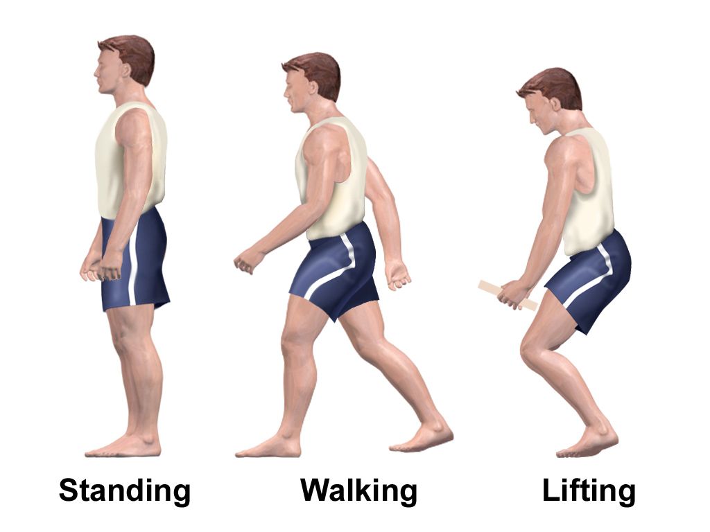
Mechanics: stand facing the patient with your feet apart, one foot slightly forward, knees and hips bent, back straight, your centre of gravity low and close to theirs. Block the patient’s weak knee with your knee, or brace their feet with yours. Have the patient scoot to the edge, place both feet flat and slightly back under the knees, lean forward — nose over toes — and push up from the mattress or armrests rather than pulling on you or on the walker. Rise, pause and let blood pressure catch up, then step and turn. Do not twist your spine; move your feet. If the patient begins to fall, do not fight it. Widen your stance, pull them toward you, protect the head, and ease them down your leg to the floor.
Modern practice is not to depend on human strength at all. Safe patient handling guidance sets the maximum recommended manual lift at roughly 35 pounds under ideal conditions; almost no adult patient qualifies. Use the equipment: friction-reducing slide sheets and air-assist mattresses for lateral transfers, sit-to-stand powered lifts for the patient who has some weight-bearing and can follow directions, full-body ceiling or floor lifts for the dependent patient, and a transfer (slide) board for the seated patient with strong arms — the classic technique for spinal cord injury at C7 and below. Bariatric patients require bariatric-rated equipment and more helpers, never more effort. The nurse who injures their back does not get to help anyone tomorrow.
Canes, walkers and crutches
Fit first. For a cane, with the patient standing upright in their shoes and arms relaxed, the handgrip should sit at the level of the greater trochanter, which is roughly the wrist crease, producing about 20 to 30 degrees of elbow flexion. The cane is held in the hand opposite the affected leg, because that reproduces normal reciprocal gait and widens the base of support toward the weak side. A quad cane gives more support than a single-point cane; position the base so the two short legs sit closest to the patient and the two longer legs face away from the foot, or the patient trips over the wide side of the base. For a walker, the same wrist-crease rule applies. A standard pick-up walker gives the most stability and the slowest gait; a two-wheeled rolling walker suits the patient who cannot lift; a four-wheeled rollator with hand brakes and a seat suits the endurance-limited patient with good cognition but is not appropriate for someone who leans hard on the frame, because it will roll away.
For axillary crutches, the top pad sits two to three finger-widths — about 1.5 to 2 inches — below the axilla, with the handgrips again at wrist-crease height and 20 to 30 degrees of elbow flexion, and the crutch tips resting about 6 inches lateral and 6 inches anterior to the foot. The weight goes through the hands, never the armpit. Weight borne on the axillary pads compresses the brachial plexus and produces “crutch palsy” — wrist drop and numbness. Teach patients to check tips for wear and to keep both hands free by using a backpack.
| Device / gait | Who it is for | Sequence |
|---|---|---|
| Cane | Mild unilateral weakness, balance | Cane in hand opposite the weak leg. Cane and weak leg advance together, then strong leg |
| Walker | Bilateral weakness, poor balance, partial weight bearing | All four points down, advance walker one step length, then weak leg, then strong leg. Never on stairs; never pull on it to stand |
| Four-point gait | Weight bearing on both legs; weak or unsteady; slowest and most stable | Right crutch, left foot, left crutch, right foot — three points on the ground at all times |
| Two-point gait | Partial weight bearing both legs; faster, mimics normal walking | Right crutch with left foot together, then left crutch with right foot together |
| Three-point gait | One leg non-weight-bearing or partial (fracture, new amputation); needs strong arms | Both crutches and the affected leg advance together, then the strong leg swings through |
| Swing-to / swing-through | Bilateral lower-limb paralysis with braces | Both crutches forward, then both legs swing to the crutches (swing-to) or past them (swing-through) |
| Stairs — UP | All devices except walker | Strong leg steps up FIRST, then crutches or cane with the weak leg |
| Stairs — DOWN | All devices except walker | Crutches or cane with the weak leg go down FIRST, then the strong leg |
The mnemonic students remember at three in the morning is “up with the good, down with the bad” — the good leg goes up to heaven, the bad leg goes down to the other place. The logic is mechanical: climbing requires the leg that can generate force, so the strong leg lifts the body up; descending is controlled lowering, so the weak leg and the device absorb the descent while the strong leg controls it from above. When a handrail is available, use it — both crutches go into the opposite hand held horizontally, one hand on the rail. Teach patients to back a wheelchair down a ramp or curb and to approach the seat until the backs of both legs touch it before sitting.
When a patient falls
Falls are the most common adverse event in rehabilitation because the whole enterprise is deliberately pushing the edge of safety. Screen everyone with a validated tool (Morse, Hendrich II or STEADI), and treat the modifiable causes: orthostatic hypotension, polypharmacy — especially benzodiazepines, sedative-hypnotics, opioids, anticholinergics, antihypertensives and hypoglycaemics — vision, footwear, urinary urgency, delirium and environmental hazards. Bed alarms and non-skid socks are the least effective interventions on the list; scheduled toileting, hourly rounding and a bedside commode are among the best.
If a fall happens, the sequence is assessment before movement. Do not lift the patient up first. Assess responsiveness, airway, breathing and circulation, then check for head strike, neck or back pain, and obvious deformity or shortening and external rotation of a leg, which suggests hip fracture. Take vital signs and a neurological check on the floor. Only when you have ruled out spinal or fracture injury do you move the patient, with enough people and usually with a lift. Then notify the provider, notify the family, perform neurological checks per protocol — more urgently if the patient is anticoagulated, because a delayed intracranial bleed is the classic killer — document objectively what you found, and file the incident report separately from the chart.
Fit the device, use the gait belt at the waist and never under the axillae, remember cane opposite the weak leg and up with the good, down with the bad — and reach for a mechanical lift rather than your own back.
Section 3Spinal Cord Injury: Levels, Syndromes and Retained Function
The first hours: spinal shock and neurogenic shock
Two different phenomena share a confusing name. Spinal shock is a neurological event: immediately after cord injury, everything below the lesion goes flaccid and areflexic — no deep tendon reflexes, no bulbocavernosus reflex, atonic bladder and bowel, loss of temperature regulation. It is not a circulatory state and it tells you nothing about final prognosis, because a completely transected cord and a badly bruised one look identical during it. Spinal shock resolves over days to weeks, and the return of reflexes — classically the bulbocavernosus — marks the end. Only then can the injury be accurately classified as complete or incomplete, which is why families should be counselled that the early picture is not the final one.
Neurogenic shock is a circulatory event, and it occurs with injuries at roughly T6 and above, where sympathetic outflow is interrupted. The result is unopposed vagal tone: vasodilation with pooled blood, and — critically — the heart cannot mount a compensatory tachycardia. The presentation is hypotension with bradycardia and warm, dry, flushed skin below the lesion. Contrast that with the trauma patient’s haemorrhagic shock: hypotension with tachycardia and cool, clammy, pale skin. A trauma patient can have both at once, so never assume the low blood pressure is “just neurogenic” until bleeding is excluded. Management is measured fluid resuscitation (over-resuscitation causes pulmonary oedema in a patient with a compromised cough), a vasopressor that also supports heart rate such as norepinephrine or dopamine — avoid pure alpha-agonists such as phenylephrine, which trigger reflex bradycardia in a heart that already cannot speed up — and atropine for symptomatic bradycardia, with a common target of maintaining mean arterial pressure around 85 to 90 mmHg for the first week to perfuse the injured cord.
The other first-hours priorities follow airway-breathing-circulation exactly. Injuries at C3 to C5 threaten the diaphragm — the old teaching “C3, 4 and 5 keep the diaphragm alive” is worth the rote memorisation. Watch for a rising respiratory rate with falling tidal volume, a weakening cough, paradoxical abdominal movement, and a fall in vital capacity; ascending oedema in the first 24 to 72 hours can convert a C5 patient who was breathing into a C4 patient who is not. Keep the neck immobilised until cleared, log-roll with enough staff, watch for ileus and gastric distension (an NG tube early is common), and expect poikilothermia — these patients take on the room’s temperature and will become hypothermic on a cold trauma bay table. One thing you should not expect to see is routine high-dose methylprednisolone: prophylactic steroids are no longer recommended in acute spinal cord injury, because the infection, pneumonia, gastrointestinal bleeding and wound complications outweigh any functional gain.
Level by level: what is retained
The neurological level of injury is the most caudal segment with normal sensation and with motor strength of at least 3 out of 5 (antigravity), provided the segment above is normal. Function is predicted by what is preserved at and above that level. Tetraplegia (formerly quadriplegia) means all four limbs and the trunk are involved, from a cervical lesion; paraplegia means the trunk and legs, from a thoracic, lumbar or sacral lesion. Learn the table below the way you learn cranial nerves — it is the backbone of every SCI question you will be asked.
| Level | Key muscle gained | Practical function |
|---|---|---|
| C1–C3 | Neck accessory muscles only | Ventilator dependent (phrenic outflow lost). Sip-and-puff or chin-controlled power chair. Total assist for ADLs |
| C4 | Diaphragm, trapezius | May wean from ventilator; shoulder shrug; head and neck control; power chair with chin or head control. Total assist for ADLs |
| C5 | Deltoid, biceps (elbow flexion, shoulder abduction) | Can feed and groom with a mobile arm support and universal cuff; no wrist or hand function; power chair; dependent for transfers |
| C6 | Wrist extensors | Tenodesis grasp — wrist extension passively closes the fingers. Dressing upper body, self-catheterisation with adaptations, slide-board transfers with assist, drives with hand controls |
| C7–C8 | Triceps (elbow extension), then finger flexors | The independence level. Can push up, do independent transfers, propel a manual wheelchair, perform most ADLs and self-care, live independently with adaptations |
| T1–T6 | Full arms and hands; intercostals begin | Fully independent in self-care and manual wheelchair. At risk for autonomic dysreflexia. Limited trunk stability; weak cough |
| T7–T12 | Abdominals, trunk extensors | Good trunk control and cough; independent transfers including floor-to-chair; standing frame or braces for exercise |
| L1–L2 | Hip flexors | Household ambulation with long leg braces (KAFOs) and crutches; wheelchair for community distances |
| L3–L4 | Quadriceps (knee extension) | Functional ambulation with ankle-foot orthoses and canes or crutches |
| L5–S1 | Dorsiflexion, then plantar flexion | Ambulates with an AFO or minimal device; footdrop pattern if L4–L5 involved |
| S2–S4 | Bowel, bladder, sexual reflex arcs | Ambulation preserved; lesions here cause a flaccid bowel and bladder and saddle anaesthesia |
Complete, incomplete and the cord syndromes
The ASIA Impairment Scale grades severity. A is complete: no motor or sensory function in the lowest sacral segments S4–S5. B is sensory incomplete: sensation but no motor function below the level, including at S4–S5. C is motor incomplete with more than half the key muscles below the level weaker than grade 3. D is motor incomplete with at least half the key muscles at grade 3 or better — most of these patients walk. E is normal. Everything turns on sacral sparing, which is why the examination always ends with light touch and pinprick at the anal mucocutaneous junction, deep anal pressure and voluntary anal contraction. A patient with any sacral sparing is incomplete and has a materially better prognosis, so never document “complete” without that part of the exam.
Four incomplete patterns recur. Central cord syndrome is the most common: a hyperextension injury in an older adult with cervical spondylosis, producing weakness that is worse in the arms than the legs — the patient walks in with useless hands. Anterior cord syndrome follows flexion injury or anterior spinal artery infarct: motor function and pain and temperature are lost below the lesion while proprioception and vibration are preserved through the intact posterior columns; prognosis is the worst of the group. Brown-Séquard syndrome follows hemisection, classically penetrating trauma: ipsilateral loss of motor function, proprioception and vibration with contralateral loss of pain and temperature beginning a segment or two below. Cauda equina and conus medullaris syndromes involve the lumbosacral roots and produce flaccid weakness, saddle anaesthesia, and areflexic bowel and bladder with urinary retention and overflow — a new saddle numbness with retention in a back-pain patient is a surgical emergency that needs imaging within hours, not a rehabilitation referral.
Predict function from the neurological level — C4 diaphragm, C6 tenodesis, C7 independence, T6 and above autonomic dysreflexia risk — and never call an injury complete until sacral sparing has been checked after spinal shock resolves.
Section 4Autonomic Dysreflexia and Neurogenic Bladder and Bowel
Autonomic dysreflexia: the emergency you must never miss
Autonomic dysreflexia occurs in patients with spinal cord injury at T6 or above — occasionally as low as T8 — once spinal shock has resolved. A noxious stimulus below the level of injury (a distended bladder, an impacted rectum, a pressure injury, an ingrown toenail, a tight leg bag strap, labour contractions, even a fracture the patient cannot feel) fires afferent signals into the cord. Below the lesion, a massive sympathetic discharge produces intense vasoconstriction and blood pressure rises steeply. Baroreceptors in the carotid and aortic arch detect the surge and send a parasympathetic correction — but that correction can only reach the body above the lesion, because the descending inhibitory signal cannot cross the injured segment. The result is a split body, and it is exactly what you see on examination.
Above the lesion: pounding headache, flushed and sweating face and neck, nasal congestion, blurred vision or spots, anxiety and a sense of doom, and a bradycardia that is often the giveaway. Below the lesion: pale, cold, dry skin with piloerection — goosebumps. The blood pressure is the diagnosis. Baseline systolic pressure in a person with a high cord injury is often only 90 to 110 mmHg, so a rise of 20 to 40 mmHg above their own baseline is significant even when the number would look unremarkable in another patient. Untreated, this is a hypertensive emergency that causes intracerebral haemorrhage, seizure, retinal haemorrhage, myocardial ischaemia and death. Some patients have no headache at all — silent dysreflexia — so any unexplained bradycardia, sweating or blood pressure jump in a T6-or-above patient is dysreflexia until you have excluded it.
The order of actions is a favourite examination item because it is counter-intuitive: this is the one hypertensive patient you sit up. Gravity is the fastest antihypertensive you own.
| Step | Action | Why |
|---|---|---|
| 1 | Sit the patient upright, head of bed high, legs dependent — do not lay them flat | Orthostatic pooling drops blood pressure within seconds |
| 2 | Loosen or remove anything constrictive: abdominal binder, compression stockings, leg-bag straps, tight clothing, shoes | Removes the stimulus and reduces venous return |
| 3 | Check blood pressure every 2–5 minutes and stay with the patient; call for help | Blood pressure is the whole monitor here |
| 4 | Bladder first. Check the catheter for kinks, obstruction and a full bag. If blocked, irrigate gently with 10–15 mL warm saline or replace the catheter. If no catheter, instil 2 percent lidocaine jelly and wait 2 minutes before catheterising; drain slowly | Bladder distension or blockage causes the large majority of episodes |
| 5 | Bowel second. If bladder is clear, instil lidocaine jelly into the rectum, wait 2–5 minutes, then gently check for and remove impaction. Stop if the blood pressure climbs further | Disimpaction is itself a noxious stimulus; anaesthetise first |
| 6 | Skin third. Head-to-toe survey for pressure injury, burn, ingrown toenail, fracture, tight dressing, sunburn | Any nociceptive input below the lesion will do it |
| 7 | If systolic pressure remains at or above about 150 mmHg, give a rapid-onset short-acting antihypertensive per order — immediate-release nifedipine (bite and swallow), nitroglycerin paste applied above the level of injury, or sublingual captopril | Short-acting and, for paste, removable — wipe it off when pressure falls |
| 8 | Monitor for at least 2 hours after resolution | Rebound hypotension and recurrence are both common |
Two drug warnings. Never give nitrates — paste, sublingual or otherwise — if the patient has taken a phosphodiesterase-5 inhibitor (sildenafil, vardenafil within 24 hours; tadalafil within 48 hours). Many men with spinal cord injury use these; ask before you reach for the nitroglycerin, because the combination causes refractory, potentially fatal hypotension. And note that the once-taught “bite and squirt under the tongue” nifedipine technique is no longer recommended; current guidance is bite the capsule and swallow. Prevention is where nursing actually wins: a bladder programme that never lets volumes exceed roughly 500 mL, a bowel programme that prevents impaction, meticulous skin surveillance, and a wallet card so that the emergency department, the dentist and the labour and delivery unit all know what this patient is at risk for.
Neurogenic bladder
Where the lesion sits relative to the sacral micturition centre at S2–S4 decides the bladder’s personality. An upper motor neuron (reflexic, spastic) bladder follows a lesion above the conus: the reflex arc is intact but disconnected from the brain, so the detrusor contracts involuntarily at small volumes and often fights a sphincter that does not relax — detrusor-sphincter dyssynergia — generating dangerously high storage pressures that back up into the kidneys. A lower motor neuron (areflexic, flaccid) bladder follows conus or cauda equina injury: the arc itself is destroyed, so the bladder fills passively, distends enormously and leaks by overflow. The clinical distinction is not academic. The first bladder is treated by relaxing it; the second is treated by emptying it.
Clean intermittent catheterisation is the standard of care for both, and it is superior to an indwelling catheter for infection, stones, urethral erosion and quality of life. Catheterise every four to six hours, adjusting frequency and fluid so that drained volumes stay under about 400 to 500 mL — the number to remember is that consistently exceeding 500 mL means catheterise more often or drink less, not use a bigger catheter. Total fluid around 2 litres a day, spread evenly and tapered in the evening. Home technique is clean, not sterile; in hospital, follow your facility’s policy. Pharmacology supports it: antimuscarinics such as oxybutynin or tolterodine quiet the overactive detrusor (watch anticholinergic burden in older adults — dry mouth, constipation, blurred vision, urinary retention, confusion); the beta-3 agonist mirabegron does the same without antimuscarinic activity, which is why it is often preferred in older adults, but it has its own parameter — monitor blood pressure and avoid it in severe uncontrolled hypertension; alpha-blockers such as tamsulosin lower outlet resistance (watch orthostatic hypotension and floppy iris at cataract surgery); onabotulinumtoxinA injected into the detrusor is used for refractory overactivity and requires the patient be willing and able to catheterise afterwards.
Teach the surveillance points. Asymptomatic bacteriuria is nearly universal in people who catheterise and should not be treated — treating it breeds resistance without helping the patient. Treat when there are symptoms, and remember that in spinal cord injury the symptoms are atypical: fever, cloudy or foul urine, new or worsening spasticity, new incontinence between catheterisations, malaise, flank or suprapubic discomfort, or an episode of autonomic dysreflexia. Long-term risks needing annual follow-up are hydronephrosis and reflux, stones, and — with decades of indwelling catheterisation — bladder cancer.
Neurogenic bowel
The same upper-versus-lower logic applies, and the bowel programme is arguably the single greatest determinant of whether a person with spinal cord injury leaves the house. A reflexic (UMN) bowel has an intact anal reflex and a tight sphincter, so it responds to digital stimulation: a gloved, well-lubricated finger inserted and rotated gently against the rectal wall for 15 to 20 seconds, repeated every 5 to 10 minutes, triggers reflex peristalsis and evacuation. Combine it with a bisacodyl or glycerin suppository, and schedule the whole programme 20 to 30 minutes after a meal to exploit the gastrocolic reflex, ideally with the patient sitting upright on a padded commode so gravity helps. Daily or every-other-day, at the same time, every time. An areflexic (LMN) bowel has a patulous, flaccid sphincter and does not respond to digital stimulation at all; it needs gentle manual evacuation, usually daily, with the stool kept firm rather than soft so it does not simply leak out.
Support the programme with the boring things that make it work: 15 to 30 g of fibre daily, adequate fluid, activity as tolerated, a stool softener such as docusate, a stimulant such as senna, and polyethylene glycol titrated to a formed but soft stool. Avoid chronic enemas and avoid escalating laxatives to fix a timing problem. The measurable goals are a complete evacuation in under about 60 minutes, no more than one unplanned stool a month, and no impaction. In older adults, review the medication list first — opioids, anticholinergics, calcium-channel blockers, iron and calcium supplements sabotage more bowel programmes than any dietary failure. And note the safety limit: digital stimulation and manual evacuation in a T6-or-above patient can themselves precipitate autonomic dysreflexia, which is why lidocaine jelly and a blood-pressure check belong in the routine, not just in the emergency.
In a patient injured at T6 or above, sudden hypertension with bradycardia and a pounding headache means autonomic dysreflexia — sit them upright first, loosen everything constrictive, then hunt the trigger starting with the bladder, then the bowel, then the skin.
Section 5Preventing Immobility Complications and Living with Chronic Illness
The immobility syndrome and how to interrupt it
Immobility is a disease with predictable organ-by-organ complications, and almost all of them are preventable by nursing action rather than by prescription. Contractures begin within days: soft tissue shortens in whatever position the limb is left. Perform passive range of motion through the full arc at least twice daily unless contraindicated, position deliberately — a trochanter roll to stop hip external rotation, no pillow under the knee, a resting hand splint to preserve the functional arch, an ankle-foot orthosis or a footboard to prevent plantar flexion contracture and footdrop, prone lying to stretch hip flexors in the patient who can tolerate it. Beware the exception: in a patient with a C6 injury who relies on tenodesis, the finger flexors are deliberately allowed to tighten a little, so follow the therapist’s hand-positioning plan rather than stretching them out.
Venous thromboembolism risk is highest in the first three months after spinal cord injury and is high after any major orthopaedic or neurological event. Pharmacological prophylaxis with low-molecular-weight heparin is generally started within the first 72 hours once bleeding risk allows, combined with sequential compression devices, and continued for weeks to months by risk. Remember that the patient cannot feel a calf. Measure thigh and calf circumference daily, look for unilateral swelling, warmth and a low-grade fever, and treat unexplained shortness of breath or a drop in oxygen saturation as pulmonary embolism until proven otherwise. Orthostatic hypotension is managed with an abdominal binder and compression stockings applied before sitting up, gradual tilt-table or reclining-chair progression, adequate salt and fluid, and, when needed, midodrine or fludrocortisone.
| System | Complication | Prevention and nursing action |
|---|---|---|
| Musculoskeletal | Contracture, footdrop | PROM twice daily, splints, AFO, trochanter roll, no knee pillow, prone positioning |
| Musculoskeletal | Disuse osteoporosis, fragility fracture; hypercalcaemia in young men early after SCI | Weight-bearing/standing frame, vitamin D, hydration; watch for lethargy, nausea, confusion |
| Musculoskeletal | Heterotopic ossification (warm, swollen joint with lost ROM, raised alkaline phosphatase) | Gentle ROM, report early; NSAIDs or bisphosphonates, surgery only after maturation |
| Vascular | DVT and pulmonary embolism | LMWH plus SCDs, daily limb measurement, mobilise early |
| Cardiovascular | Orthostatic hypotension, deconditioning | Binder and stockings before sitting, gradual upright progression, midodrine |
| Integumentary | Pressure injury | Braden on admission and per policy (18 or below at risk); turn every 2 h in bed; weight shift every 15–30 min in the chair; pressure-redistributing cushion, never a donut; heels floated; head of bed 30 degrees or less; protein 1.25–1.5 g/kg/day when healing |
| Respiratory | Atelectasis, pneumonia, retained secretions | Incentive spirometry, assisted (quad) cough, upright positioning, vital capacity trending |
| Genitourinary | Stones, UTI, reflux | Catheterisation schedule, fluid 2 L/day, annual upper-tract surveillance |
| Gastrointestinal | Constipation, impaction, ileus | Scheduled bowel programme, fibre, fluid, medication review |
| Metabolic | Insulin resistance, weight gain, negative nitrogen balance | Dietitian referral, adjusted calorie needs, resistance exercise |
| Neuropsychiatric | Depression, delirium, sleep disruption, isolation | PHQ-9 screening, sleep protection, peer support, remove tethers, mobilise |
Spasticity, pain and the messages they carry
Spasticity emerges after spinal shock resolves and is not purely an enemy — a degree of tone helps with transfers, standing and venous return, and preserves muscle bulk. It becomes a target when it interferes with function, positioning, sleep, hygiene or skin. First-line management is non-pharmacological: daily stretching, positioning, weight-bearing, cold, and above all removal of noxious triggers. Drugs are added when that is not enough. Baclofen, a GABA-B agonist, is the workhorse; oral doses are titrated slowly and cause sedation and weakness. An intrathecal baclofen pump gives far greater effect at a fraction of the dose, and it carries one lesson you must not forget: abrupt intrathecal baclofen withdrawal — from a pump failure, empty reservoir, catheter kink or missed refill — produces high fever, rebound spasticity far worse than baseline, altered mental status, rhabdomyolysis, and death. It mimics sepsis and malignant hyperthermia. Treat it as an emergency: escalate immediately, get the pump interrogated urgently, restart baclofen by mouth and give a benzodiazepine as ordered as a bridging GABA agent while intrathecal delivery is restored, and never let a refill appointment be treated as optional.
Tizanidine, an alpha-2 agonist, sedates and drops blood pressure, so warn about orthostasis and check liver enzymes. Dantrolene works at the muscle rather than the cord and is the one with dose-related hepatotoxicity — baseline and serial liver function tests are mandatory. Diazepam helps nocturnal spasms but adds falls, sedation and dependence, particularly in older adults. Botulinum toxin targets a focal problem muscle without systemic sedation.
Here is the clinical reflex that matters more than any of those drugs: a sudden increase in spasticity is a symptom, not a diagnosis. In a patient whose sensation is absent, escalating tone may be the only way the body can report a urinary tract infection, a full bladder, an impacted rectum, a developing pressure injury, an ingrown toenail, a fracture or an acute abdomen. Late in the course, ascending sensory loss with new pain and rising spasticity months to years after injury suggests post-traumatic syringomyelia and needs MRI. Investigate the change before you increase the dose. The same applies to pain: neuropathic, burning, below-level pain responds to gabapentin or pregabalin (renally dosed, sedating, watch for oedema and dizziness) or duloxetine, while nociceptive shoulder pain in a manual wheelchair user is an overuse injury that needs a seating and propulsion review, not an opioid.
The psychosocial work of chronic illness
Every patient in this chapter is being asked to do something harder than any exercise on the therapy schedule: to become a different person than the one who woke up on the day of the injury or the diagnosis. Grief here is real but is not tidy. The old stage models are useful only as vocabulary; adjustment is non-linear, and chronic sorrow — recurring waves of grief triggered by milestones, anniversaries, a friend’s wedding, a child’s first steps — is a normal response to a permanent loss, not a failure of coping. Depression is genuinely common after stroke, spinal cord injury, amputation and progressive neurological disease, at roughly a fifth to a third of patients, and it is undertreated because staff mistake it for realism. Screen formally with the PHQ-9, ask directly about suicidal thinking, and treat it, because untreated depression is the strongest predictor of failed rehabilitation participation.
The clinical model to work from is Corbin and Strauss’s idea that chronic illness has a trajectory with phases — onset, crisis, stable, unstable, downward, comeback — and that the patient and family are doing three simultaneous jobs: managing the illness, managing their everyday life, and managing the identity change. Self-management education, in the Lorig tradition, targets all three: problem-solving skills, decision-making, resource use, partnership with clinicians, and above all self-efficacy. Practically, that means teaching in short chunks, at a fifth-to-eighth-grade reading level, using teach-back (“show me how you would do your catheterisation tonight”), building an action plan the patient writes in their own words, and using motivational interviewing rather than lecturing when behaviour is the target. Address sexuality explicitly rather than waiting to be asked: reflexogenic erections are often preserved in upper motor neuron injury, PDE-5 inhibitors are commonly used, women usually resume menses within six months and remain fertile, and pregnancy in a woman with a T6-or-above injury carries a real risk of autonomic dysreflexia in labour that the obstetric team must know about in advance.
Do not treat the family as visitors. Caregiver strain is measurable, predicts placement and predicts the caregiver’s own morbidity; ask the caregiver how they are sleeping, whether they get any time away, and what they are afraid of. Train them in the actual tasks — transfers, bowel programme, catheterisation, skin checks, medication schedules — with return demonstration before discharge, not with a handout on the day. Connect patients with peer mentors, because a person who has lived ten years with the same injury teaches things no professional can. Anticipate the financial and vocational dimensions: refer to vocational rehabilitation early, know that reasonable workplace accommodation is a legal right, and get the social worker involved with the durable medical equipment and benefits paperwork long before the discharge date.
Immobility complications are prevented by nursing routines — range of motion, pressure redistribution, VTE prophylaxis, bowel and bladder programmes — while the harder half of rehabilitation is helping a person rebuild an identity, a household and a role around a condition that is not going away.
The chapter in one breath
Rehabilitation is the branch of med-surg nursing that measures success in function. Start by scoring it: ADLs and IADLs, Barthel, FIM or Section GG, Timed Up and Go, and chart the exact level of assist rather than an adjective, because the difference between supervision and hands-on assist decides where the patient lives next. Move patients safely — check weight-bearing status and orthostatics first, use a gait belt at the waist, never lift by the axillae, use mechanical equipment rather than your back, put the chair on the strong side. Fit devices correctly: cane opposite the weak leg, walker never on stairs, crutch pads two to three finger-widths below the axilla, four-point for stability, three-point for a non-weight-bearing leg, and up with the good, down with the bad. In spinal cord injury, predict function from level — C4 diaphragm, C6 tenodesis, C7 independence — distinguish neurogenic shock (hypotension, bradycardia, warm dry skin) from haemorrhage, and treat autonomic dysreflexia by sitting the patient up first, then hunting the trigger: bladder, bowel, skin. Build bowel and bladder programmes around the upper-versus-lower motor neuron distinction. Prevent contractures, VTE and pressure injury by routine. Then do the real work: identity, family, money and role.
Words worth knowing
| Term | What it means |
|---|---|
| Activities of daily living (ADLs) | The six basic self-care tasks — bathing, dressing, toileting, transferring, continence, feeding. |
| Instrumental ADLs (IADLs) | Household and community tasks — phone, shopping, cooking, housekeeping, laundry, transport, medications, finances. These fail first. |
| Functional Independence Measure (FIM) | Eighteen items scored 1 to 7 (total 18–126); 7 is complete independence, 1 is total assistance. The 5-to-4 boundary separates hands-off from hands-on help. |
| Barthel Index | Ten ADL items scored to 100; higher is more independent, but 100 does not equal safe to live alone. |
| Timed Up and Go (TUG) | Rise, walk 3 metres, turn, return, sit. Twelve seconds or more flags increased fall risk. |
| Gait belt | A webbed belt around the waist over clothing, two fingers of slack, used to guide and lower — never a hoist, and never applied under the axillae. |
| Three-point gait | Crutch pattern for a non-weight-bearing leg: both crutches and the affected leg advance together, then the strong leg swings through. |
| Tenodesis grasp | Passive finger closure produced by active wrist extension; the functional hand pattern of a C6 injury. |
| Neurological level of injury | The most caudal cord segment with normal sensation and at least antigravity (3 out of 5) motor strength, with normal function above it. |
| Spinal shock | Transient flaccid paralysis and areflexia below the lesion after acute cord injury; ends with the return of reflexes and says nothing about prognosis. |
| Neurogenic shock | Distributive shock from lost sympathetic outflow at T6 or above: hypotension with bradycardia and warm, dry skin below the lesion. |
| Autonomic dysreflexia | Uninhibited sympathetic surge from a noxious stimulus below a T6-or-above lesion causing dangerous hypertension, bradycardia, headache, flushing above and pallor below. Sit the patient up first. |
| ASIA Impairment Scale | A (complete) through E (normal), determined by motor and sensory testing and by sacral sparing at S4–S5. |
| Central cord syndrome | Hyperextension injury in cervical spondylosis producing weakness worse in the arms than the legs. |
| Brown-Séquard syndrome | Cord hemisection: ipsilateral loss of motor function, proprioception and vibration; contralateral loss of pain and temperature. |
| Cauda equina syndrome | Lumbosacral root compression with saddle anaesthesia, flaccid bowel and bladder and urinary retention — a surgical emergency. |
| Neurogenic bladder (UMN vs LMN) | Reflexic and spastic with small capacity above the conus; areflexic and flaccid with overflow at or below S2–S4. |
| Digital stimulation | Gentle rotation of a lubricated gloved finger in the rectum for 15 to 20 seconds to trigger reflex evacuation in an upper motor neuron bowel. |
| Heterotopic ossification | Bone formation in soft tissue around a joint after injury; warm swollen joint, lost range, elevated alkaline phosphatase. |
| Chronic sorrow | Recurring, situation-triggered waves of grief over a permanent loss — a normal response, not failed coping. |