The Axial Skeleton
A column of bone runs down the center of your body. It holds you upright, guards your most delicate organs, and gives your muscles something to pull on. This core is the axial skeleton, made of the skull, the spine, and the rib cage, with eighty bones lined up along the long axis of your body. In this chapter, we will take that core apart piece by piece. You will name the bones of the skull and stack the twenty-six bones of the backbone. You will look closely at one vertebra to learn the plan they all share. Then you will build the cage that guards the heart and lungs. Last, you will trace the gentle S-curve that lets your spine carry your weight without bending in. By the end, a chest X-ray or a skull drawing will read like a map you already know.
By the end of this chapter you can
- Split the skeleton into its axial and appendicular parts, and list what belongs in each.
- Name the eight cranial and fourteen facial bones, and find the main sutures (seams) of the skull.
- List the five regions of the backbone and the number of bones in each.
- Label the parts of a typical vertebra, and tell what the body, arch, and processes each do.
- Sort the ribs into true, false, or floating, and name the three parts of the breastbone.
- Tell primary spine curves from secondary ones, and spot kyphosis, lordosis, and scoliosis.
- Link each axial bone to the organ or job it protects or supports.
Section 1Axial vs appendicular
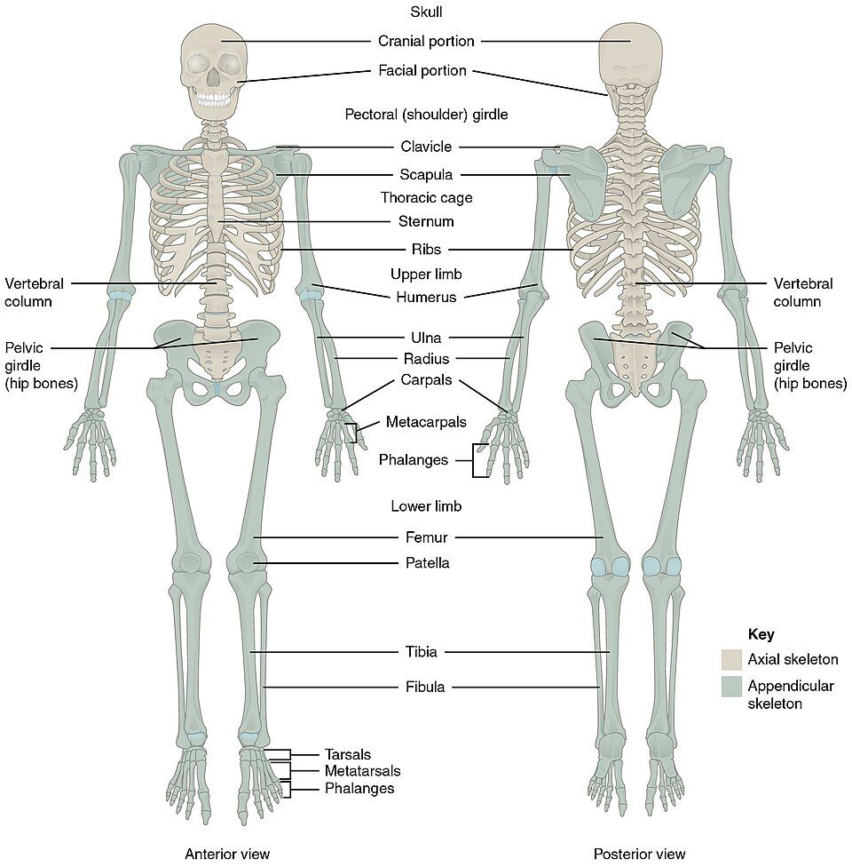
Two skeletons in one
An adult body has 206 bones. Anatomists sort every one of them into two groups. The axial skeleton forms the center line, or axis, of your body. This line runs from the top of the skull, down the spine, and out through the ribs. The axial skeleton has 80 bones. Its job is protection and support. It holds the brain, the spinal cord, the heart, and the lungs, and it gives your trunk a firm core.
The appendicular skeleton is everything that hangs off that center line. These are the appendages, or limbs. It has 126 bones, which make up your arms and legs, plus the two girdles (shoulder and hip) that hold the limbs on. Its job is movement. Here is a quick way to tell them apart. If a bone sits on the midline or wraps around an organ, it is axial. If it helps you reach, walk, or grip, it is appendicular.
What the axial 80 are made of
The eighty axial bones fall into just three groups, and this whole chapter is a tour of them. The skull and the bones that go with it make up twenty-nine. The vertebral column, or backbone, has twenty-six. The thoracic cage, or rib cage, has twenty-five. See how each group matches the organ it guards. The skull wraps the brain, the vertebrae wrap the spinal cord, and the rib cage wraps the heart and lungs. In a real sense, the axial skeleton is your body's suit of armor.
| Region | Bones in it | Count |
|---|---|---|
| Skull | 8 cranial + 14 facial bones | 22 |
| Bones that go with the skull | 6 ear bones (auditory ossicles) + 1 hyoid bone | 7 |
| Vertebral column | 24 vertebrae + sacrum + coccyx | 26 |
| Thoracic cage | 24 ribs (12 pairs) + 1 sternum | 25 |
| Axial total | — | 80 |
Switch between the two skeletons below. Watch the axial and appendicular bones light up in turn.
- The skeleton splits into two parts. The axial part has 80 bones on the center line, for protection and support.
- The appendicular part has 126 bones in the limbs and girdles, for movement.
- The axial skeleton has three parts: the skull, vertebral column, and thoracic cage.
- Axial bones sit on the midline. They guard the brain, spinal cord, heart, and lungs. 80 + 126 = 206.
Section 2The skull
Eight bones cradle the brain
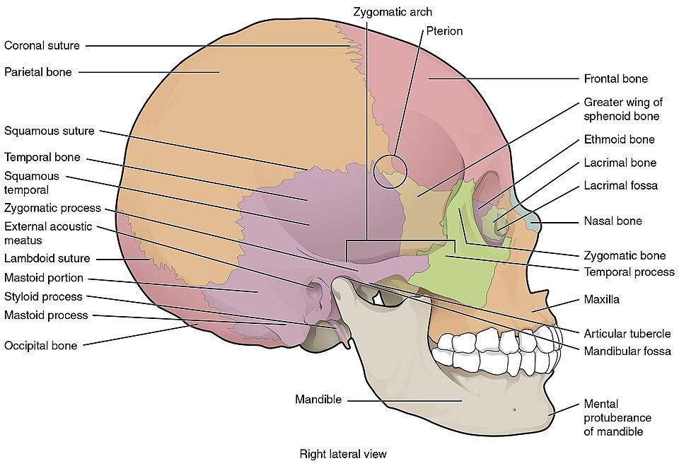
The skull has 22 bones, and the first split is between the part around the brain and the part that builds the face. The cranium is the braincase, and it is made of eight cranial bones. Four of them are single bones on the midline:
The frontal bone is your forehead. The occipital bone is the back and base of the skull. It has a large hole, the foramen magnum, where the spinal cord leaves the skull. The sphenoid bone is shaped like a bat. It spans the floor of the skull and locks the other bones together, like the keystone of an arch. The ethmoid bone is a thin, fragile bone between the eyes. It forms part of the nose cavity.
The other two come in pairs. The two parietal bones form the roof and upper sides. The two temporal bones sit low on each side and hold the ears. Together these bones form a hard dome whose only job is to keep the brain safe.
Fourteen bones build the face
The 14 facial bones give your face its shape, and they frame the openings for seeing, smelling, and eating. Most come in pairs:
The two maxillae form the upper jaw. They join at the midline. The two zygomatic bones are your cheekbones. The two nasal bones form the bridge of the nose. The two lacrimal bones are tiny bones at the inner corner of each eye. They help drain tears. The two palatine bones form the back of the hard palate, the roof of your mouth. The two inferior nasal conchae are curled shelves inside the nose.
Two facial bones are single. The vomer forms the lower part of the wall between your nostrils (the nasal septum). The mandible is the lower jaw. The mandible is special, because it is the only freely movable bone in the skull. It meets the temporal bone at the jaw joint (the temporomandibular joint), which lets you chew and speak.
| Group | Single bones | Paired bones |
|---|---|---|
| Cranial (8) | Frontal, occipital, sphenoid, ethmoid | Parietal (2), temporal (2) |
| Facial (14) | Mandible, vomer | Maxilla (2), zygomatic (2), nasal (2), lacrimal (2), palatine (2), inferior nasal concha (2) |
Sutures, sinuses, and the infant skull
The cranial bones do not meet at smooth joints. Instead, they lock together along jagged seams that cannot move. These seams are called sutures, and they fit together like the teeth of a zipper. Four are worth naming:
The coronal suture runs between the frontal and parietal bones. The sagittal suture runs front to back between the two parietal bones. The lambdoid suture runs between the parietal bones and the occipital bone. The squamous suture runs along the top of each temporal bone.
In a newborn, these seams have not closed yet. The gaps between them are the fontanelles, or "soft spots." They let the skull bend a little during birth and grow as the brain grows.
Some skull bones are also hollowed out by air-filled spaces. These are the paranasal sinuses, the air pockets around the nose. They make the skull lighter and help your voice ring and carry.
Tap any bone on the side view to find it and name it.
- The skull has 22 bones: 8 cranial bones around the brain and 14 facial bones that shape the face.
- The single cranial bones are the frontal, occipital, sphenoid, and ethmoid. The parietal and temporal bones come in pairs.
- The mandible is the only skull bone that moves freely.
- Cranial bones meet at sutures that cannot move. In babies these are still open as fontanelles.
Section 3The vertebral column
Twenty-six bones, five regions
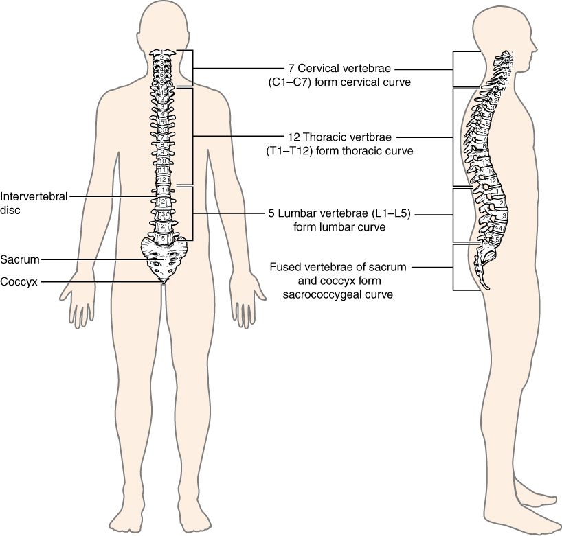
The vertebral column is your backbone, a stack of 26 bones that runs from the base of the skull to the hips. It does three big jobs at once. It supports your head and trunk. It protects the spinal cord that runs down its center. And it bends enough to let you bend and twist.
Each bone in the stack is a vertebra, and together they are called vertebrae. They are grouped into five regions from top to bottom. A classic memory trick links the counts to mealtimes. Eat breakfast at 7, lunch at 12, and dinner at 5:
| Region | Bones | Where it is & notes |
|---|---|---|
| Cervical | 7 (C1–C7) | The neck. C1 is the atlas, and C2 is the axis. |
| Thoracic | 12 (T1–T12) | The chest. Each one holds a pair of ribs. |
| Lumbar | 5 (L1–L5) | The lower back. These are the largest and carry the most weight. |
| Sacral | 1 (5 fused) | The sacrum. It joins the hip bones. |
| Coccygeal | 1 (4 fused) | The coccyx, or tailbone. |
A child actually starts with 33 separate vertebrae. As you grow, five of them fuse into one sacrum and four more fuse into the coccyx, so the adult total ends up at 26. Notice a pattern, too. The vertebrae get steadily larger as you move down the column. That is because each lower bone carries the weight of everything above it. The lumbar vertebrae hold up the whole upper body, so they are the biggest and bulkiest of all.
Discs, the cushions between
From C2 downward, a pad called an intervertebral disc sits between each pair of vertebrae. It has a tough outer ring of fibers, the anulus fibrosus. That ring wraps around a soft, jelly-like center, the nucleus pulposus. Think of a jelly donut, and you have 23 of them in your spine.
The discs do two jobs. They act as shock absorbers that soften the jolt of every step. They also form joints that move a little, so the spine can bend. Sometimes the soft center pushes out through a weak spot in the ring. This is a herniated ("slipped") disc. It can press on a nearby spinal nerve and send pain shooting down an arm or leg.
Tap each region to pick it out and see its vertebrae.
- The column has 26 bones in five regions: 7 cervical, 12 thoracic, 5 lumbar, plus the fused sacrum and coccyx. Remember 7–12–5.
- Vertebrae get larger as you go down, because they carry more weight. The lumbar vertebrae are the largest.
- Intervertebral discs cushion the spine and let it move. A herniated disc can press on a spinal nerve.
Section 4A typical vertebra
The body: the weight-bearer
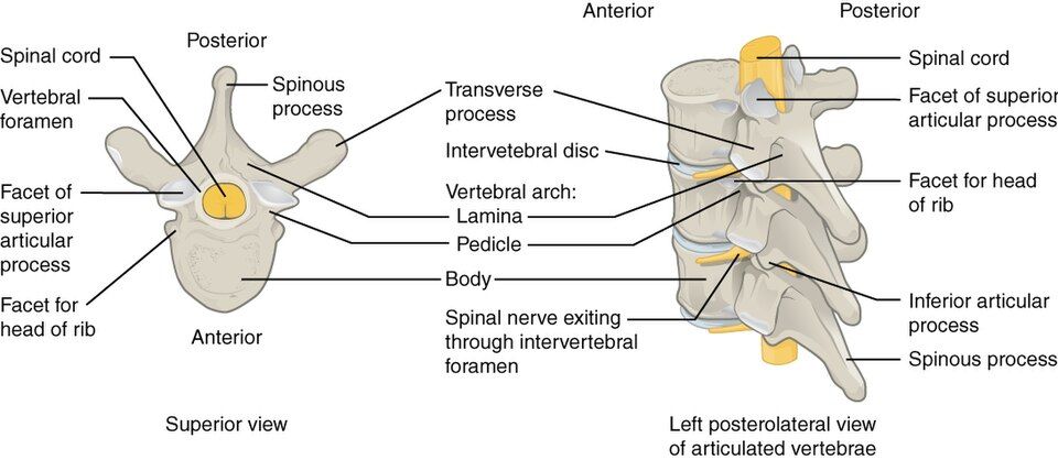
Vertebrae look different from region to region, but they all share one basic plan. The front part of every vertebra is the body, also called the centrum. It is a thick, round block of bone that faces the front (anterior) and does the heavy lifting. The bodies stack on top of each other, with discs between them. Together they form a sturdy pillar that carries the weight of your head and trunk. This is why the bodies get bigger toward the lower back. The lower you go, the more weight there is to hold up.
The arch: the shield
Behind the body is the vertebral arch, a ring of bone that sticks out toward the back. Two short walls rise from the body, and these are the pedicles. Two flat plates form the roof and meet in the middle, and these are the laminae.
The body in front and the arch behind surround a hole. This hole is the vertebral foramen. When all the vertebrae are stacked, these holes line up to form one long tunnel, the vertebral canal. This bony tunnel shelters the spinal cord along its whole length. So the arch works like the spine's version of the skull, and its job is pure protection.
The processes: the handles
Seven bony knobs called processes stick out from the arch, and they work as levers and joints. The single spinous process points straight back. These are the bumps you feel down the middle of your back. The two transverse processes stick out to the sides. Muscles and ligaments grab onto the spinous and transverse processes.
The last four are the paired superior and inferior articular processes. "Superior" means upper, and "inferior" means lower. Each has a smooth, flat surface called a facet. The facets form joints with the vertebra just above and just below, which links the whole column together.
| Part | Number | Job |
|---|---|---|
| Body (centrum) | 1 | Carries weight. Faces the front. |
| Pedicles + laminae | 2 + 2 | Form the vertebral arch |
| Vertebral foramen | 1 | Hole that the spinal cord runs through |
| Spinous process | 1 | Points back. Muscles and ligaments attach here. |
| Transverse processes | 2 | Point to the sides. Muscles and ligaments attach here. |
| Articular processes | 4 | Form joints with the vertebrae above and below |
Look straight down on a vertebra. Tap each part to name it.
- Every vertebra has a weight-bearing body in front. Behind it is a protective arch, made of pedicles and laminae.
- The body and arch surround the vertebral foramen. Stacked up, these holes form the vertebral canal, which holds the spinal cord.
- The spinous and transverse processes hold muscles. The articular processes form joints with the vertebrae next to them.
Section 5The thoracic cage
The sternum: the breastplate
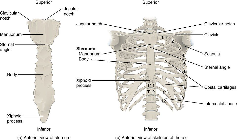
The front of the chest is held in place by the sternum, or breastbone. It is a flat bone shaped like a dagger, with three parts. The wide manubrium sits on top and joins the collarbones. The long body forms the middle. The small xiphoid process is the pointed tip at the bottom, and it starts out as cartilage.
Where the manubrium meets the body, there is a small ridge. This is the sternal angle, also called the angle of Louis. You can feel it through your skin, and it lines up with the second rib. So doctors and nurses use it as a starting point for counting ribs and placing a stethoscope.
Twelve pairs of ribs: true, false, and floating
You have 12 pairs of ribs, which curve from the spine in back around to the front of the chest. Every rib joins a thoracic vertebra in back. But ribs differ in how they reach the sternum in front, or if they reach it at all. That is how we sort them:
The true ribs are pairs 1–7. Each one joins the sternum directly by its own strip of costal cartilage. This is a strip of bendy tissue that links a rib to the breastbone. The false ribs are pairs 8–12. They do not reach the sternum directly. Pairs 8–10 join the costal cartilage of the rib just above them. The last two pairs, 11 and 12, have no front attachment at all. They are called floating ribs, and they end freely in the muscles of the back wall.
| Type | Rib pairs | How they attach in front |
|---|---|---|
| True ribs | 1–7 | Straight to the sternum, each by its own costal cartilage |
| False ribs | 8–10 | Not directly. They join the costal cartilage of the rib above. |
| Floating ribs | 11–12 | None. They do not attach to the sternum. |
A cage that breathes
The thoracic cage is not a solid box. It is a bendable basket that moves with every breath. When you breathe in, muscles lift the ribs up and out. This makes your chest bigger, so the lungs have room to fill with air. When you breathe out, the ribs drop back down. All the while, the cage shields the heart, lungs, and great vessels (the largest blood vessels) inside it. It also gives your breathing and upper-body muscles a frame to pull on, so it is both armor and machine in one.
Tap the sternum, the spine, and each group of ribs to see how the cage fits together.
- The sternum has three parts: the manubrium, body, and xiphoid process. The sternal angle marks the second rib.
- Ribs 1–7 are true ribs that join the sternum directly. Ribs 8–10 are false ribs that join through the rib above. Ribs 11–12 are floating ribs with no front attachment.
- The cage protects the heart and lungs. It expands with each breath.
Section 6Spinal curvatures
Four curves, two born and two learned
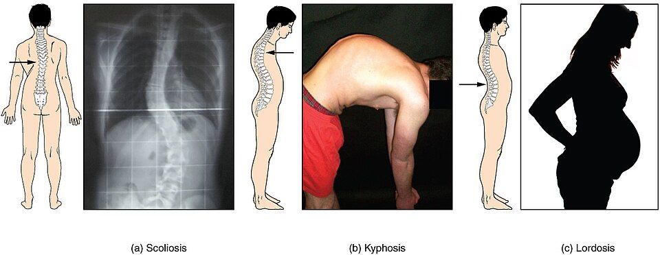
Seen from the side, a healthy spine is not straight. It makes a gentle S-shape with four curves. These curves balance your weight over your hips, and they act like a spring that softens every step.
Two of them are primary curves, which you have at birth. They match the curled-up shape of a baby in the womb. They are the thoracic curve (in the chest) and the sacral curve (in the pelvis). Both bend away from the front, so they are hollow in front.
The other two are secondary curves. They form after birth, as a baby learns to hold up its head and then to walk. The cervical curve appears when a baby starts to lift its head. The lumbar curve appears when a toddler stands and walks. Together, the four curves let the spine carry a heavy head and trunk without tipping over.
| Curve | Type | When it appears |
|---|---|---|
| Cervical | Secondary | When a baby lifts its head |
| Thoracic | Primary | There at birth |
| Lumbar | Secondary | When a toddler stands and walks |
| Sacral | Primary | There at birth |
When curves go wrong
The curves are healthy when they are gentle. But a curve can become too deep or bend the wrong way, and then it gets a medical name.
Kyphosis is a thoracic curve that is too deep. It gives a rounded, hunched upper back, sometimes called "hunchback." It is common in older adults with osteoporosis (weak, thin bones). Lordosis is a lumbar curve that is too deep. The lower back sways inward too much. This is called "swayback." Scoliosis is different from both. It is a sideways (lateral) curve of the spine. Seen from behind, the spine bends into a C or an S shape.
Children are checked for scoliosis at school. Finding it early, while the skeleton is still growing, gives the best chance to fix it.
Compare the normal S-curve with kyphosis, lordosis, and scoliosis side by side.
- The spine has four curves. The primary curves (thoracic, sacral) are there at birth.
- The secondary curves (cervical, lumbar) form later, as a baby lifts its head and learns to walk.
- Kyphosis is a thoracic curve that is too deep. Lordosis is a lumbar curve that is too deep. Both are front-to-back curves.
- Scoliosis is an abnormal lateral (sideways) curve. Children are checked for it while they grow.
The chapter in one breath
- The skeleton splits in two. The axial part has 80 bones in the skull, spine, and rib cage, for protection and support. The appendicular part has 126 bones in the limbs and girdles, for movement.
- The skull has 22 bones: 8 cranial bones around the brain and 14 facial bones. The mandible is the only one that moves. The others are joined by sutures that do not move.
- The vertebral column has 26 bones in five regions: 7 cervical, 12 thoracic, 5 lumbar, plus the fused sacrum and coccyx. Discs cushion the bones.
- Every vertebra has a weight-bearing body. It has an arch that surrounds the vertebral foramen. It also has processes for muscles and joints.
- The thoracic cage is the three-part sternum plus 12 pairs of ribs. The ribs are true, false, and floating. The cage guards the heart and lungs and moves with each breath.
- The spine's four curves form a springy S. The thoracic and sacral curves are primary. The cervical and lumbar curves are secondary. Problem curves are kyphosis, lordosis, and the sideways scoliosis.
- One theme runs through it all. The axial skeleton is armor for your most vital organs.
ReferenceWords worth knowing
Here is a quick list of the terms in this chapter. Skim it now, or come back when a word trips you up.
- Appendicular skeleton
- The 126 bones of the limbs and the two girdles (shoulder and hip) that hold them on. They are built for movement.
- Atlas (C1)
- The first neck vertebra. It holds up the skull and lets your head nod "yes."
- Axial skeleton
- The 80 bones along the center line of the body: the skull, spine, and rib cage. They protect and support.
- Axis (C2)
- The second neck vertebra. It has a peg that points up and lets your head turn to say "no."
- Coccyx
- The small tailbone at the bottom of the spine. It is made of four fused vertebrae.
- Costal cartilage
- The strip of cartilage that links a rib to the sternum. It joins directly, or through the rib above.
- Cranial bones
- The eight skull bones that surround the brain. They are the frontal, occipital, sphenoid, and ethmoid, plus the paired parietals and temporals.
- Facial bones
- The fourteen skull bones that shape the face. They frame the eyes, nose, and mouth.
- Floating ribs
- Rib pairs 11 and 12. They attach only to the vertebrae in back and have no front connection.
- Fontanelle
- A soft gap of tough tissue between a baby's skull bones. It lets the skull bend at birth and grow with the brain.
- Foramen magnum
- The large hole in the occipital bone. Here the spinal cord joins the brain.
- Intervertebral disc
- A pad of tough cartilage between two vertebral bodies. It absorbs shock and lets the spine move.
- Kyphosis
- A thoracic curve that bends back too far. It gives a hunched upper back.
- Lordosis
- A lumbar curve that bends forward too far. It gives an overdone swayback.
- Mandible
- The lower jaw. It is the only skull bone that moves freely.
- Manubrium
- The wide top part of the sternum. It forms joints with the collarbones (clavicles) and the first ribs.
- Sacrum
- A triangle-shaped bone made of five fused vertebrae. It joins the spine to the hip bones.
- Scoliosis
- An abnormal sideways (lateral) curve of the spine.
- Sternal angle
- The ridge you can feel where the manubrium meets the body of the sternum. It lines up with the second rib.
- Suture
- A joint between two cranial bones. The bones lock together and cannot move.
- Vertebral foramen
- The central hole of a vertebra, formed by the body and arch. The spinal cord runs through it.
- Xiphoid process
- The small pointed tip at the bottom of the sternum. It is a key landmark for CPR.