The Reproductive System
Every other system in this book works to keep you alive. The reproductive system is the one that looks past you — its job is to make a new person. To do that, the body must accomplish something no other tissue attempts: it must build a special cell, a gamete, that carries exactly half of your genetic instructions, so that when a sperm meets an egg the full set is restored. This chapter walks both anatomies, the male path from testis to exit and the female path from ovary to uterus. It follows the two production lines — spermatogenesis and oogenesis — that turn stem cells into gametes, and it traces the hormonal tide of the menstrual cycle. Underneath all of it sits one elegant kind of cell division, meiosis, which we save for last because once you see it, the rest of the chapter clicks into place.
By the end of this chapter you can
- Trace the path of sperm from the seminiferous tubules to the urethra, and name the glands that add fluid along the way.
- Describe spermatogenesis — how a diploid stem cell becomes four haploid sperm — and where and how long it takes.
- Identify the major female reproductive organs and give the function of the ovary, uterine tube, uterus, and their layers.
- Explain oogenesis and how its unequal divisions and lifelong arrest differ from sperm production.
- Sequence the phases of the ovarian and uterine cycles and match each to its dominant hormone (FSH, LH, estrogen, progesterone).
- Distinguish meiosis from mitosis and explain how crossing over and independent assortment create genetic variety.
- Define diploid and haploid and explain why halving the chromosome number is essential to sexual reproduction.
Section 1Male reproductive anatomy
Two jobs: make sperm, deliver sperm
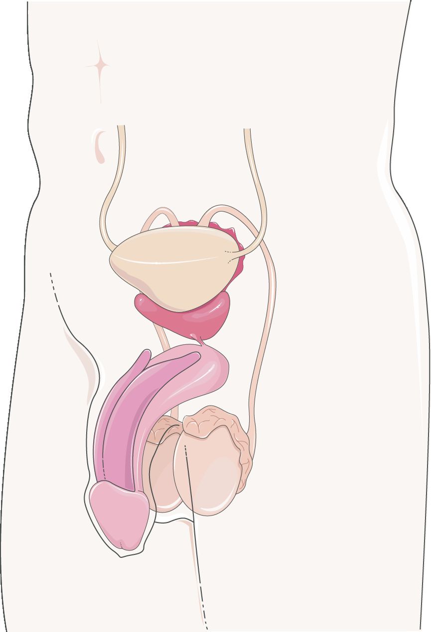
The male system has a wonderfully simple mandate — produce sperm and deliver them, suspended in a nourishing fluid, to the female tract. Everything in the anatomy serves one of those two aims. Sperm are manufactured in the paired testes, which hang outside the body wall inside the scrotum. That external location is not an accident: sperm production needs a temperature a few degrees below core body heat, and the scrotum, with the help of a small muscle that raises and lowers the testes, acts as a thermostat. Inside each testis are tightly coiled seminiferous tubules, and it is along the walls of these tubules that sperm are born.
Following the road out
Once made, sperm are not yet able to swim or to fertilize. They travel first into the epididymis, a comma-shaped coil of tubing draped over the back of each testis, where over roughly two weeks they mature and gain motility and are stored. From there they enter the ductus (vas) deferens, a muscular tube that climbs out of the scrotum, loops over the bladder, and joins the duct of the seminal vesicle to form the ejaculatory duct. That short duct passes through the prostate and empties into the urethra, the final common passage that carries semen out through the penis. Notice that in the male the urethra is shared plumbing — it carries both urine and semen, though never at the same time.
| Structure | Role in the journey | Note |
|---|---|---|
| Seminiferous tubules | Site of sperm production | Coiled inside each testis |
| Epididymis | Sperm mature, gain motility, and are stored | ~6 m of coiled tube on the testis |
| Ductus (vas) deferens | Propels sperm toward the urethra | Cut and tied in a vasectomy |
| Ejaculatory duct | Joins sperm with seminal vesicle fluid | Passes through the prostate |
| Urethra | Carries semen (and urine) out | Shared passage in the male |
The glands that make semen
Sperm make up only a small fraction of semen — most of the volume is fluid added by three accessory glands, and each fluid has a purpose. The paired seminal vesicles contribute the largest share: a thick, sugar-rich fluid whose fructose fuels the sperm's tails. The single prostate gland, which encircles the urethra just below the bladder, adds a milky, slightly alkaline fluid that helps sperm survive the acidic environment of the female tract. Finally, the small bulbourethral glands release a clear pre-ejaculate that lubricates and neutralizes traces of acidic urine in the urethra. Together these secretions activate the sperm, feed them, and protect them for the trip ahead.
Tap each structure in the interactive to trace the sperm's route from where it is made to the exit.
- Sperm are made in the seminiferous tubules of the testes, which sit in the scrotum to stay slightly cooler than the body.
- The road out runs testis → epididymis → ductus deferens → ejaculatory duct → urethra.
- Three glands add fluid: seminal vesicles (fructose), prostate (alkaline), and bulbourethral (lubricating).
- The male urethra is shared plumbing for both urine and semen.
Section 2Spermatogenesis
Turning a stem cell into a swimmer
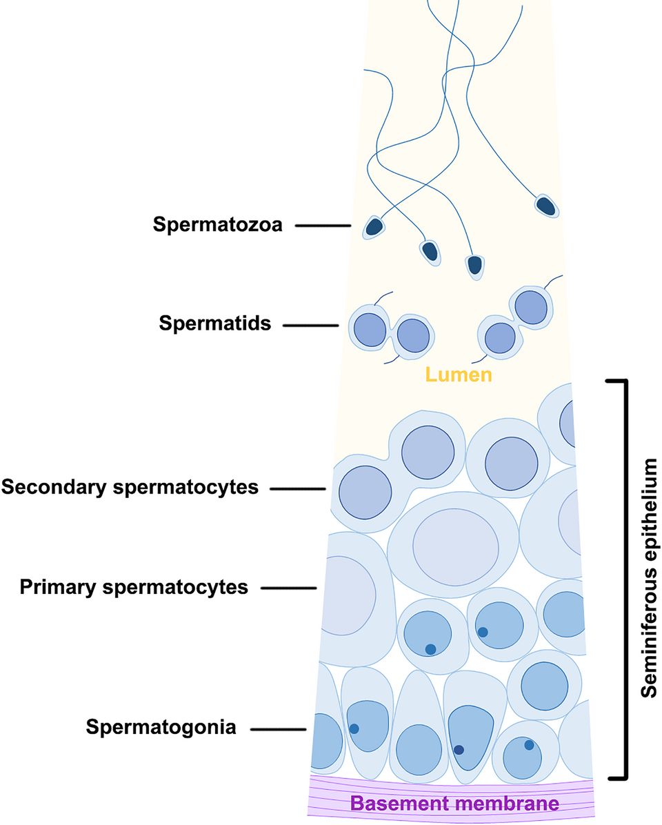
Spermatogenesis is the assembly line that converts a diploid stem cell into mature sperm, and it runs continuously along the walls of the seminiferous tubules from puberty onward. It begins with a spermatogonium, a stem cell carrying the full 46 chromosomes (the diploid, or 2n, number). One daughter of each division stays behind as a stem cell — which is why a male never runs out — while the other, now a primary spermatocyte, commits to becoming sperm. That primary spermatocyte enters meiosis, the special division that halves the chromosome count.
Two divisions, four cells
The math of spermatogenesis is clean. The primary spermatocyte completes meiosis I to become two secondary spermatocytes, each now carrying the haploid (n = 23) number. Each of those completes meiosis II, yielding a total of four spermatids. Unlike egg production, the divisions here are equal — every cell gets a fair share of cytoplasm, so one starting cell produces four usable products. The spermatids are round and immobile; they still must be remodeled into streamlined sperm in a final step called spermiogenesis, in which the cell sheds excess cytoplasm, packs its DNA into a compact head, caps it with an enzyme-filled acrosome, and grows the whip-like flagellum (tail) it will swim with. A midpiece stuffed with mitochondria supplies the energy for the trip.
| Stage | Chromosomes | What happens |
|---|---|---|
| Spermatogonium | Diploid (2n) | Stem cell; one daughter self-renews |
| Primary spermatocyte | Diploid (2n) | Enters and completes meiosis I |
| Secondary spermatocyte | Haploid (n) | Two cells; complete meiosis II |
| Spermatid | Haploid (n) | Four cells; round and non-motile |
| Sperm (spermatozoon) | Haploid (n) | Head + acrosome + midpiece + tail |
Helped along by nurse cells
The developing cells do not fend for themselves. Tucked among them are tall Sustentacular (Sertoli) cells, sometimes called nurse cells, that nourish the maturing sperm and form the blood–testis barrier, a protective wall that keeps the immune system from attacking sperm as if they were foreign. Between the tubules sit the interstitial (Leydig) cells, which produce testosterone — the hormone that drives sperm production and the male secondary sex characteristics. The whole cycle, from spermatogonium to finished sperm, takes about 64 to 74 days, and a healthy male makes hundreds of millions of sperm every single day.
Step through each division in the interactive to watch a stem cell become four swimming sperm.
- A diploid spermatogonium yields four haploid sperm through two equal meiotic divisions.
- Spermiogenesis remodels round spermatids into streamlined sperm with an acrosome, midpiece, and tail.
- Sertoli cells nurse the sperm and form the blood–testis barrier; Leydig cells make testosterone.
- The full cycle takes about 64–74 days, and production is continuous.
Section 3Female reproductive anatomy
A place to make an egg and a place to grow a baby
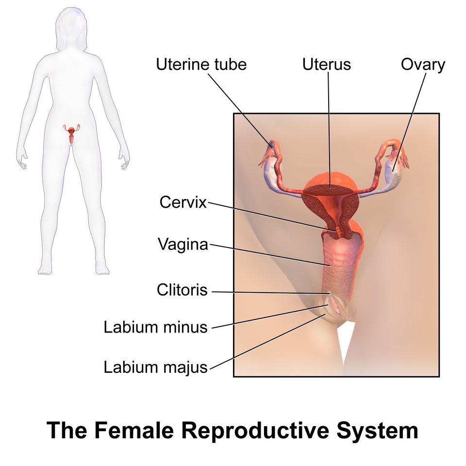
The female system does two things the male system does not: it not only makes the gamete, it also provides the place where a fertilized egg can implant and develop for nine months. The paired ovaries, each about the size of an almond, sit low in the pelvis and hold the lifetime supply of eggs. Once a month an ovary releases a mature egg in ovulation. Reaching toward each ovary is a uterine (fallopian) tube, whose fringed, finger-like end — the fimbriae — sweeps the released egg into the tube. Fertilization, when it happens, usually takes place here, in the outer third of the tube.
The uterus and its layers
The tube carries the egg toward the uterus, a muscular, pear-shaped organ built to house a growing fetus. Its wall has three layers worth knowing. The inner endometrium is the lining that thickens each month to receive an embryo and, if none arrives, is shed as the menstrual period. The thick middle myometrium is smooth muscle that will contract powerfully during labor. The thin outer perimetrium is the serous covering. The lower neck of the uterus, the cervix, opens into the vagina, the muscular canal that serves as the birth canal and receives sperm during intercourse.
| Organ | Function | Note |
|---|---|---|
| Ovary | Produces eggs and the hormones estrogen & progesterone | Holds all eggs from birth |
| Uterine (fallopian) tube | Carries the egg toward the uterus; usual site of fertilization | Fimbriae sweep in the egg |
| Uterus | Houses and nourishes the developing fetus | Endometrium / myometrium / perimetrium |
| Cervix | Neck of the uterus; opens into the vagina | Dilates in labor |
| Vagina | Birth canal; receives semen | Muscular, distensible |
The ovary is also a gland
It is easy to think of the ovary as just an egg store, but it is equally an endocrine organ. Each month a cluster of cells around the maturing egg — the follicle — pumps out estrogen, and after ovulation the leftover follicle transforms into the corpus luteum, which secretes progesterone. These two hormones drive the changes in the uterine lining and, working with the brain, run the whole monthly cycle we will meet in Section 5. So the ovary does double duty: it releases the gamete and orchestrates the tissue that will receive it.
Tap each structure in the interactive to explore the female organs from the front.
- The ovaries store eggs and act as glands, making estrogen and progesterone.
- The uterine tube catches the ovulated egg with its fimbriae; fertilization usually occurs in its outer third.
- The uterus wall has three layers: endometrium (shed lining), myometrium (labor muscle), perimetrium (serous cover).
- The cervix opens into the vagina, the birth canal.
Section 4Oogenesis
The same tool, used very differently
Egg production, or oogenesis, uses the very same meiosis as sperm production, but the female body runs the process on a completely different schedule and with a completely different goal. Where the male makes four small sperm from one cell and does it endlessly, the female makes one large egg from one cell and does it on a strict, once-a-month timetable. The reason for the difference is investment: an egg must carry enough cytoplasm, nutrients, and organelles to launch a new organism, so the female strategy is to pour almost everything into a single winner.
Unequal divisions on purpose
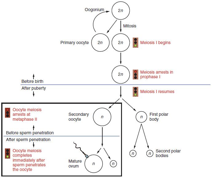
Here is the twist. When a primary oocyte completes meiosis I, it does not split its cytoplasm evenly. Instead it divides unequally: one daughter, the secondary oocyte, keeps nearly all the cytoplasm, while the other becomes a tiny polar body that gets almost nothing and eventually degenerates. The same lopsided split happens again at meiosis II — but note that in humans this second division only completes if a sperm fertilizes the egg. The result of the whole process is one functional egg (ovum) plus two or three doomed polar bodies. One cell in, one usable egg out — the mirror image of the male's one-in-four.
| Feature | Oogenesis (egg) | Spermatogenesis (sperm) |
|---|---|---|
| Products per starting cell | One egg (+ polar bodies) | Four sperm |
| Cytoplasm split | Unequal — egg keeps it all | Equal |
| Timing | Begins before birth; one per cycle | Continuous from puberty |
| Completion of meiosis II | Only if fertilized | Always completed |
| Lifetime output | ~400 eggs ovulated | Trillions of sperm |
A process paused for decades
Oogenesis also has the strangest timeline in the body. A female is born with all the eggs she will ever have — roughly one to two million primary oocytes, already begun on meiosis I and then frozen partway through, arrested before birth. They wait, paused, for years. Beginning at puberty, one oocyte finishes meiosis I each month and is ovulated; the rest keep waiting. This means an egg ovulated at age forty has been sitting in suspended animation for four decades — a delay that helps explain why the chance of chromosomal errors, such as those causing Down syndrome, rises with maternal age. Supply dwindles over the reproductive years until, at menopause, only about a thousand follicles remain — too few and too unresponsive to keep the hormonal cycle going — and the cycles stop.
Step through the interactive to watch the unequal divisions concentrate everything into one egg.
- Oogenesis makes one egg (plus polar bodies) per starting cell, through unequal cytoplasm divisions.
- All eggs begin meiosis before birth and arrest; one finishes per cycle, and meiosis II completes only at fertilization.
- A female is born with her lifetime egg supply, which declines to a critically low level — on the order of a thousand follicles — by menopause, at which point cycling stops.
- Long meiotic arrest helps explain the rising rate of chromosomal errors with maternal age.
Section 5The menstrual cycle
Two cycles running as one
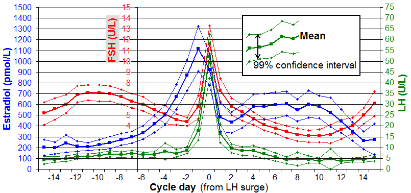
The roughly 28-day menstrual cycle is really two synchronized cycles — one in the ovary and one in the uterus — conducted by four hormones. Two come from the brain's pituitary gland: follicle-stimulating hormone (FSH) and luteinizing hormone (LH). Two come from the ovary itself: estrogen and progesterone. The ovarian cycle tracks what is happening to the egg and its follicle; the uterine cycle tracks the building and shedding of the endometrial lining. They are timed so that a fresh lining is ready exactly when an egg might arrive.
Follicular phase, ovulation, luteal phase
The cycle opens with menstruation — the shedding of last month's lining — on day 1. During the follicular phase that follows, FSH coaxes several follicles to grow; one becomes dominant and pours out estrogen, which rebuilds the endometrium. Rising estrogen eventually triggers a dramatic surge of LH around day 14, and that LH surge causes ovulation — the dominant follicle bursts and releases its egg. In the luteal phase afterward, the emptied follicle becomes the corpus luteum, which secretes progesterone to maintain and enrich the lining, readying it for a possible implantation. If no pregnancy occurs, the corpus luteum withers, progesterone and estrogen fall, and the unsupported lining sheds — and the next cycle begins.
| Phase (days) | Dominant hormone | What is happening |
|---|---|---|
| Menstruation (1–5) | Estrogen and progesterone low; FSH rising | Old endometrium is shed while the next follicles are recruited |
| Follicular (1–13) | FSH, then estrogen | A follicle ripens; lining rebuilds |
| Ovulation (~14) | LH surge | Follicle bursts; egg released |
| Luteal (15–28) | Progesterone | Corpus luteum enriches the lining |
Feedback that flips
What makes this cycle beautiful is that the same hormone can push in opposite directions depending on timing. For most of the cycle, estrogen exerts negative feedback, quietly restraining the pituitary. But as the dominant follicle matures, estrogen climbs to a high, sustained level, and at that threshold the feedback flips to positive — high estrogen now drives the pituitary to release the LH surge that triggers ovulation. It is one of the body's few examples of positive feedback, and it exists for exactly the reason positive feedback always does: to produce a decisive, one-time event. After ovulation, progesterone from the corpus luteum re-establishes negative feedback and calms everything down.
Tap a phase in the interactive to see the hormones, the ovary, and the lining move together.
- Four hormones run the cycle: FSH and LH from the pituitary, estrogen and progesterone from the ovary.
- The phases are menstruation → follicular → ovulation (LH surge, ~day 14) → luteal.
- Follicles make estrogen (rebuild lining); the corpus luteum makes progesterone (maintain lining).
- A high estrogen level flips to positive feedback to trigger the ovulatory LH surge.
Section 6Meiosis
Why half is the whole point
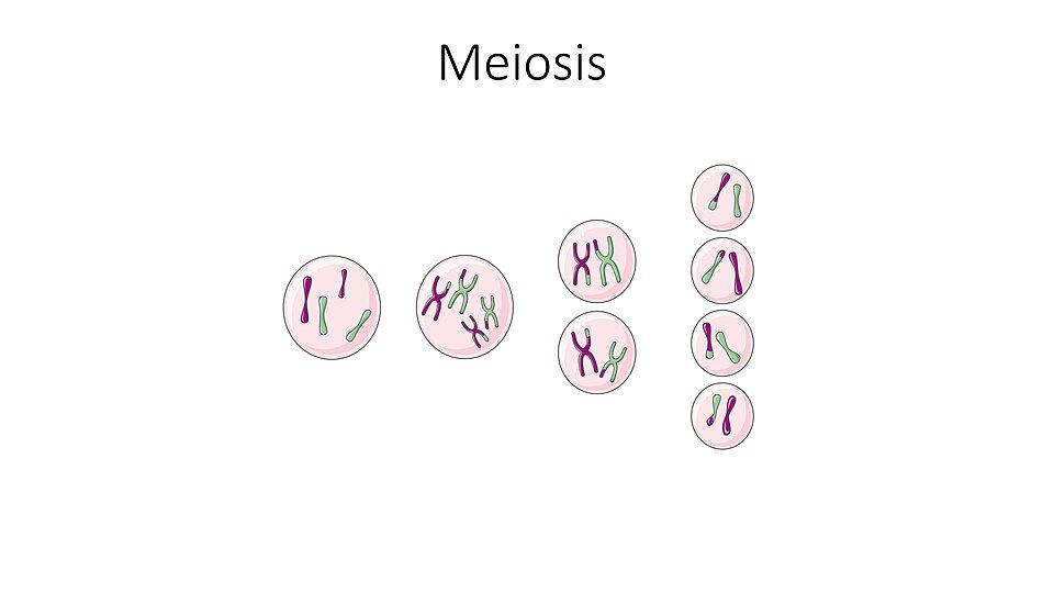
We have saved the engine for last. Every body cell you own is diploid — it carries 46 chromosomes, arranged as 23 pairs, one member of each pair from each of your parents. If a sperm with 46 chromosomes fertilized an egg with 46, the child would have 92, and the number would double every generation. Nature's solution is meiosis, a special division that makes haploid gametes with just 23 chromosomes. When a haploid sperm meets a haploid egg, the full diploid set of 46 is restored — and the count stays stable forever. Halving the chromosome number is not a side effect of gamete production; it is the reason meiosis exists.
Two divisions, not one
Meiosis reaches the haploid number through two back-to-back divisions after a single DNA copy. In meiosis I, the matched pairs of chromosomes (called homologous pairs) line up and are separated — this is the reduction step that takes the cell from diploid to haploid. In meiosis II, which looks much like an ordinary mitosis, the two copies of each chromosome finally split apart. The end result is four haploid cells (four sperm, or one egg plus polar bodies). Compare that with mitosis, the ordinary division that builds and repairs the body: mitosis is one division that makes two identical diploid cells.
| Feature | Mitosis | Meiosis |
|---|---|---|
| Purpose | Growth and repair | Make gametes |
| Divisions | One | Two |
| Daughter cells | 2 | 4 |
| Chromosome number | Diploid (2n) — unchanged | Halved to haploid (n) |
| Genetically | Identical clones | Unique — all different |
Shuffling the deck
Meiosis does more than halve — it reshuffles your genes so that no two gametes are alike. It does this two ways. In crossing over, homologous chromosomes swap matching segments while paired up in meiosis I, blending the versions you inherited from each parent. In independent assortment, each pair lines up and separates at random, so which grandparent's chromosome ends up in a given gamete is a coin flip — done 23 times over. Together these produce millions of genetically distinct gametes from one person, which is why siblings differ and why sexual reproduction generates the variety that evolution acts on. When something goes wrong here and a pair fails to separate — nondisjunction — a gamete ends up with the wrong number of chromosomes, the cause of conditions such as trisomy 21.
Step through both divisions in the interactive to watch the chromosomes halve and the genes shuffle.
- Meiosis halves the chromosome number from diploid (46) to haploid (23) so fertilization can restore the full set.
- It uses two divisions: meiosis I separates homologous pairs (the reduction step); meiosis II splits the sister copies.
- Crossing over and independent assortment make every gamete genetically unique.
- Nondisjunction — a pair failing to separate — gives a gamete the wrong chromosome number.
The chapter in one breath
- The reproductive system exists to make a new person, which requires haploid gametes that combine to restore the full chromosome set.
- In the male, sperm made in the seminiferous tubules travel epididymis → ductus deferens → ejaculatory duct → urethra, mixed with fluid from the seminal vesicles, prostate, and bulbourethral glands.
- Spermatogenesis makes four sperm per stem cell through equal divisions, continuously, over ~64–74 days.
- In the female, the ovary both stores eggs and makes hormones; the uterine tube catches the egg and the uterus (endometrium, myometrium, perimetrium) houses a pregnancy.
- Oogenesis makes one egg plus polar bodies through unequal divisions, starts before birth, and completes meiosis II only at fertilization.
- The menstrual cycle is run by FSH, LH, estrogen, and progesterone: follicles make estrogen, an LH surge triggers ovulation, and the corpus luteum makes progesterone.
- Meiosis halves the chromosomes in two divisions — meiosis I reduces, meiosis II divides — and crossing over plus independent assortment make every gamete unique.
ReferenceWords worth knowing
A quick reference for the terms in this chapter — skim it now, or circle back whenever a word trips you up.
- Acrosome
- The enzyme-filled cap over a sperm's head that helps it penetrate the egg's outer coat.
- Corpus luteum
- The structure formed from the emptied follicle after ovulation; it secretes progesterone to maintain the uterine lining.
- Diploid (2n)
- Having the full set of chromosomes — 46 in humans, arranged as 23 pairs; the state of body cells.
- Ductus (vas) deferens
- The muscular tube that propels sperm from the epididymis toward the urethra; cut in a vasectomy.
- Endometrium
- The inner lining of the uterus that thickens each cycle to receive an embryo and is shed as menstruation if none implants.
- Epididymis
- The coiled tube on the back of the testis where sperm mature, gain motility, and are stored.
- Estrogen
- The ovarian hormone, made mainly by the growing follicle, that rebuilds the uterine lining and triggers the LH surge.
- Follicle
- The cluster of ovarian cells surrounding a developing egg; it secretes estrogen and releases the egg at ovulation.
- FSH (follicle-stimulating hormone)
- A pituitary hormone that stimulates follicle growth in the ovary and sperm production in the testis.
- Gamete
- A haploid reproductive cell — a sperm or an egg — that fuses with another at fertilization.
- Haploid (n)
- Having half the chromosome set — 23 in humans; the state of gametes.
- LH (luteinizing hormone)
- A pituitary hormone whose mid-cycle surge triggers ovulation and forms the corpus luteum.
- Meiosis
- The two-division process that halves the chromosome number to make haploid gametes and shuffles genes.
- Menopause
- The permanent end of menstrual cycles, reached when the ovaries’ remaining follicles are too few and too unresponsive to sustain the hormonal cycle — the supply runs critically low rather than to zero.
- Nondisjunction
- The failure of a chromosome pair to separate during meiosis, giving a gamete the wrong chromosome number.
- Oogenesis
- The production of eggs; unequal meiotic divisions yield one ovum and two or three polar bodies.
- Ovulation
- The release of a mature egg from the ovary, triggered by the mid-cycle LH surge.
- Polar body
- A small cell produced by the unequal divisions of oogenesis that receives little cytoplasm and degenerates.
- Progesterone
- The hormone made by the corpus luteum that maintains and enriches the uterine lining after ovulation.
- Semen
- The fluid ejaculated from the male tract, made of sperm plus secretions of the seminal vesicles, prostate, and bulbourethral glands.
- Seminiferous tubules
- The coiled tubes inside the testis where sperm are produced.
- Spermatogenesis
- The production of sperm; equal meiotic divisions yield four haploid sperm from one stem cell.