Blood Vessels & Circulation
The heart is only the pump — the real work of the circulation happens in the tubes. In this chapter you will trace a single red blood cell from the moment it is flung out of the heart, through arteries that branch into ever-finer channels, into capillaries so thin that oxygen and nutrients leak across their walls into your tissues, and then back up through the veins against gravity to start the loop again. Along the way you will learn why an artery wall is thick and a capillary wall is one cell thick, what blood pressure really measures, how fluid decides whether to leave the blood or return to it, and the clever tricks your body uses to lift blood out of your feet. Master the vessels and you will understand not just how blood moves, but why swollen ankles, varicose veins, and a stroke all trace back to the same set of rules.
By the end of this chapter you can
- Name the three tunics of a blood vessel wall and describe the tissue and job of each.
- Compare arteries, veins, and capillaries by wall thickness, lumen size, pressure, and function.
- Define blood pressure, distinguish systolic from diastolic, and calculate pulse pressure and mean arterial pressure.
- Explain how pressure and resistance shape blood flow, and why the arterioles are the body's main resistance vessels.
- Diagram capillary exchange as a tug-of-war between hydrostatic and colloid osmotic pressure, and explain how the balance can produce edema.
- Distinguish the pulmonary and systemic circuits and describe special routes such as the hepatic portal system.
- List the mechanisms of venous return — the skeletal muscle pump, the respiratory pump, and one-way valves — and connect their failure to real disease.
Section 1The wall of a blood vessel
Three layers around a hollow center
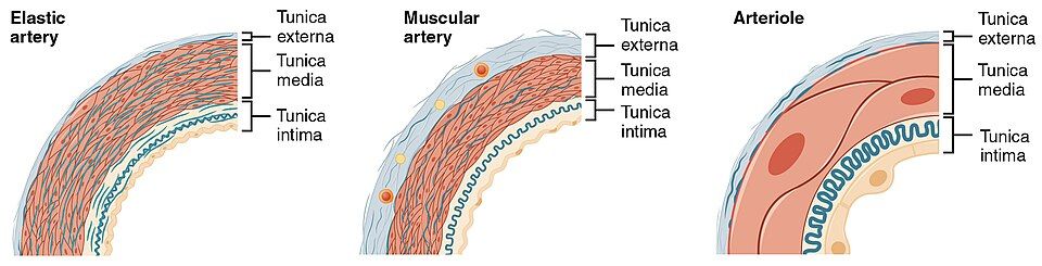
Every artery and vein is built the same way: as a set of concentric tunics (from the Latin for "coats") wrapped around a hollow channel called the lumen, the space the blood actually flows through. Reading from the inside out, the three layers are the tunica intima, the tunica media, and the tunica externa. The layers are the same in every vessel; what changes from an artery to a vein to a capillary is simply how thick each layer is and how much muscle or elastic tissue it contains. Learn the three coats once and you can read the wall of any vessel in the body.
Intima, media, externa — from lining to anchor
The innermost coat, the tunica intima, is dominated by a single sheet of flat cells called the endothelium that lines the entire cardiovascular system in one continuous surface. This lining is far more than wallpaper: it is smooth enough that blood cells glide past without clotting, and it releases chemical signals (such as nitric oxide) that tell the muscle underneath to relax or contract. Beneath it sits a thin basement membrane and, in arteries, a springy sheet called the internal elastic lamina.
The middle coat, the tunica media, is usually the thickest and is the muscle of the vessel. It is made of smooth muscle arranged in rings, mixed with sheets of elastic fibers. When the smooth muscle tightens, the lumen narrows — vasoconstriction; when it relaxes, the lumen widens — vasodilation. Because this layer is controlled by sympathetic nerves and by hormones, the tunica media is the dial your nervous system turns to route blood where it is needed and to set blood pressure. The outermost coat, the tunica externa (also called the tunica adventitia), is a tough sleeve of collagen fibers that anchors the vessel to surrounding tissue and keeps it from over-stretching. In large vessels it even carries its own tiny blood supply, the vasa vasorum — "vessels of the vessels" — because the wall is too thick to feed itself by diffusion.
| Tunic | Main tissue | Job |
|---|---|---|
| Tunica intima | Endothelium + basement membrane (+ internal elastic lamina in arteries) | Smooth, non-stick lining; releases signals that relax or constrict the wall |
| Tunica media | Smooth muscle + elastic fibers | Vasoconstriction and vasodilation; sets vessel diameter and blood pressure |
| Tunica externa | Collagen (and elastic) connective tissue | Anchors and protects the vessel; carries vasa vasorum in large vessels |
Peel the wall apart layer by layer in the interactive below.
- Every vessel wall has three tunics around a central lumen: intima, media, externa.
- The tunica intima's endothelium is a continuous, non-stick lining that signals the wall to relax or constrict.
- The tunica media (smooth muscle) sets vessel diameter through vasoconstriction and vasodilation.
- The tunica externa (collagen) anchors the vessel; atherosclerosis begins in the injured intima.
Section 2Arteries, veins & capillaries
Arteries: the high-pressure pipes
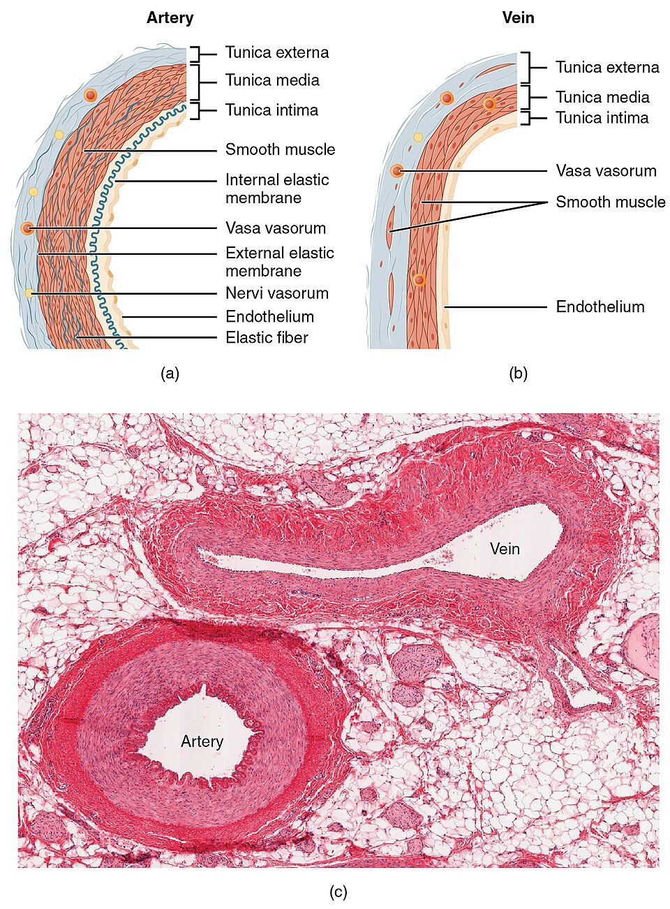
An artery is any vessel that carries blood away from the heart — a definition based on direction, not on oxygen content, which matters later. Because arteries receive blood at high pressure with every heartbeat, they have a thick, muscular tunica media and strong elastic walls. The largest arteries near the heart, such as the aorta, are elastic (conducting) arteries: their walls stretch as the ventricle ejects blood and recoil between beats, smoothing the pulsing flow into a steadier stream. Farther out, muscular (distributing) arteries deliver blood to specific organs, and the smallest branches, the arterioles, act as adjustable nozzles that control how much blood enters each capillary bed.
Veins: the low-pressure reservoirs
A vein carries blood back toward the heart. By the time blood reaches the veins it has spent nearly all its pressure crossing the capillaries, so veins do not need thick muscular walls. Instead they have a thin media, a large floppy lumen, and a prominent tunica externa. Their most distinctive feature is a set of one-way valves — flaps of intima that snap shut to stop blood from sliding backward. Because veins are so distensible, they act as the body's blood reservoir: at rest, roughly two-thirds of your total blood volume is sitting in the veins, ready to be squeezed back into circulation when demand rises. For this reason veins are also called capacitance vessels.
Capillaries: where the exchange happens
Between the smallest arteriole and the smallest venule lie the capillaries — the reason the whole cardiovascular system exists. A capillary wall is nothing but a single layer of endothelium on a thin basement membrane: just the tunica intima, stripped down to its essentials. That paper-thinness is the point, because it lets oxygen, carbon dioxide, glucose, and wastes slip across the wall between blood and tissue. Capillaries are so numerous that no cell in the body sits more than about a tenth of a millimeter from one. They come in three grades of leakiness — continuous (tightest, as in muscle and brain), fenestrated (with pores, as in the kidney and intestine), and sinusoidal (widest gaps, as in the liver and bone marrow) — each tuned to how much needs to cross.
| Feature | Artery | Capillary | Vein |
|---|---|---|---|
| Direction | Away from heart | Through tissues | Toward heart |
| Wall | Thick, muscular, elastic | One endothelial cell thick | Thin, large lumen |
| Pressure | High | Falling | Low |
| Valves | None (aortic/pulmonary valves are in the heart) | None | Yes — one-way |
| Main job | Carry & distribute pressurized blood | Exchange O₂, CO₂, nutrients, wastes | Return blood; store volume (reservoir) |
Switch between the three vessel types and watch their jobs change.
- Arteries carry blood away from the heart under high pressure and have thick, elastic, muscular walls.
- Veins carry blood toward the heart, have thin walls, one-way valves, and hold most of the blood as a reservoir.
- Capillaries are one cell thick — the only place O₂, CO₂, and nutrients are exchanged.
- Artery vs. vein is defined by direction of flow, not by oxygen content.
Section 3Blood pressure & the pressure gradient
Systolic over diastolic
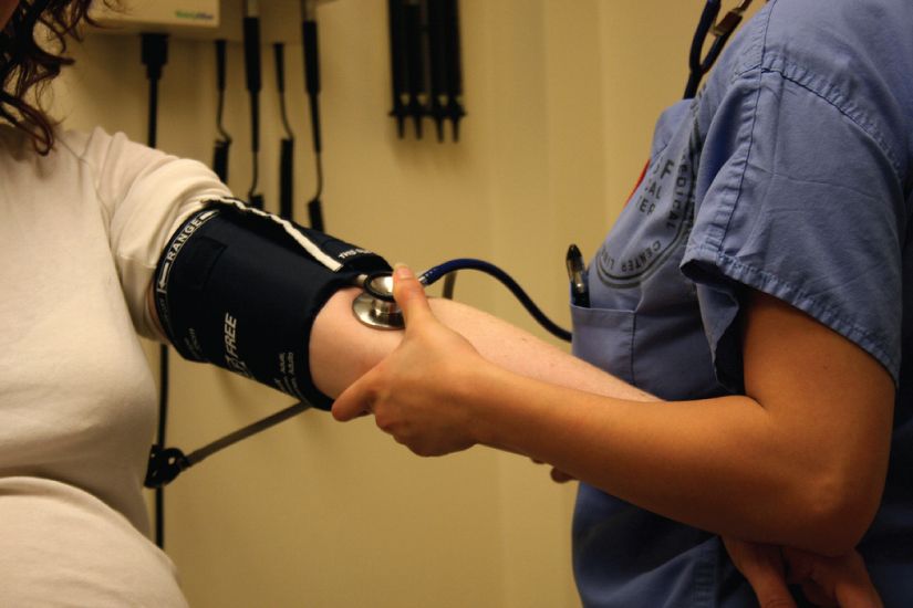
Blood pressure is simply the force that flowing blood pushes against the vessel walls, measured in millimeters of mercury (mm Hg). In the big arteries this pressure is not steady — it rises and falls with each heartbeat. When the ventricles contract and eject blood, pressure peaks at the systolic pressure; when the ventricles relax and refill, pressure sags to its low point, the diastolic pressure. A typical healthy reading is about 120/80 mm Hg — systolic over diastolic. The gap between them, systolic minus diastolic, is the pulse pressure (here 40 mm Hg), the surge you feel as a pulse at your wrist.
Two numbers worth calculating
Because the heart spends more of each cycle in diastole than in systole, the true average pressure driving blood forward is not the simple midpoint. This average is the mean arterial pressure (MAP), estimated as the diastolic pressure plus one-third of the pulse pressure. MAP matters clinically because it is the pressure that actually perfuses your organs; a MAP that falls much below about 60 mm Hg starves the brain and kidneys. For a 120/80 reading, MAP is roughly 80 + (40 ÷ 3) ≈ 93 mm Hg.
Pressure falls, and the arterioles set the toll
Follow the blood outward and its pressure drops the whole way — that steady decline is the pressure gradient, and it is precisely what keeps blood flowing (fluid always moves from higher to lower pressure). Pressure is high in the aorta, still pulsatile in the large arteries, and then falls off a cliff across the arterioles. Blood flow through the system follows a simple rule: flow = pressure difference ÷ resistance. Resistance depends most of all on vessel radius, and it varies inversely with the fourth power of that radius (R ∝ 1 ÷ r⁴) — so as a vessel narrows, resistance climbs steeply: halve the radius and resistance rises sixteen-fold, while doubling it cuts resistance to one-sixteenth. Since arterioles have muscular walls that can constrict and dilate on command, they are the body's chief resistance vessels, the adjustable faucets that set both organ blood flow and overall blood pressure. By the time blood reaches the venae cavae, pressure is near zero.
| Location | Approx. pressure | What is happening |
|---|---|---|
| Aorta / large arteries | ~120/80 mm Hg | High and pulsatile; elastic recoil smooths flow |
| Arterioles | Sharp drop | The biggest fall — the main resistance vessels |
| Capillaries | ~35 → 17 mm Hg | Low and gentle — protects the thin walls; drives exchange |
| Venules / veins | Low | Little pressure left; other pumps take over |
| Venae cavae | Near 0 mm Hg | Blood arrives back at the heart |
Tap each segment of the journey to see the pressure fall.
- Blood pressure = force of blood on the wall; reported as systolic/diastolic (e.g. 120/80 mm Hg).
- Pulse pressure = systolic − diastolic; MAP ≈ diastolic + ⅓ of pulse pressure, and it perfuses your organs.
- Blood flows down a pressure gradient; flow = ΔP ÷ resistance, and resistance varies inversely with r⁴ (R ∝ 1 ÷ r⁴), so a narrower vessel resists far more.
- Arterioles are the main resistance vessels — the dial for both organ flow and blood pressure.
Section 4Capillary exchange
A tug-of-war between two pressures
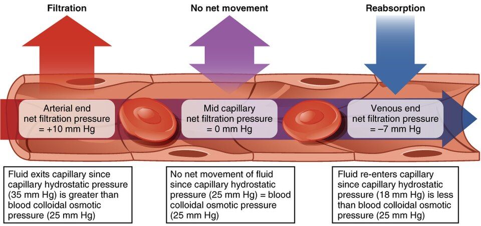
The entire cardiovascular system is built to deliver blood to the capillaries, and here the crucial trade happens across that one-cell-thick wall. Small molecules cross easily; what is trickier is the movement of fluid in and out, which is governed by a contest between two opposing pressures. The first is hydrostatic pressure — the blood pressure inside the capillary, which physically pushes fluid out through the wall and into the tissue spaces. The second is colloid osmotic pressure (also called oncotic pressure), created mainly by the large plasma protein albumin, which is too big to leave the blood. Because these proteins stay behind, they pull water back in by osmosis. Fluid moves whichever way the stronger pressure dictates.
Out at the start, back in at the end
What makes a capillary elegant is that the balance flips along its length. At the arteriolar end, hydrostatic pressure is relatively high (around 35 mm Hg) and easily overpowers the roughly 25 mm Hg of osmotic pull, so the net force pushes fluid out — this is filtration, delivering water, oxygen, and nutrients to the cells. As blood creeps along the capillary, its hydrostatic pressure bleeds away while the osmotic pull stays steady. By the venule end, hydrostatic pressure has fallen below the osmotic pressure, so the net force now draws fluid back in — this is reabsorption, collecting carbon dioxide and wastes on the way. The net difference between the pushing and pulling forces at any point is called the net filtration pressure.
The lymphatic safety net — and edema
The books do not quite balance: capillaries filter out a little more fluid than they reabsorb, roughly 85% back into the blood and about 15% left behind in the tissues. That leftover fluid is not lost — it is mopped up by the lymphatic vessels and eventually returned to the bloodstream. When any part of this system fails, fluid piles up in the tissues and you get edema, or swelling. Too much hydrostatic pressure (as in heart failure or a standing leg), too little albumin to pull fluid back (as in liver or kidney disease or malnutrition), or blocked lymphatic drainage will each tip the balance toward accumulation.
| Force | Source | Direction |
|---|---|---|
| Hydrostatic pressure | Blood pressure inside the capillary | Pushes fluid out of the blood |
| Colloid osmotic (oncotic) pressure | Plasma proteins, mainly albumin | Pulls fluid into the blood |
| Net result — arteriolar end | Hydrostatic > osmotic | Filtration (fluid leaves) |
| Net result — venule end | Osmotic > hydrostatic | Reabsorption (fluid returns) |
Tap each end of the capillary to watch the tug-of-war play out.
- Fluid exchange is a contest between hydrostatic pressure (pushes out) and colloid osmotic pressure (pulls in).
- Filtration dominates at the arteriolar end; reabsorption dominates at the venule end.
- Capillaries filter slightly more than they reabsorb; the lymphatics return the surplus.
- Imbalance — high hydrostatic pressure, low albumin, or blocked lymphatics — causes edema.
Section 5The circulatory routes
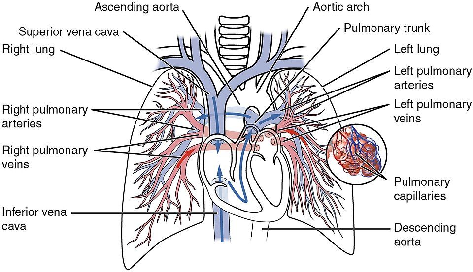
Two great circuits
Blood does not wander — it runs in fixed loops called circuits, and the whole system is really two circuits joined at the heart. The pulmonary circuit is the short loop to the lungs: the right side of the heart pumps oxygen-poor blood out through the pulmonary arteries, the blood picks up oxygen and dumps carbon dioxide in the lung capillaries, and the pulmonary veins carry the now oxygen-rich blood back to the left side of the heart. The systemic circuit is the long loop to everything else: the left side of the heart pumps oxygen-rich blood out through the aorta to every organ and limb, and the veins bring the oxygen-poor blood back to the right side to begin again. The two circuits run in series, so the same volume must pass through both — the right and left hearts pump in perfect lockstep.
Portal systems: two capillary beds in a row
Most blood passes through only one capillary bed between leaving and returning to the heart. A portal system is the exception: blood flows through two capillary beds in series, linked by a vein. The most important example is the hepatic portal system. Blood draining the stomach and intestines is loaded with freshly absorbed nutrients (and any swallowed drugs or toxins). Rather than heading straight back to the heart, it is funneled through the hepatic portal vein into a second capillary bed inside the liver, which stores nutrients, detoxifies chemicals, and adjusts blood sugar before releasing the blood onward. Only then does it join the general circulation through the hepatic veins.
Special deliveries
A few organs have their own dedicated arrangements. The coronary circulation is the heart feeding itself, branching off the aorta the instant blood leaves the heart. The cerebral circulation supplies the brain through a protective ring of connected arteries, the circle of Willis, so that if one supply artery is blocked, others can reroute blood around the gap. Such connections between vessels are called anastomoses — natural detours that provide a backup blood supply. Where anastomoses are sparse, a blockage is far more dangerous, which is one reason a clot in a coronary artery so reliably causes a heart attack.
| Feature | Pulmonary circuit | Systemic circuit |
|---|---|---|
| Pumped by | Right side of the heart | Left side of the heart |
| Sends blood to | The lungs | The rest of the body |
| Blood leaving heart | Oxygen-poor (in pulmonary arteries) | Oxygen-rich (in the aorta) |
| Blood returning | Oxygen-rich (in pulmonary veins) | Oxygen-poor (in the venae cavae) |
| Main purpose | Load O₂, unload CO₂ | Deliver O₂ & nutrients, collect wastes |
Tap each route to trace where the blood goes.
- The pulmonary circuit (right heart → lungs) loads O₂; the systemic circuit (left heart → body) delivers it.
- In the pulmonary circuit, arteries carry oxygen-poor blood and veins carry oxygen-rich blood.
- The hepatic portal system routes gut blood through the liver — two capillary beds in series — before it returns to the heart.
- Anastomoses (as in the circle of Willis) give backup routes; where they are sparse, a blockage is dangerous.
Section 6Venous return
An uphill problem
By the time blood reaches the veins, it has almost no pressure left — often around 15 mm Hg, falling toward zero at the heart. Yet the blood in your feet must somehow climb more than a meter, against gravity, back to the right side of the heart. The volume of blood returning to the heart each minute is called the venous return, and over any stretch of time it must exactly equal the cardiac output — whatever the heart pumps out, the veins must bring back. With so little pressure to work with, the veins cannot do this alone. They rely on a set of external pumps and clever one-way plumbing.
The muscle pump and the valves
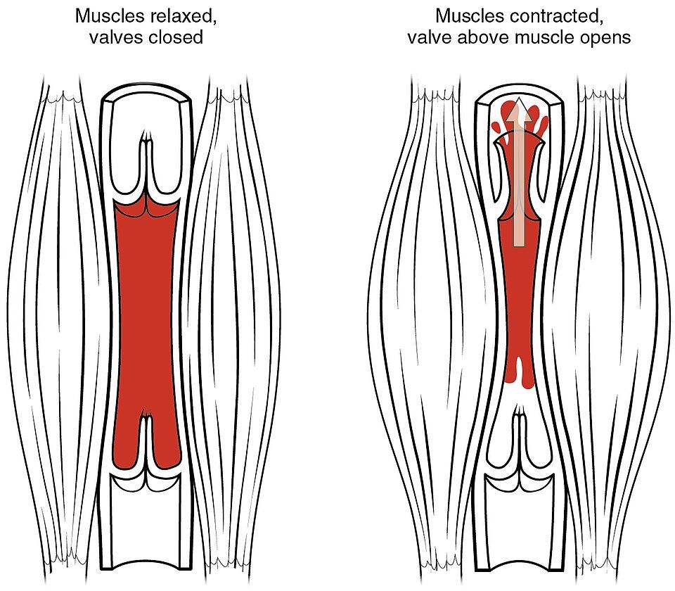
The workhorse is the skeletal muscle pump. Deep veins run between your muscles, so every time you contract a muscle — each step you take — it squeezes the veins and forces blood along. The trick that makes the squeeze productive is the one-way valves: when a muscle presses on a vein, the valve above snaps open to let blood rush upward while the valve below slams shut to stop it falling back. Contract, and blood is pushed toward the heart; relax, and the valves hold the ground you gained. Walk across a room and your calf muscles are, quite literally, a second heart pumping blood up your legs.
The respiratory pump, and what fails
A second helper is the respiratory pump. Each time you inhale, the chest cavity expands and its pressure drops while pressure in the abdomen rises; this squeezes abdominal veins and draws blood upward into the low-pressure thoracic veins toward the heart. A third contributor is venoconstriction — the sympathetic nervous system tightening the vein walls to push stored blood back into circulation when demand rises. When these mechanisms falter, the consequences are familiar. Stand still for a long time and the muscle pump stops, blood pools in the legs, and you may faint. Weakened or leaky valves let blood collect and stretch the veins into bulging varicose veins. And blood that sits motionless — on a long flight, or after surgery — can clot in a deep vein, a dangerous deep vein thrombosis.
| Mechanism | How it works | When it matters most |
|---|---|---|
| Skeletal muscle pump | Contracting muscles squeeze deep veins; valves keep flow one-way | Walking, running — any leg movement |
| Respiratory pump | Inhaling lowers chest pressure and raises abdominal pressure, drawing blood upward | Every breath; deeper with exercise |
| Venous valves | One-way flaps prevent backflow between pumps | Standing upright, against gravity |
| Venoconstriction | Sympathetic tightening of vein walls pushes reserve blood forward | Exercise, blood loss, standing up |
Watch the muscle pump and valves lift blood against gravity in the interactive.
- Venous return is the blood coming back to the heart; over time it must equal cardiac output.
- With little pressure left, veins rely on the skeletal muscle pump, the respiratory pump, and one-way valves.
- Venoconstriction pushes reserve blood forward during exercise or blood loss.
- Failure produces fainting, varicose veins, and deep vein thrombosis — which is why movement matters.
The chapter in one breath
- Every vessel wall has three tunics — intima, media, externa — around a central lumen; the endothelium lining is where vascular disease begins.
- Arteries carry blood away under high pressure with thick elastic walls; veins return it under low pressure with valves and hold most of the blood; capillaries are one cell thick and do all the exchanging.
- Blood pressure is reported as systolic/diastolic; pulse pressure is their difference and MAP perfuses the organs.
- Blood flows down a pressure gradient (flow = ΔP ÷ resistance); the muscular arterioles are the main resistance vessels because resistance scales with 1 ÷ r⁴.
- Capillary exchange is a tug-of-war between hydrostatic (pushes out) and colloid osmotic (pulls in) pressure — filtration at the arteriolar end, reabsorption at the venule end; imbalance causes edema.
- Blood runs in a pulmonary circuit (to the lungs) and a systemic circuit (to the body); special routes like the hepatic portal system send blood through two capillary beds.
- Venous return against gravity depends on the skeletal muscle pump, the respiratory pump, and one-way valves — failure brings fainting, varicose veins, and clots.
ReferenceWords worth knowing
A quick reference for the terms in this chapter — skim it now, or circle back whenever a word trips you up.
- Arteriole
- A very small artery with a muscular wall that regulates flow into a capillary bed; the body's main resistance vessel.
- Artery
- A vessel that carries blood away from the heart, defined by direction rather than by oxygen content.
- Blood pressure
- The force that flowing blood exerts against the vessel wall, measured in millimeters of mercury (mm Hg).
- Capillary
- The smallest vessel, one endothelial cell thick, where oxygen, carbon dioxide, nutrients, and wastes are exchanged with tissues.
- Colloid osmotic (oncotic) pressure
- The inward pull created by plasma proteins (mainly albumin) that draws water back into the capillary.
- Diastolic pressure
- The lower arterial pressure, measured while the ventricles relax and refill.
- Edema
- Swelling from excess fluid accumulating in the tissues when capillary filtration and reabsorption fall out of balance.
- Endothelium
- The single layer of flat cells lining the entire cardiovascular system; the inner surface of the tunica intima.
- Filtration
- The movement of fluid out of a capillary into the tissues, dominant at the arteriolar end where hydrostatic pressure is highest.
- Hepatic portal system
- A route in which nutrient-rich blood from the gut passes through the liver's capillaries before returning to the heart.
- Hydrostatic pressure
- The blood pressure inside a capillary that pushes fluid outward through the wall.
- Mean arterial pressure (MAP)
- The average pressure driving blood through the systemic circuit; roughly diastolic pressure plus one-third of the pulse pressure.
- Pulmonary circuit
- The loop that carries blood from the right heart to the lungs and back, loading oxygen and unloading carbon dioxide.
- Pulse pressure
- The difference between systolic and diastolic pressure; the pressure surge felt as a pulse.
- Reabsorption
- The return of fluid into a capillary from the tissues, dominant at the venule end where osmotic pressure exceeds hydrostatic pressure.
- Resistance
- The opposition to blood flow, determined mainly by vessel radius: it varies inversely with the fourth power of the radius (R ∝ 1 ÷ r⁴), rising steeply as the vessel narrows.
- Skeletal muscle pump
- The squeezing of deep veins by contracting muscles, which — with one-way valves — drives blood back toward the heart.
- Systemic circuit
- The loop that carries blood from the left heart to the whole body and back, delivering oxygen and collecting wastes.
- Systolic pressure
- The higher arterial pressure, measured while the ventricles contract and eject blood.
- Tunica media
- The middle vessel coat of smooth muscle and elastic fibers that sets vessel diameter through vasoconstriction and vasodilation.
- Vein
- A vessel that carries blood toward the heart; thin-walled, valved, and the body's main blood reservoir.
- Venous return
- The volume of blood returning to the heart per minute; over time it must equal cardiac output.