The Autonomic Nervous System
Right now, without a single deliberate thought, your heart is beating, your gut is churning breakfast, your pupils are adjusting to the light, and your blood pressure is being nudged up and down a dozen times a minute. All of that quiet housekeeping belongs to the autonomic nervous system — the part of you that runs the organs while your conscious mind is busy elsewhere. In this chapter you will meet its two divisions, the sympathetic "fight or flight" system and the parasympathetic "rest and digest" system, and see how they pull most organs in opposite directions to keep the body tuned. You will learn the two-neuron wiring that makes autonomic pathways different from every other motor route, the two chemicals that carry every autonomic message, and the elegant reflex that stops you from fainting each time you stand up.
By the end of this chapter you can
- Define the autonomic nervous system and explain how it differs from the somatic (voluntary) motor system.
- Contrast the sympathetic and parasympathetic divisions by their overall job, their outflow from the CNS, and their effects on target organs.
- Describe the two-neuron autonomic pathway and locate the ganglia of each division.
- Explain dual innervation and autonomic tone, and predict the net effect when one division dominates.
- Trace the whole-body fight-or-flight response organ by organ and describe the parasympathetic return to calm.
- Match the autonomic neurotransmitters (acetylcholine, norepinephrine) to their receptors and predict the effect at each target.
- Diagram the baroreceptor reflex as a negative feedback loop that defends blood pressure.
Section 1Two divisions, opposite jobs
A motor system for the organs you never think about
The nervous system has two motor outputs. The somatic system carries your voluntary commands to skeletal muscle — the decision to lift a cup. The autonomic nervous system (ANS) does everything else: it is the involuntary, visceral motor system that controls smooth muscle, cardiac muscle, and glands. It sets your heart rate, the width of your pupils and airways, the pace of your digestion, the tone of your blood vessels, and the activity of your sweat and salivary glands — all automatically, below the level of awareness. You feel its work only indirectly, as a pounding heart before a test or a dry mouth when you are nervous.
Two divisions that pull opposite ways
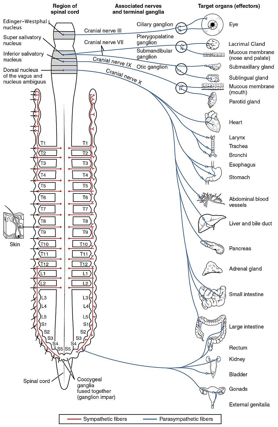
The ANS is built from two divisions that usually act as opponents. The sympathetic division is the body's accelerator — the "fight or flight" system that ramps you up to meet a threat. The parasympathetic division is the brake — the "rest and digest" system that runs the body during calm, restful conditions and handles the unglamorous business of digestion, waste elimination, and energy storage. Most organs receive a nerve supply from both divisions, an arrangement called dual innervation, and the two supplies push the organ in opposite directions. The heart is the clearest example: sympathetic input speeds it up, parasympathetic input slows it down, and your actual heart rate at any moment is the running balance between the two.
Where each division leaves the central nervous system
The two divisions even exit the CNS from different places, which is the easiest way to tell them apart. Sympathetic fibers all emerge from the thoracolumbar region — the lateral horns of spinal segments T1 through L2 — so the sympathetic division is often called the thoracolumbar outflow. Parasympathetic fibers emerge from the two ends of the CNS: from the brainstem in four cranial nerves (III, VII, IX, and the far-reaching vagus, X) and from the sacral spinal cord (segments S2–S4). That top-and-bottom pattern gives the parasympathetic division its nickname, the craniosacral outflow.
| Target organ | Sympathetic ("fight or flight") | Parasympathetic ("rest and digest") |
|---|---|---|
| Heart | Faster rate, stronger beat | Slower rate |
| Pupil | Dilates (widens) | Constricts (narrows) |
| Airways (bronchi) | Dilate — more air | Constrict |
| Digestive tract | Slows; blood diverted away | Speeds up; secretions flow |
| Salivary glands | Scant, thick saliva (dry mouth) | Copious, watery saliva |
| Urinary bladder | Relaxes (holds urine) | Contracts (empties) |
| Liver | Releases glucose to the blood | Stores glucose |
Tap any organ in the interactive to line up its two opposing nerve supplies side by side.
- The autonomic nervous system is the involuntary motor system for smooth muscle, cardiac muscle, and glands.
- Its two divisions are opponents: sympathetic = fight or flight (accelerator); parasympathetic = rest and digest (brake).
- Sympathetic outflow is thoracolumbar (T1–L2); parasympathetic outflow is craniosacral (CN III, VII, IX, X and S2–S4).
- Most organs have dual innervation and are steered by shifting the balance of ongoing autonomic tone.
Section 2The two-neuron pathway
Two neurons in a relay, not one
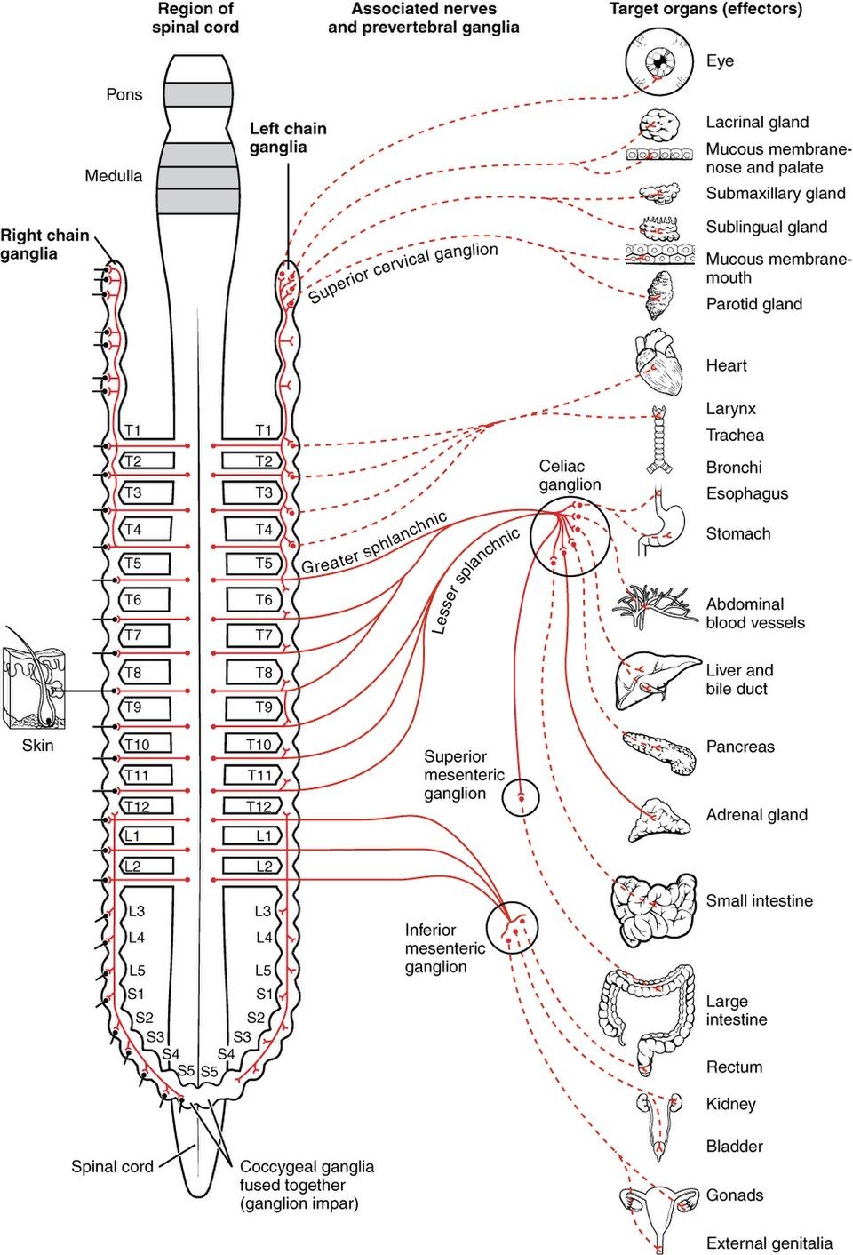
Here is the single structural fact that defines the whole autonomic system: it takes two neurons to carry a signal from the CNS to a visceral target, and they meet at a relay station called a ganglion. The first neuron, the preganglionic neuron, has its cell body inside the CNS; its axon runs out to a ganglion and synapses there. The second neuron, the postganglionic neuron, has its cell body in the ganglion and sends its axon the rest of the way to the organ. Compare this to the somatic system, where a single motor neuron reaches all the way from the spinal cord to the muscle with no relay in between. That extra synapse in the ganglion is where the two divisions do their most distinctive wiring.
Sympathetic wiring: short first, long second, and it spreads
In the sympathetic division, the ganglia sit close to the spinal cord. Most form the sympathetic chain ganglia (the paravertebral ganglia) — a beaded double column running alongside the vertebral column that you can see flanking the cord in the figure. A few others, the prevertebral (collateral) ganglia such as the celiac and mesenteric ganglia, sit out in front of the aorta and are reached by the splanchnic nerves. Because the relay is near the cord, sympathetic preganglionic fibers are short and postganglionic fibers are long. Crucially, each preganglionic fiber synapses on many postganglionic neurons — a fan-out called divergence — which is exactly why a sympathetic response is not local but sweeps the whole body at once.
Parasympathetic wiring: long first, short second, and it stays local
The parasympathetic division reverses the geometry. Its ganglia sit in or right beside the target organ (terminal or intramural ganglia), so preganglionic fibers are long and postganglionic fibers are very short. There is little divergence, so a parasympathetic signal produces a discrete, localized effect on one organ rather than a body-wide surge — you can slow the heart without also cramping the gut. This precision fits the calm, one-thing-at-a-time character of rest and digest.
| Feature | Sympathetic | Parasympathetic |
|---|---|---|
| CNS outflow | Thoracolumbar (T1–L2) | Craniosacral (brainstem + S2–S4) |
| Ganglion location | Near the spinal cord (chain & prevertebral) | Near or in the target organ |
| Preganglionic fiber | Short | Long |
| Postganglionic fiber | Long | Short |
| Divergence | High → widespread response | Low → localized response |
Switch between the two divisions in the interactive to watch the ganglion move from beside the cord to beside the organ.
- Every autonomic route uses two neurons — preganglionic and postganglionic — relaying at a ganglion.
- Sympathetic: ganglia near the cord (chain & prevertebral), short pre / long post fibers, high divergence → body-wide response.
- Parasympathetic: ganglia at the organ, long pre / short post fibers, low divergence → localized response.
- The adrenal medulla is a modified sympathetic ganglion that releases epinephrine as a hormone.
Section 3Fight or flight, rest and digest
One switch, and the whole body answers
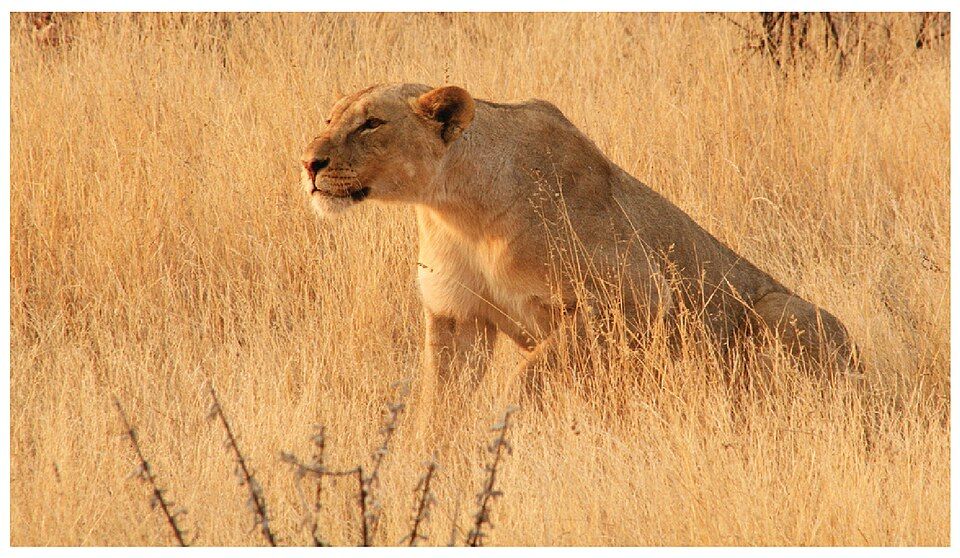
Because sympathetic fibers diverge so widely and the adrenal medulla adds a hormonal boost, the sympathetic division works as a coordinated, all-at-once package. Imagine the moment you step off a curb and a car horn blares. Before you can think, your heart pounds, your breathing deepens, your eyes go wide, your palms sweat, and your stomach seems to drop. That is the fight-or-flight response — the sympathetic division reprioritizing the entire body toward one goal: sudden, intense physical exertion to escape or confront a threat. Every change is purposeful, and each one shifts resources away from long-term maintenance and toward immediate muscle power.
The sympathetic surge, organ by organ
Follow the surge through the body and every effect earns its place. The heart beats faster and harder to move oxygen quickly. The airways widen so more air reaches the lungs. The pupils dilate to take in more of the scene. The liver pours stored glucose into the blood to fuel muscle. Blood vessels constrict in the gut and skin but dilate in skeletal muscle, shunting the blood where it is needed. Digestion and urination pause — no time for either. Sweat glands switch on to pre-cool the body for exertion. It is a beautifully logical package: everything that helps you run or fight is turned up, and everything that can wait is turned down.
| Fight-or-flight change | Why it helps survival |
|---|---|
| Heart rate and force rise | Delivers oxygen and fuel to muscle faster |
| Bronchioles dilate | More airflow, more oxygen uptake |
| Pupils dilate | Sharper, wider vision of the threat |
| Liver releases glucose | Ready fuel for a burst of effort |
| Blood shifts to skeletal muscle | Power where it is needed, not the gut |
| Digestion and urination halt | Saves energy for the emergency |
| Sweating increases | Cools the body before heavy exertion |
Rest and digest: paying the debt back
When the threat is gone, the parasympathetic division takes the body back to baseline and then to the quiet work the emergency interrupted. The heart slows, the airways and pupils return to normal, blood flows back to the gut, and digestion, absorption, and elimination resume. This is the state you spend most of your life in — the body building and storing rather than spending. The two divisions are not enemies so much as a well-matched pair: the sympathetic system is the sprint, the parasympathetic system is the recovery, and health depends on being able to swing smoothly between them.
Flip the switch in the interactive to trip the sympathetic surge, then let the parasympathetic division bring the body back down.
- The fight-or-flight response is a coordinated, body-wide sympathetic package aimed at intense physical effort.
- It raises heart rate, airflow, pupil size, and blood glucose and shifts blood to muscle, while pausing digestion and urination.
- The parasympathetic division restores baseline and resumes rest-and-digest housekeeping.
- Circulating epinephrine outlasts the nerve signals, which is why arousal fades slowly after a scare.
Section 4The chemistry: acetylcholine and norepinephrine
Only two chemicals do all the talking
For a system that controls so much, the ANS speaks a remarkably small vocabulary: nearly every autonomic message is carried by just two neurotransmitters. A neuron that releases acetylcholine (ACh) is called cholinergic; one that releases norepinephrine (NE) is called adrenergic. Where in the pathway each chemical is used follows a simple pattern. All preganglionic neurons of both divisions are cholinergic — they release ACh in the ganglion. The postganglionic neurons then split: parasympathetic postganglionic fibers release ACh onto the organ, while sympathetic postganglionic fibers release NE. (Two tidy exceptions: sympathetic fibers to sweat glands use ACh, and the adrenal medulla releases epinephrine into the blood.)
The receptor decides the effect, not the chemical
Here is the idea students most need to grasp: the same neurotransmitter can excite one organ and inhibit another, because the outcome depends on the receptor the chemical lands on. Cholinergic receptors come in two flavors — nicotinic receptors (found in all autonomic ganglia and at the skeletal neuromuscular junction) and muscarinic receptors (on the organs that parasympathetic fibers control). Adrenergic receptors also come in families — alpha and beta subtypes. A catecholamine acting on alpha-1 receptors constricts blood vessels, while the same chemical family acting on beta-2 receptors relaxes the airways. Norepinephrine is the alpha-1 workhorse; it binds beta-2 only weakly, so the bronchodilating job falls to circulating epinephrine from the adrenal medulla (and to beta-2 drugs such as albuterol). One chemical family, opposite effects — the target cell's receptor writes the instruction.
| Fiber | Neurotransmitter | Receptor on target |
|---|---|---|
| Preganglionic (both divisions) | Acetylcholine | Nicotinic |
| Parasympathetic postganglionic | Acetylcholine | Muscarinic |
| Sympathetic postganglionic (most) | Norepinephrine | Adrenergic (α or β) |
| Sympathetic to sweat glands | Acetylcholine | Muscarinic |
| Adrenal medulla → blood | Epinephrine (hormone) | Adrenergic (α or β) |
A loose spray, not a tight synapse
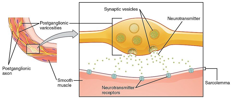
Notice in the figure that the postganglionic axon does not end in a single neat terminal. Instead it is studded with beads called varicosities that release neurotransmitter in a diffuse spray over a wide patch of smooth muscle. This is why one autonomic fiber can influence a broad sheet of tissue at once — ideal for setting the tone of an entire vessel wall or gut segment rather than commanding a single cell.
Tap each receptor in the interactive to see how the same two chemicals produce different effects.
- All preganglionic fibers and all parasympathetic postganglionic fibers release acetylcholine (cholinergic); most sympathetic postganglionic fibers release norepinephrine (adrenergic).
- The receptor — nicotinic vs muscarinic, alpha vs beta — determines whether a target is excited or inhibited.
- Postganglionic axons release transmitter from varicosities, spraying a whole patch of tissue.
- Receptor subtypes are the reason autonomic drugs (beta-blockers, albuterol, atropine) can be so targeted.
Section 5The baroreceptor reflex
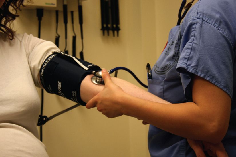
The body's fastest blood-pressure guard
The autonomic system does more than react to emergencies — it runs continuous, silent reflexes that keep vital numbers steady. The most important of these is the baroreceptor reflex, the second-by-second guardian of blood pressure. It is a textbook negative feedback loop, and once you see its three parts you will recognize the same machinery you met in Chapter 1: a sensor, a control center, and effectors.
Sense, compare, correct
The sensors are baroreceptors — stretch receptors packed into the walls of the carotid sinus (where blood heads to the brain) and the aortic arch. When blood pressure rises, these vessel walls stretch more and the receptors fire faster; when pressure falls, they fire less. Their reports travel along cranial nerves IX (glossopharyngeal) and X (vagus) to the cardiovascular center in the medulla of the brainstem — the control center that compares the signal to the desired pressure. The medulla then adjusts its two autonomic effectors: it dials parasympathetic (vagal) and sympathetic output up or down to change heart rate, contractility, and vessel diameter. Because the response always opposes the disturbance, pressure is pushed back toward its set point.
| Blood pressure | Baroreceptor firing | Autonomic response | Result |
|---|---|---|---|
| Rises too high | Increases | Parasympathetic up, sympathetic down | Heart slows, vessels dilate → BP falls |
| Falls too low | Decreases | Sympathetic up, parasympathetic down | Heart speeds, vessels constrict → BP rises |
Standing up without fainting
You depend on this reflex every time you stand. When you rise, gravity pools blood in your legs and pressure at the head momentarily drops. Baroreceptors instantly sense the fall, the medulla ramps up sympathetic drive, your heart quickens and your vessels tighten, and pressure is restored before the brain is ever short-changed — all in a single heartbeat or two. When this reflex is sluggish (from aging, dehydration, or certain drugs), the correction lags and you feel lightheaded on standing, a condition called orthostatic hypotension.
Knock the pressure off-target in the interactive and watch the reflex pull it home.
- The baroreceptor reflex is a negative feedback loop that stabilizes blood pressure moment to moment.
- Baroreceptors in the carotid sinus and aortic arch sense stretch and report via CN IX and X to the medulla.
- The medulla corrects pressure by shifting the sympathetic/parasympathetic balance to heart rate, force, and vessel diameter.
- A slow reflex causes orthostatic hypotension — lightheadedness on standing.
Section 6Somatic versus autonomic
Two ways the brain moves the body
It is worth putting the autonomic system side by side with its sibling, the somatic motor system, because the contrast makes both clearer. The somatic system carries voluntary commands to skeletal muscle. It uses a single motor neuron from the CNS straight to the muscle, that neuron releases acetylcholine onto nicotinic receptors, and its effect is always the same: excitation — a somatic motor neuron can only tell a muscle to contract, never to relax. To stop a movement, the CNS simply stops firing.
Where the autonomic system differs
The autonomic system differs on nearly every count. It is involuntary; it targets smooth muscle, cardiac muscle, and glands; it uses a two-neuron chain relaying at a ganglion; it uses ACh or NE depending on the fiber; and it can be either excitatory or inhibitory at the target — the parasympathetic vagus can actively slow the heart, something no somatic neuron can do to a muscle. That ability to both stimulate and inhibit, division by division, is what lets the ANS fine-tune organs in both directions.
| Feature | Somatic | Autonomic |
|---|---|---|
| Control | Voluntary | Involuntary |
| Effector (target) | Skeletal muscle | Smooth & cardiac muscle, glands |
| Neurons to target | One | Two (relay at a ganglion) |
| Neurotransmitter | Acetylcholine only | Acetylcholine or norepinephrine |
| Effect on target | Always excitatory | Excitatory or inhibitory |
A third player: the gut's own brain
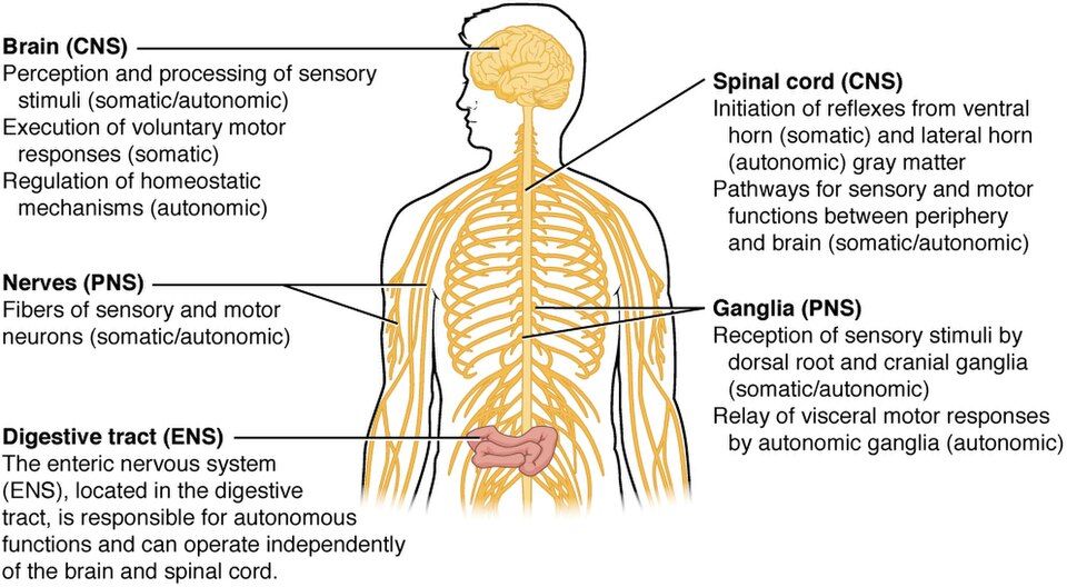
The figure also names a quieter third division, the enteric nervous system (ENS) — a vast web of neurons embedded in the wall of the digestive tract. So numerous that it is nicknamed the "second brain," the ENS can run local digestive reflexes on its own, coordinating the muscle contractions and secretions of the gut even when its links to the CNS are cut. The autonomic divisions still modulate it — parasympathetic input revs digestion, sympathetic input suppresses it — but the day-to-day churning of your intestines is largely self-governed.
Switch between the somatic and autonomic pathways in the interactive to compare their wiring and behavior.
- The somatic system is voluntary, uses one neuron to skeletal muscle, releases only ACh, and is always excitatory.
- The autonomic system is involuntary, uses a two-neuron chain to smooth/cardiac muscle and glands, uses ACh or NE, and can excite or inhibit.
- The enteric nervous system is a self-governing "second brain" in the gut wall, modulated but not commanded by the ANS.
The chapter in one breath
- The autonomic nervous system is the involuntary motor system that runs smooth muscle, cardiac muscle, and glands.
- Its two divisions oppose each other: sympathetic (fight or flight, thoracolumbar) and parasympathetic (rest and digest, craniosacral), with most organs under dual innervation and steered by shifting tone.
- Every autonomic route is a two-neuron chain relaying at a ganglion — sympathetic ganglia sit near the cord and diverge widely; parasympathetic ganglia sit at the organ and stay local.
- The fight-or-flight response is a coordinated, body-wide sympathetic surge; the parasympathetic division restores calm, aided by the slow clearance of adrenal epinephrine.
- Just two neurotransmitters — acetylcholine and norepinephrine — run the system, and the receptor decides whether a target is excited or inhibited.
- The baroreceptor reflex is a negative feedback loop that defends blood pressure by balancing sympathetic and parasympathetic output through the medulla.
- Compared with the somatic system (voluntary, one neuron, ACh, always excitatory), the autonomic system is involuntary, two-neuron, and can both excite and inhibit; the enteric system runs the gut semi-independently.
ReferenceWords worth knowing
A quick reference for the terms in this chapter — skim it now, or circle back whenever a word trips you up.
- Acetylcholine (ACh)
- The neurotransmitter released by all autonomic preganglionic fibers and by parasympathetic postganglionic fibers; neurons that use it are called cholinergic.
- Adrenal medulla
- The inner core of the adrenal gland; a modified sympathetic ganglion that releases epinephrine into the blood, giving fight or flight a hormonal, longer-lasting phase.
- Adrenergic
- Describing a neuron or receptor that uses norepinephrine (or epinephrine); adrenergic receptors are grouped into alpha and beta subtypes.
- Autonomic nervous system
- The involuntary motor division of the nervous system that controls smooth muscle, cardiac muscle, and glands.
- Autonomic tone
- The steady background firing of both divisions, so an organ is regulated by shifting the balance between them rather than switching one on from off.
- Baroreceptor
- A stretch receptor in the carotid sinus and aortic arch that senses blood pressure by how much the vessel wall is stretched.
- Baroreceptor reflex
- The negative feedback loop that stabilizes blood pressure by adjusting sympathetic and parasympathetic output to the heart and vessels.
- Cholinergic
- Describing a neuron or receptor that uses acetylcholine; cholinergic receptors are either nicotinic or muscarinic.
- Craniosacral outflow
- The origin of parasympathetic fibers from the brainstem (cranial nerves III, VII, IX, X) and sacral spinal cord (S2–S4).
- Dual innervation
- The supply of a single organ by both the sympathetic and parasympathetic divisions, which act on it in opposite directions.
- Enteric nervous system
- The extensive network of neurons in the wall of the digestive tract that can run local gut reflexes independently of the brain and spinal cord.
- Fight-or-flight response
- The coordinated, body-wide sympathetic reaction that prepares the body for intense physical exertion in the face of a threat.
- Ganglion (autonomic)
- The relay station outside the CNS where a preganglionic neuron synapses on a postganglionic neuron.
- Muscarinic receptor
- A cholinergic receptor on the target organs of parasympathetic (and sweat-gland) fibers; its effect can be excitatory or inhibitory depending on the organ.
- Nicotinic receptor
- A cholinergic receptor found in all autonomic ganglia and at the skeletal neuromuscular junction; always excitatory.
- Norepinephrine (NE)
- The neurotransmitter released by most sympathetic postganglionic fibers; it acts on adrenergic receptors.
- Parasympathetic division
- The "rest and digest" division of the ANS; craniosacral in origin, it conserves energy and runs digestion and elimination.
- Postganglionic neuron
- The second neuron of an autonomic pathway, with its cell body in the ganglion and its axon running to the effector organ.
- Preganglionic neuron
- The first neuron of an autonomic pathway, with its cell body in the CNS and its axon running to the ganglion.
- Sympathetic chain ganglia
- The paired columns of paravertebral ganglia alongside the vertebral column that house most sympathetic postganglionic cell bodies.
- Sympathetic division
- The "fight or flight" division of the ANS; thoracolumbar in origin, it mobilizes the body for exertion.
- Thoracolumbar outflow
- The origin of sympathetic fibers from the lateral horns of spinal segments T1 through L2.