The Nervous System & Nervous Tissue
Every thought you have ever had, every muscle you have ever moved, and every sensation you have ever felt was carried by a wave of electricity racing along a living wire. This chapter is the story of that wire — the neuron — and the tissue it lives in. We will map how the whole nervous system is organized, meet the loyal support cells that keep neurons alive, and then get to the heart of the matter: how a neuron builds an electrical charge, fires an action potential, sends it flying down a myelinated axon, and hands the message to the next cell across a microscopic gap. Master these ideas and the rest of neuroscience — the brain, the spinal cord, reflexes, the senses — becomes a set of variations on one theme.
By the end of this chapter you can
- Divide the nervous system into its central and peripheral parts and into its sensory, motor, somatic, and autonomic pathways.
- State the nervous system's three core jobs — sensory input, integration, and motor output — and give an example of each.
- Name the parts of a neuron and the six neuroglia, and say what each glial cell does and where it lives.
- Explain how the resting membrane potential of about −70 mV is created and maintained.
- Walk through the phases of an action potential and explain why it is all-or-nothing and one-way.
- Contrast continuous and saltatory conduction and list the two factors that set conduction speed.
- Describe the steps of chemical synaptic transmission and tell an excitatory signal from an inhibitory one.
- Connect each mechanism to a real clinical picture — multiple sclerosis, local anesthetics, nerve gas, and more.
Section 1How the nervous system is organized

One control center, wires everywhere
The nervous system is the body's fast communication network, and anatomists split it two different ways — by where the parts are and by which direction the traffic flows. By location there are just two divisions. The central nervous system (CNS) is the brain and spinal cord — the headquarters where information is processed and decisions are made. The peripheral nervous system (PNS) is everything else: the nerves and ganglia that fan out from the CNS to reach every corner of the body. Think of the CNS as the computer and the PNS as the cables running to every sensor and every motor.
Three jobs: sense, decide, act
No matter how complicated a nervous pathway looks, it is always doing one of three things. First comes sensory input — receptors detect a change, inside the body or out, and send that news toward the CNS. Second is integration — the CNS processes and interprets the incoming signals and decides what, if anything, to do. Third is motor output — a command travels away from the CNS to an effector (a muscle or a gland) that carries out the response. Touch a hot stove and all three fire in a heartbeat: skin receptors sense heat (input), the spinal cord decides "pull back" (integration), and arm muscles yank the hand away (output).
The words for these directions are worth memorizing because they appear on every diagram. Neurons carrying signals toward the CNS are afferent (sensory); neurons carrying commands away from the CNS are efferent (motor). The efferent side splits again by target. The somatic nervous system carries voluntary commands to skeletal muscle — the movements you choose. The autonomic nervous system (ANS) runs the automatic housekeeping, sending signals to cardiac muscle, smooth muscle, and glands without your conscious say-so. And the ANS itself has two arms that usually oppose each other: the sympathetic division (the "fight-or-flight" accelerator) and the parasympathetic division (the "rest-and-digest" brake).
| Division | What it includes | Direction / job |
|---|---|---|
| Central (CNS) | Brain and spinal cord | Integration — processes input, issues commands |
| Peripheral (PNS) | Nerves and ganglia | Carries signals to and from the CNS |
| Sensory (afferent) | Sensory neurons | Toward the CNS — input |
| Motor (efferent) | Motor neurons | Away from the CNS — output |
| Somatic | Motor neurons to skeletal muscle | Voluntary movement |
| Autonomic (ANS) | Motor neurons to cardiac/smooth muscle & glands | Involuntary control |
| Sympathetic | Thoracolumbar outflow | Fight-or-flight (accelerate) |
| Parasympathetic | Craniosacral outflow | Rest-and-digest (calm) |
Tap through each division — central and peripheral, sensory and motor, somatic and autonomic — in the interactive below.
- By location: the CNS is the brain and spinal cord; the PNS is the nerves and ganglia.
- By function: afferent (sensory) signals arrive at the CNS; efferent (motor) signals exit toward effectors.
- The motor side divides into somatic (voluntary, skeletal muscle) and autonomic (involuntary), and the autonomic into sympathetic and parasympathetic.
- Every nervous act reduces to three steps: sensory input → integration → motor output.
Section 2Neurons and their support crew
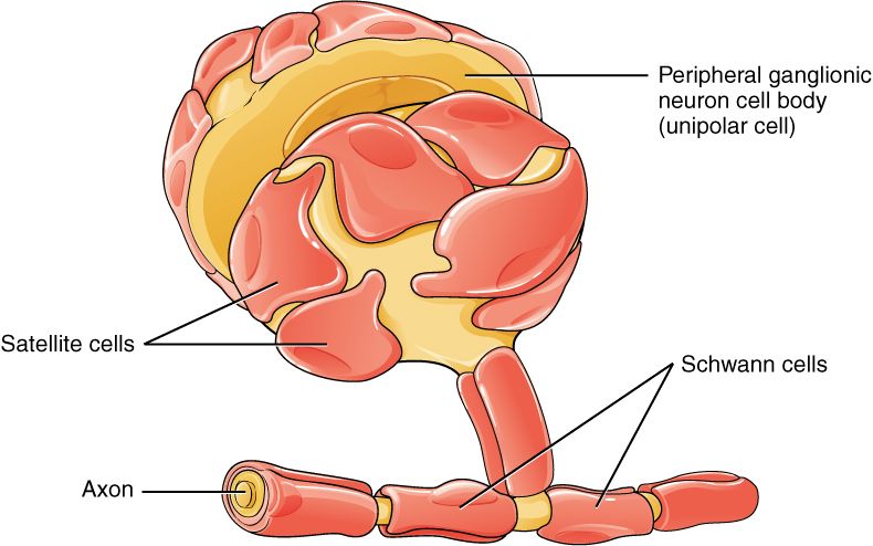
The neuron: a cell built to signal
Before we meet the support cells, meet the star. A neuron is an excitable cell shaped for one purpose — to receive a signal at one end and pass it along from the other. Three parts do the work. The dendrites are branching antennae that receive incoming signals. The cell body (or soma) holds the nucleus and does the cell's living and housekeeping; it is also where incoming signals are added up. The axon is a single long fiber that carries the outgoing signal away, often wrapped in a fatty myelin sheath and ending in axon terminals that hand the message to the next cell. Neurons are also amitotic — most cannot divide to replace themselves — which is exactly why damage to the brain and spinal cord is so hard to repair.
The support crew: six kinds of glia
For every neuron there are roughly as many, or more, neuroglia (or glial cells) — the crew that feeds, insulates, cleans, and protects. Glia do not fire action potentials, but without them neurons would starve, short-circuit, and drown in debris. There are six types: four live in the CNS and two in the PNS.
| Glial cell | Where | Job |
|---|---|---|
| Astrocyte | CNS | Most abundant; anchors neurons to blood supply, controls the chemical environment, helps form the blood–brain barrier |
| Oligodendrocyte | CNS | Wraps CNS axons in myelin (one cell myelinates many axons) |
| Microglia | CNS | The brain's immune cells — engulf pathogens and dead cells |
| Ependymal cell | CNS | Line the brain's cavities; circulate cerebrospinal fluid |
| Satellite cell | PNS | Surround and support neuron cell bodies in ganglia |
| Schwann cell | PNS | Wraps PNS axons in myelin (one cell myelinates one segment) |
Two cells make myelin — keep them straight
The two myelinating cells cause endless confusion, so pin them down now. In the CNS, an oligodendrocyte reaches out several arms and myelinates many different axons at once. In the PNS, a Schwann cell wraps itself around a single segment of one axon like a jelly roll. Both make the same fatty insulation, but the difference matters clinically: diseases that attack CNS myelin (multiple sclerosis) and those that attack PNS myelin (Guillain–Barré syndrome) are entirely separate conditions with different courses.
Tap each of the six glial cells to see where it sits and what it does.
- A neuron has three parts: dendrites (receive), cell body/soma (integrate and live), and axon (transmit).
- The six glia: CNS — astrocyte, oligodendrocyte, microglia, ependymal; PNS — satellite, Schwann.
- Oligodendrocytes myelinate many CNS axons; Schwann cells each myelinate one PNS segment.
- Microglia are the CNS's immune cells; astrocytes are the most abundant glia and help form the blood–brain barrier.
Section 3The resting membrane potential
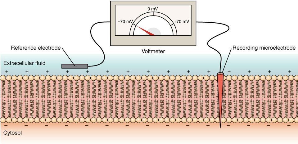
A charged battery, waiting
A neuron at rest is not doing nothing — it is armed. Across its membrane sits a tiny voltage called the resting membrane potential, about −70 mV, meaning the inside of the cell is 70 millivolts negative compared with the outside. A membrane that holds this separation of charge is said to be polarized, like a charged battery ready to be tapped. Everything a neuron does — every signal — is a brief, controlled discharge of that stored charge. So the first question of all neurophysiology is simply: how does the cell build the charge in the first place?
Why the inside is negative
The answer is unequal ion distribution. The cell carefully keeps different ions at different concentrations inside versus outside. Sodium (Na⁺) is piled up outside the cell; potassium (K⁺) is concentrated inside. In addition, the inside is stuffed with large negatively charged proteins and phosphates that are simply too big to leave. Now add the membrane's channels. At rest the membrane is far leakier to potassium than to sodium — there are many more open leak channels for K⁺. So potassium trickles out down its gradient, carrying positive charge with it and leaving the inside more negative. That potassium leak, working against the trapped internal anions, is the main reason the resting voltage lands near −70 mV.
| Ion | Concentrated where | Role at rest |
|---|---|---|
| Sodium (Na⁺) | Outside the cell | Poised to rush in; membrane mostly closed to it at rest |
| Potassium (K⁺) | Inside the cell | Leaks out through many open channels — sets the resting voltage |
| Chloride (Cl⁻) | Outside the cell | Contributes to the negative interior |
| Large anions (proteins) | Trapped inside | Cannot cross — hold negative charge in |
The pump that never sleeps
Leaks would eventually run the gradients down — sodium would trickle in, potassium would drift out, and the battery would go flat. To prevent that, the membrane runs the sodium–potassium pump (Na⁺/K⁺ ATPase) around the clock. Burning ATP for fuel, it pumps 3 Na⁺ out for every 2 K⁺ in, restoring both gradients continuously. Because it moves three positives out for every two brought in, the pump also removes a little net positive charge, nudging the interior slightly more negative. This pump is so essential that it consumes a large share of the body's resting energy budget — a big reason the brain is such a hungry organ.
Explore the ion gradients, the leak channels, and the tireless pump in the interactive below.
- The resting membrane potential is about −70 mV, inside negative — the membrane is polarized.
- It arises from unequal ion distribution: Na⁺ high outside, K⁺ high inside, large anions trapped inside.
- Potassium leaking out through open channels is the main setter of the resting voltage.
- The Na⁺/K⁺ pump (3 out, 2 in, using ATP) maintains the gradients and adds a small negative bias.
Section 4The action potential
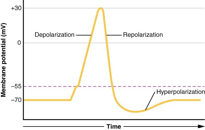
All or nothing
An action potential is the neuron's signal — a brief, dramatic reversal of the membrane voltage that shoots down the axon. It does not happen unless the membrane is pushed from its resting −70 mV up to a critical threshold of about −55 mV. Below threshold, nothing fires. Reach threshold and a full-sized spike goes off every time — the same height whether the stimulus was barely enough or enormous. This is the famous all-or-nothing law: a neuron cannot fire a partial action potential any more than a gun can fire a partial bullet. Strength is coded not by bigger spikes but by how often the neuron fires.
The upstroke: sodium floods in
The whole event is run by voltage-gated channels — ion gates that open in response to voltage. When threshold is reached, voltage-gated Na⁺ channels snap open and sodium, which was piled up outside and pulled inward by both its concentration gradient and the negative interior, floods in. Each entering positive charge makes the inside less negative, which opens still more sodium gates — a fast, self-amplifying rush called depolarization that drives the voltage from −55 all the way up to about +30 mV. For an instant, the inside of the cell is actually positive.
The downstroke and the reset
The spike ends as fast as it began. At the peak the Na⁺ channels inactivate (they auto-close), and now the slower voltage-gated K⁺ channels finish opening. Potassium rushes out, carrying positive charge away and dropping the voltage back down — this is repolarization. The potassium gates are sluggish to close, so a little too much K⁺ leaves and the membrane briefly dips below rest to about −80 mV, a hyperpolarization or undershoot. Finally the leak channels and the Na⁺/K⁺ pump restore the tidy −70 mV. During the recovery there is a refractory period when the axon either cannot fire at all (absolute) or needs an unusually strong stimulus (relative).
| Phase | What the channels do | Voltage |
|---|---|---|
| Resting | Only leak channels open | −70 mV |
| Threshold | Enough Na⁺ gates begin to open | −55 mV |
| Depolarization | Voltage-gated Na⁺ channels open; Na⁺ rushes in | rising to +30 mV |
| Repolarization | Na⁺ channels inactivate; K⁺ channels open, K⁺ exits | falling back down |
| Hyperpolarization | K⁺ channels close slowly — slight overshoot | ~−80 mV |
| Return to rest | Leak channels + Na⁺/K⁺ pump restore balance | −70 mV |
Hit "Stimulate" and follow the voltage through every phase in the interactive below.
- An action potential fires only if the membrane reaches threshold (~−55 mV), and then it is all-or-nothing.
- Depolarization = voltage-gated Na⁺ channels open, Na⁺ floods in, voltage climbs to ~+30 mV.
- Repolarization = Na⁺ channels inactivate and K⁺ channels open, K⁺ exits, voltage falls; a brief hyperpolarization follows.
- The refractory period keeps the signal moving one way and limits firing rate; stimulus strength is coded by frequency.
Section 5Saltatory conduction & speed
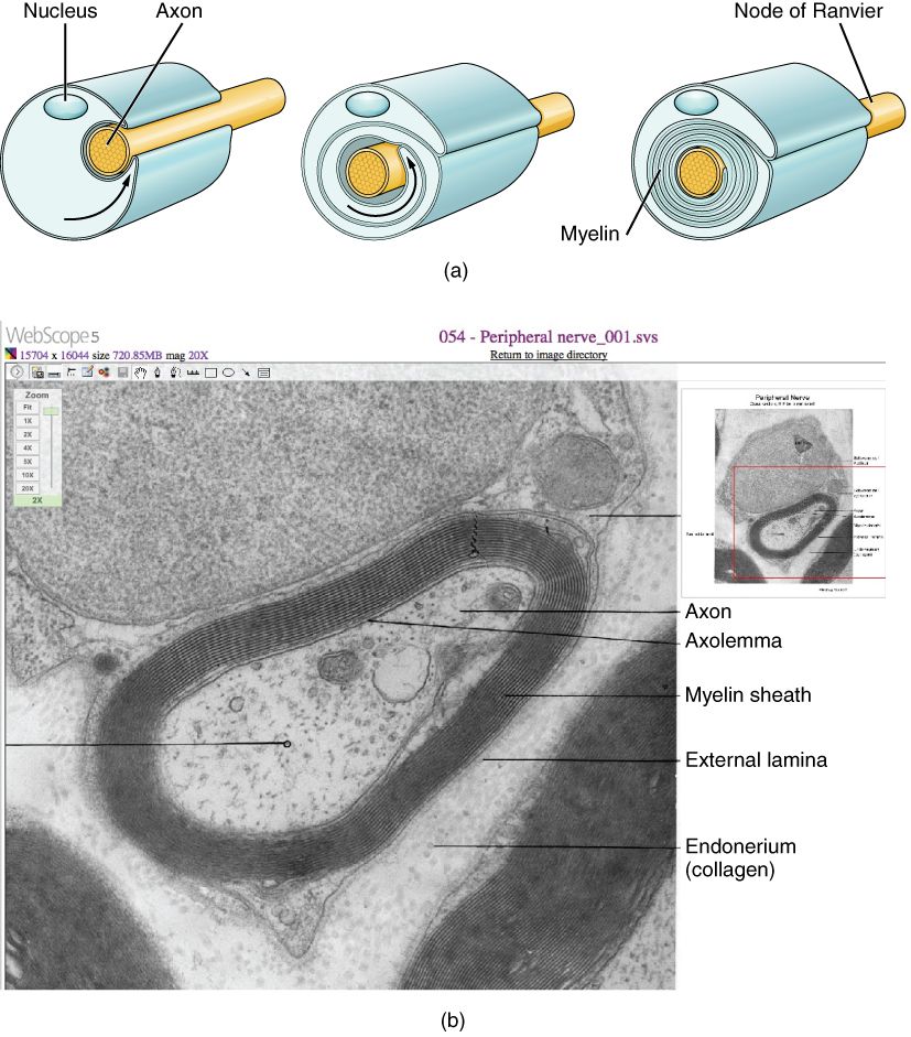
Crawling versus leaping
Once an action potential is born, it has to travel the length of the axon, and there are two ways to do that. On a bare, unmyelinated axon the spike regenerates itself at every adjacent patch of membrane, marching along step by step — continuous conduction. It works, but it is slow. On a myelinated axon the fatty sheath insulates long stretches so the current cannot leak out there; the action potential can only regenerate at the bare gaps between myelin segments. So the signal appears to jump from gap to gap, skipping the insulated lengths in between. This leaping is saltatory conduction, from the Latin saltare, "to leap," and it is dramatically faster.
Nodes of Ranvier
The bare gaps have a name worth knowing: the nodes of Ranvier. These little exposed rings are packed with voltage-gated sodium channels, so the action potential is regenerated fresh and full-strength at each node. Between the nodes, the insulated segment (the internode) carries the current quickly and passively, with no channels to slow it down. The result is that the impulse effectively hops from node to node — regenerate, glide, regenerate, glide — covering far more ground per unit time than a continuous crawl.
What sets conduction speed
Two features determine how fast a fiber conducts. The first is myelination: a myelinated axon massively outpaces an unmyelinated one of the same size. The second is axon diameter: fatter axons conduct faster because they offer less resistance to current, the same way a wide pipe moves water faster than a narrow one. The body uses both dials deliberately — the large, heavily myelinated fibers that carry reflexes and limb-position sense can conduct at speeds over 100 meters per second, while the thin, unmyelinated fibers that carry dull, aching pain crawl along at barely one. (Sharp, localized pain travels on thinner myelinated fibers — faster than the dull, unmyelinated kind, but far short of that top speed.)
| Factor | Faster conduction | Slower conduction |
|---|---|---|
| Myelin | Myelinated (saltatory) | Unmyelinated (continuous) |
| Axon diameter | Large diameter | Small diameter |
| Typical role | Reflexes, sharp/localized pain, position | Dull aching pain, temperature, autonomic |
Race a myelinated signal against an unmyelinated one and watch it leap the nodes in the interactive below.
- Continuous conduction regenerates the spike at every patch (slow, unmyelinated); saltatory conduction leaps between gaps (fast, myelinated).
- The bare gaps are the nodes of Ranvier, packed with Na⁺ channels; the insulated internodes carry current passively.
- Speed rises with myelination and with axon diameter.
- Demyelinating disease (MS in the CNS, Guillain–Barré in the PNS) slows or blocks conduction.
Section 6The synapse: passing the message on
Neurons don't quite touch
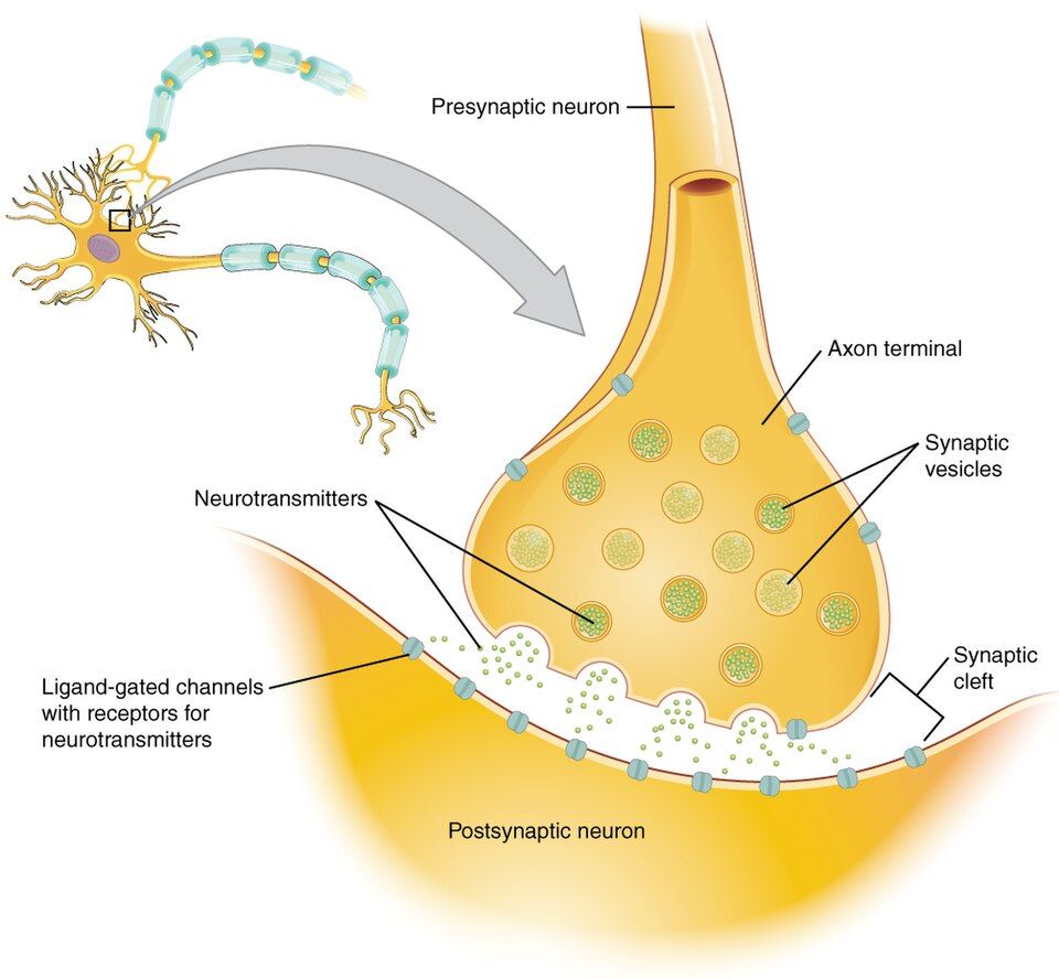
When an action potential reaches the end of an axon, it faces a problem: the next cell is not connected to it. There is a microscopic gap in between called the synaptic cleft, and the junction as a whole is the synapse. The neuron sending the message is presynaptic; the cell receiving it is postsynaptic. Because the electrical signal cannot jump the gap on its own, most synapses convert it into a chemical messenger — a neurotransmitter — that drifts across and re-creates the signal on the far side. Electrical, then chemical, then electrical again.
Six steps across the gap
Chemical transmission always follows the same choreography. (1) The action potential arrives at the presynaptic axon terminal. (2) Its voltage opens voltage-gated calcium (Ca²⁺) channels, and calcium flows into the terminal. (3) Calcium triggers tiny sacs called synaptic vesicles to fuse with the membrane and dump their contents. (4) Neurotransmitter is released by exocytosis and diffuses across the cleft. (5) It binds receptors on the postsynaptic membrane, opening ion channels that create a small graded potential. (6) The neurotransmitter is quickly cleared — broken down by enzymes, taken back up (reuptake), or diffused away — so the signal is crisp and brief.
Excitatory or inhibitory?
What the postsynaptic cell does next depends entirely on which neurotransmitter and receptor are involved. An excitatory signal depolarizes the postsynaptic membrane, nudging it toward threshold — an excitatory postsynaptic potential (EPSP). An inhibitory signal hyperpolarizes it, pushing it away from threshold — an inhibitory postsynaptic potential (IPSP). A single neuron receives thousands of these at once and sums them; only if the excitation outweighs the inhibition enough to reach threshold does it fire. The synapse, in other words, is where the nervous system actually makes decisions.
| Neurotransmitter | Typical effect | Where / note |
|---|---|---|
| Acetylcholine (ACh) | Excites skeletal muscle | Neuromuscular junction; broken down by acetylcholinesterase |
| Glutamate | Excitatory | The main excitatory transmitter of the CNS |
| GABA | Inhibitory | The main inhibitory transmitter of the CNS |
| Glycine | Inhibitory | Common in the spinal cord |
| Norepinephrine | Excites or inhibits | Sympathetic (fight-or-flight), mood, arousal |
| Dopamine | Modulatory | Reward, movement; disrupted in Parkinson disease |
Step through the electrical-to-chemical-to-electrical handoff in the interactive below.
- Neurons meet at a synapse across the synaptic cleft; the sender is presynaptic, the receiver postsynaptic.
- Transmission: AP arrives → Ca²⁺ enters → vesicles fuse → neurotransmitter released → binds receptors → cleared.
- An EPSP pushes the postsynaptic cell toward threshold; an IPSP pushes it away; the cell sums them to decide whether to fire.
- Key transmitters: ACh (muscle), glutamate (main excitatory), GABA (main inhibitory); the synapse is where most drugs act.
The chapter in one breath
- The nervous system splits into the CNS (brain and spinal cord) and PNS (nerves and ganglia), and its work is always sensory input → integration → motor output.
- Motor pathways divide into somatic (voluntary) and autonomic (involuntary, split into sympathetic and parasympathetic); afferent arrives, efferent exits.
- A neuron (dendrites, soma, axon) is supported by six neuroglia — astrocyte, oligodendrocyte, microglia, ependymal (CNS) and satellite, Schwann (PNS).
- At rest the membrane is polarized to ~−70 mV, set by K⁺ leaking out and trapped internal anions, and maintained by the Na⁺/K⁺ pump.
- Reaching threshold (~−55 mV) fires an all-or-nothing action potential: Na⁺ in (depolarize) then K⁺ out (repolarize), with a refractory period.
- Myelin lets the impulse leap node to node — saltatory conduction — and speed rises with myelination and diameter.
- At the synapse, the signal crosses as a neurotransmitter; the postsynaptic cell sums EPSPs and IPSPs to decide whether to fire.
ReferenceWords worth knowing
A quick reference for the terms in this chapter — skim it now, or circle back whenever a word trips you up.
- Action potential
- A brief, all-or-nothing reversal of membrane voltage that travels down an axon; the neuron's electrical signal.
- Afferent (sensory) neuron
- A neuron that carries signals toward the central nervous system (afferent arrives).
- All-or-nothing
- The principle that a neuron either fires a full-sized action potential or none at all; stimulus strength is coded by firing frequency.
- Astrocyte
- The most abundant CNS glial cell; anchors neurons to blood vessels, regulates the chemical environment, and helps form the blood–brain barrier.
- Autonomic nervous system
- The involuntary motor division controlling cardiac muscle, smooth muscle, and glands; split into sympathetic and parasympathetic arms.
- Axon
- The single long fiber of a neuron that carries the action potential away from the cell body.
- Depolarization
- A shift of the membrane voltage toward positive (less negative), driven mainly by Na⁺ entering the cell.
- Efferent (motor) neuron
- A neuron that carries commands away from the central nervous system to an effector (efferent exits).
- Myelin sheath
- Fatty insulation wrapped around an axon (by oligodendrocytes in the CNS, Schwann cells in the PNS) that speeds conduction.
- Neuroglia
- The support cells of nervous tissue that feed, insulate, clean, and protect neurons; six types across the CNS and PNS.
- Neuron
- The excitable signaling cell of the nervous system, with dendrites (receive), a cell body (integrate), and an axon (transmit).
- Neurotransmitter
- A chemical messenger released across a synapse to relay a signal to the next cell.
- Node of Ranvier
- A bare gap between myelin segments, rich in Na⁺ channels, where an action potential is regenerated during saltatory conduction.
- Refractory period
- The brief interval after a spike when a region of membrane cannot fire again (absolute) or needs a stronger stimulus (relative), forcing one-way conduction.
- Repolarization
- The return of membrane voltage toward its negative resting value, driven mainly by K⁺ leaving the cell.
- Resting membrane potential
- The voltage (~−70 mV, inside negative) across a resting neuron's membrane, set by ion gradients and K⁺ leak.
- Saltatory conduction
- Fast impulse transmission along a myelinated axon in which the signal leaps from node to node.
- Schwann cell
- The PNS glial cell that wraps a single axon segment in myelin.
- Sodium–potassium pump
- An ATP-powered membrane transporter that moves 3 Na⁺ out and 2 K⁺ in, maintaining the ion gradients.
- Synapse
- The junction where one neuron communicates with another cell across the synaptic cleft.
- Threshold
- The membrane voltage (~−55 mV) that must be reached to trigger an action potential.