Muscle Tissue
Muscle is the tissue that turns a decision into a movement. Every heartbeat, every blink, every stride, every push of food through your gut is a muscle cell doing the one thing muscle does supremely well — shortening on command. In this chapter we take a single skeletal muscle apart, layer by layer, until we reach the microscopic engine that actually pulls: the sarcomere. Then we watch that engine run — how a nerve signal crosses into the muscle, how calcium throws the switch, how filaments slide past one another, and how the body dials the force up from a delicate flicker to an all-out heave. By the end you will understand contraction not as magic but as a beautifully ordered chain of events you can follow from thought to motion.
By the end of this chapter you can
- Compare the three types of muscle tissue — skeletal, cardiac, and smooth — by structure, control, and location.
- Trace the connective-tissue wrappings of a skeletal muscle from epimysium down to a single fiber, and name the parts inside that fiber.
- Diagram a sarcomere and identify its bands, zones, and lines, and explain why skeletal muscle looks striped.
- Explain the sliding filament theory and the steps of the cross-bridge cycle, including the roles of ATP and calcium.
- Describe the neuromuscular junction and follow a signal from motor neuron to muscle action potential.
- Define a motor unit and explain how recruitment and summation grade muscle tension from a twitch to tetanus.
- Distinguish the three skeletal muscle fiber types and match each to the kind of work it does best.
Section 1How a muscle is built
Three kinds of muscle tissue
Before we open up one muscle, it helps to know that the body builds muscle in three different flavors, each tuned to a different job. Skeletal muscle attaches to bones and moves the body; it is under voluntary control, and under the microscope its cells are long, striped (striated), and packed with many nuclei. Cardiac muscle is found only in the heart wall; it is striated too, but it is involuntary, its cells are branched with usually a single nucleus, and they are joined end-to-end by intercalated discs that let the whole heart contract as one. Smooth muscle lines the walls of hollow organs — blood vessels, gut, bladder, airways — where it squeezes slowly and tirelessly; it is involuntary, has no stripes, and its cells are tapered with one central nucleus. This chapter concentrates on skeletal muscle, because it shows every principle most clearly, but keep the other two in mind — the contraction machinery is largely shared.
| Feature | Skeletal | Cardiac | Smooth |
|---|---|---|---|
| Location | Attached to bones | Heart wall only | Walls of hollow organs & vessels |
| Striations | Striated | Striated | Not striated |
| Control | Voluntary | Involuntary | Involuntary |
| Cell shape | Long cylinder, many nuclei | Branched, usually 1 nucleus | Tapered spindle, 1 nucleus |
| Special feature | Motor-neuron controlled | Intercalated discs; self-rhythmic | Slow, sustained, self-triggering |
Bundles inside bundles
A whole skeletal muscle — your biceps, say — is not one solid block but a nested set of bundles, each wrapped in its own sheet of connective tissue. Working from the outside in: the entire muscle is sheathed in the epimysium; inside that, fibers are gathered into bundles called fascicles, each wrapped by perimysium; and inside each fascicle, every individual muscle cell (a muscle fiber) is wrapped in a delicate endomysium. These three sheaths are continuous with one another and, at the ends of the muscle, they merge and extend beyond it as a tendon (a cord) or an aponeurosis (a flat sheet) that anchors the muscle to bone. This arrangement is not just tidy packaging: the connective tissue carries the blood vessels and nerves in to each fiber, and it transmits the pull of every cell to the tendon so the whole muscle moves as one.
Inside a single fiber
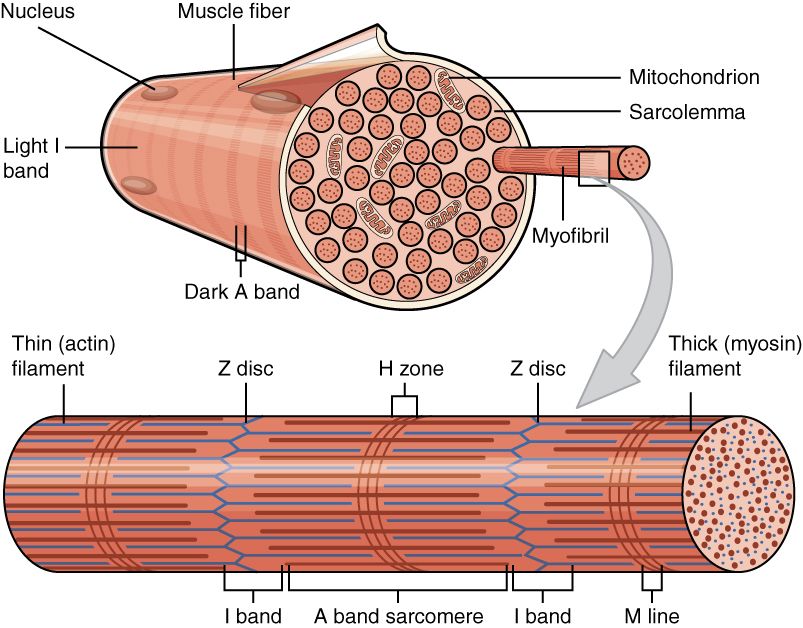
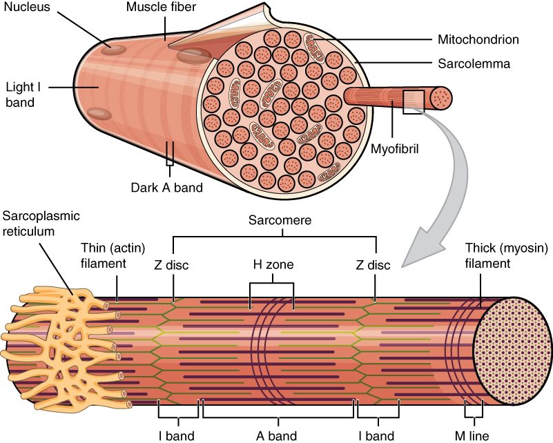
Zoom into one muscle fiber and you find a cell built for pulling. Its plasma membrane is called the sarcolemma, and its cytoplasm the sarcoplasm (the prefix sarco- means “flesh”). The sarcolemma dives deep into the cell as tunnels called transverse (T) tubules, which carry the electrical signal into the fiber’s core. Wrapped around the working machinery is a specialized smooth endoplasmic reticulum, the sarcoplasmic reticulum (SR), which stores and releases the calcium that triggers contraction. Filling most of the cell are hundreds of long threads called myofibrils, and each myofibril is a chain of the contractile units — the sarcomeres — made of myofilaments of the proteins actin and myosin. Because a muscle fiber forms during development by many cells fusing together, it holds many nuclei pushed to the edge just under the sarcolemma, and a reserve of satellite cells sits nearby to repair damage.
Follow all of this in the interactive: start at the whole muscle and zoom, wrapping by wrapping, down to the sarcomere.
- Three muscle types: skeletal (voluntary, striated, bones), cardiac (involuntary, striated, heart), smooth (involuntary, unstriated, hollow organs).
- A skeletal muscle nests bundles inside bundles: epimysium → fascicle (perimysium) → fiber (endomysium), all continuous with the tendon.
- Inside a fiber: sarcolemma, T-tubules, sarcoplasmic reticulum, myofibrils of actin and myosin, and many nuclei at the edge.
Section 2The sarcomere
Stripes with a purpose
The stripes that give skeletal and cardiac muscle their name are not decoration — they are the visible signature of astonishingly ordered machinery. Inside every myofibril, the contractile proteins are lined up in identical repeating segments, and each segment is a sarcomere, the functional unit of contraction. One sarcomere runs from one Z-disc to the next Z-disc. When millions of these little units all shorten at once, in register, the whole muscle shortens — and it is their alternating dense and light regions, stacked side by side across the fiber, that our eyes read as striations.
The bands, zones, and lines
A sarcomere is built from two kinds of filament. The thick filaments are made of myosin, each with a hinged head that sticks out; the thin filaments are made of actin. Where the two overlap, the sarcomere looks dark; where only one is present, it looks lighter. Learning the map of bands is worth the effort, because in the next section you will watch these regions change size as the muscle contracts.
| Region | What it contains | Appearance |
|---|---|---|
| A band | The full length of the thick filaments (with overlap at its edges) | Dark; does not change length in contraction |
| I band | Thin filaments only, spanning two adjacent sarcomeres | Light; narrows during contraction |
| H zone | Thick filaments only, center of the A band (no overlap) | Lighter center; narrows during contraction |
| Z-disc (Z-line) | Anchors the thin filaments; the border of the sarcomere | Dark line; Z-discs are pulled together |
| M-line | Anchors the thick filaments at the sarcomere’s center | Fine central line |
Thick, thin, and the switch proteins
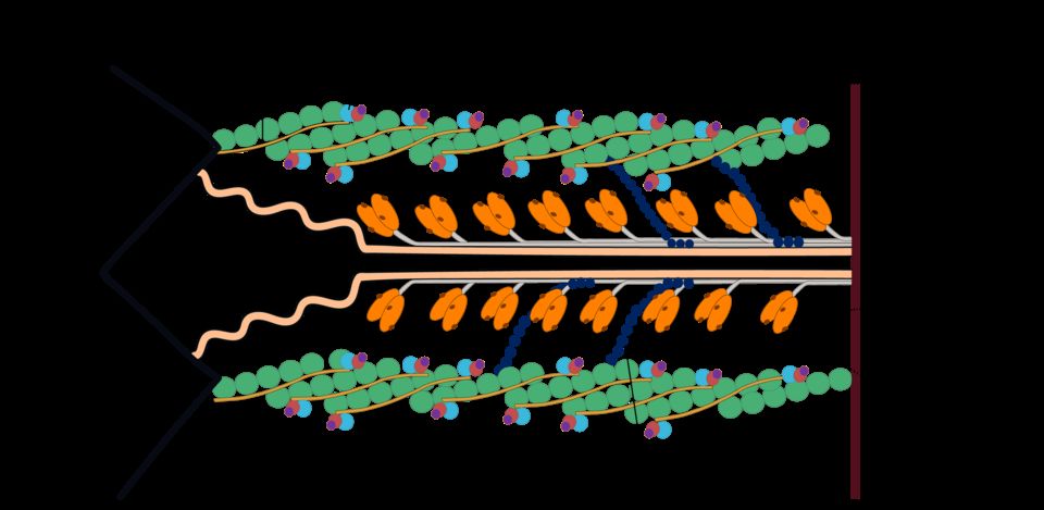
The thin filament is more than plain actin. Wrapped along it lies a ropelike protein, tropomyosin, that in a resting muscle physically covers the spots where myosin would attach — the myosin-binding sites. Holding tropomyosin in place is a three-part protein complex, troponin, one piece of which (troponin C) is a calcium receptor. This is the molecular switch: when calcium binds troponin, the whole complex shifts, tropomyosin slides aside, and the binding sites open. Two more proteins keep the sarcomere honest: giant, springy titin runs from Z-disc to M-line, keeping the thick filament centered and giving muscle its passive recoil, and nebulin runs alongside the thin filament, setting its length. Structure, once again, is built exactly for the job.
Tap each part in the interactive to see how the filaments build the stripes.
- The sarcomere (Z-disc to Z-disc) is the functional unit of contraction; ordered sarcomeres create the visible striations.
- Thick filaments are myosin (with heads); thin filaments are actin, guarded by tropomyosin and the calcium-sensing troponin.
- The A band stays constant while the I band and H zone narrow during contraction — the tell-tale of sliding filaments.
Section 3The sliding filament theory
Sliding, not shrinking
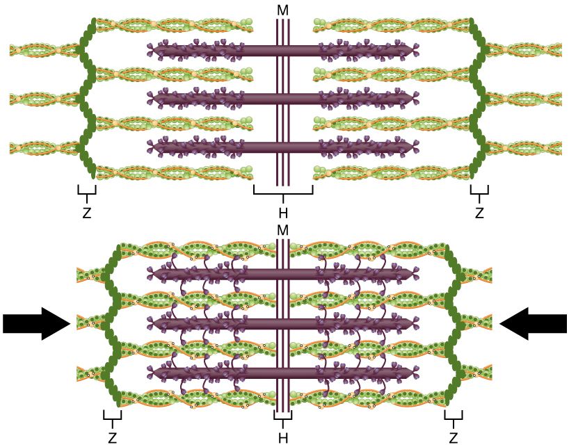
Here is the single most important idea in this chapter, and it surprises almost everyone the first time: the filaments do not get shorter. When a muscle contracts, the thick and thin filaments slide past one another, increasing their overlap and dragging the Z-discs closer together. Each filament keeps its exact length; the sarcomere is what shortens. That is why, in the reference figure, the A band (the length of the thick filament) never changes, while the I band and H zone shrink and the Z-discs march inward. Multiply that tiny slide across millions of sarcomeres in series and the whole muscle noticeably shortens and pulls on its tendon.
The cross-bridge cycle
The sliding is powered by the myosin heads, which act as tiny oars. Each head reaches out, grabs actin to form a cross-bridge, and swivels — a power stroke — that ratchets the thin filament a few nanometers toward the center. Then it lets go, re-cocks, and grabs again, farther along. The cycle repeats many times per second, and it runs on ATP:
| Step | What happens | Role of ATP / Ca²⁺ |
|---|---|---|
| 1. Cross-bridge forms | The energized myosin head binds an exposed site on actin | Requires Ca²⁺ to have opened the site |
| 2. Power stroke | The head pivots, sliding the thin filament inward; ADP & Pi (inorganic phosphate) are released | Uses energy stored from a previous ATP |
| 3. Detachment | A fresh ATP binds the myosin head, which releases from actin | ATP binding breaks the cross-bridge |
| 4. Re-cocking | ATP is split to ADP + Pi, re-energizing and re-setting the head | ATP hydrolysis re-cocks the head |
Notice the twin jobs of ATP: it is needed to release the head (step 3) and to re-cock it (step 4). This has a memorable consequence — in death, when ATP production stops, myosin heads bind actin and can no longer let go, and the body stiffens into rigor mortis.
Calcium throws the switch: excitation–contraction coupling
Cross-bridges can only form if the binding sites are uncovered, and that is calcium’s job. The link between an electrical signal and a mechanical pull is called excitation–contraction coupling. A muscle action potential races along the sarcolemma and dives down the T-tubules into the fiber. That voltage change tells the sarcoplasmic reticulum to dump its stored Ca²⁺ into the sarcoplasm. Calcium binds troponin, troponin tugs tropomyosin off the actin, the binding sites open, and cross-bridging — and sliding — begins. Relaxation is just the reverse: when the signal stops, a pump called SERCA hauls Ca²⁺ back into the SR, tropomyosin re-covers the sites, cross-bridges can no longer form, and the muscle goes slack. No calcium, no contraction.
Watch the Z-lines pull together while the filaments hold their length in the interactive.
- In the sliding filament theory, filaments keep their length; the sarcomere shortens as thick and thin filaments overlap more.
- The cross-bridge cycle — bind, power stroke, detach, re-cock — runs on ATP, which is needed to release and to re-energize the myosin head.
- Excitation–contraction coupling: an action potential → T-tubules → SR releases Ca²⁺ → troponin/tropomyosin uncover binding sites; SERCA reclaims calcium to relax.
Section 4The neuromuscular junction
From nerve to muscle
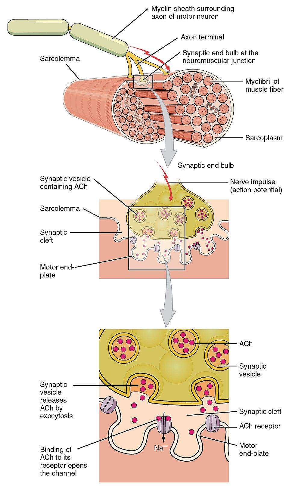
A skeletal muscle fiber will not contract on its own — it waits for the command of a nerve. That command is delivered at a specialized synapse called the neuromuscular junction (NMJ). A motor neuron reaches out from the spinal cord, and its axon terminal comes to rest just above a folded patch of sarcolemma called the motor end plate. The two never touch; a tiny gap, the synaptic cleft, separates them. To bridge that gap, the neuron uses a chemical messenger — the neurotransmitter acetylcholine (ACh).
The handoff, step by step
The transfer of the signal from nerve to muscle is one of the most reliable events in physiology — a single nerve impulse essentially always triggers a muscle impulse. Follow the chain:
| Step | Event |
|---|---|
| 1 | A nerve action potential arrives at the axon terminal. |
| 2 | Voltage-gated Ca²⁺ channels open; calcium rushes into the terminal. |
| 3 | Calcium triggers vesicles to release acetylcholine into the synaptic cleft. |
| 4 | ACh crosses the cleft and binds nicotinic receptors on the motor end plate. |
| 5 | Receptor channels open; Na⁺ floods in, creating an end-plate potential. |
| 6 | The depolarization sets off a muscle action potential that spreads along the sarcolemma and down the T-tubules — and contraction follows. |
Notice how this dovetails with the last section: step 6 is exactly the muscle action potential that starts excitation–contraction coupling. The NMJ is the doorway; the sliding filament is the room it opens onto.
Switching the signal off — and where it breaks
A signal that never ended would leave the muscle locked in contraction, so the cleft contains an enzyme, acetylcholinesterase (AChE), that chops ACh apart within milliseconds. This tidy on/off system is also where many drugs and diseases act, which makes the NMJ one of the highest-yield spots in clinical medicine. In myasthenia gravis, the immune system destroys ACh receptors, so muscles fatigue and weaken. Curare (an arrow poison) blocks the receptors, causing paralysis. Organophosphate pesticides and nerve agents inhibit AChE, so ACh piles up and muscles seize. Botulinum toxin (Botox) blocks ACh release, relaxing muscle — useful for spasms and wrinkles.
Step through the chemical handoff yourself in the interactive.
- The neuromuscular junction is the synapse where a motor neuron signals a muscle fiber using acetylcholine across the synaptic cleft.
- ACh binds nicotinic receptors on the motor end plate, letting Na⁺ in to trigger a muscle action potential; acetylcholinesterase then ends the signal.
- NMJ disorders map to the transmitter: blocking ACh → weakness (myasthenia gravis, curare, botulinum); prolonging ACh → spasm (AChE inhibitors, nerve agents).
Section 5Motor units & muscle tension
The motor unit
A single motor neuron rarely commands just one muscle fiber. Instead it branches and connects to a group of fibers, and the neuron together with all the fibers it controls is a motor unit. When that neuron fires, every fiber in its unit contracts at once — they are an all-or-nothing team. The size of the unit sets how finely a muscle can be controlled. Muscles that need delicacy, like those moving the eye or the fingers, have small motor units (a neuron to only a handful of fibers). Muscles built for power, like the quadriceps or the calf, have large motor units (a neuron to hundreds of fibers). Small units mean fine control; large units mean strong, coarse force.
Twitch, summation, tetanus
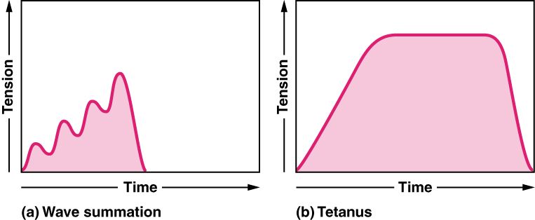
One brief stimulus produces a single, quick contraction-and-relaxation called a twitch, which has three phases: a latent period (the hidden delay while calcium is released), a contraction phase, and a relaxation phase. A twitch is a flick, not a useful pull. To get smooth, strong movement, the nervous system fires the motor neuron again before the fiber fully relaxes, so the second contraction stacks on top of the first. This stacking is wave (temporal) summation. Fire faster still and the individual twitches blur into a partly smoothed unfused (incomplete) tetanus, and faster yet into a steady, maximal, perfectly smooth fused (complete) tetanus. Nearly all normal movements use tetanus, not single twitches.
| Response | Stimulus pattern | Result |
|---|---|---|
| Twitch | One stimulus | A single brief contraction and full relaxation |
| Wave summation | A second stimulus before relaxation ends | The second contraction adds to the first — more tension |
| Unfused tetanus | Rapid, repeated stimuli | Strong, sustained pull with slight wobbles |
| Fused tetanus | Very high-frequency stimuli | Maximal, perfectly smooth, sustained contraction |
Two dials for force: recruitment and rate
The body grades how hard a muscle pulls using two controls at once. The first is recruitment — switching on more motor units. Under the size principle, small, weak, fatigue-resistant units are recruited first, and larger, more powerful ones are added only as more force is needed, so movements start smooth and controllable. The second dial is rate coding — firing each active unit faster to climb the summation-to-tetanus ladder above. Together, recruitment and rate let you lift a feather or a barbell with the same muscle. Even at rest, a few units are always active in rotation, keeping muscles firm and ready — this steady low-level tension is muscle tone.
Pick a stimulation pattern in the interactive and watch tension climb from twitch to tetanus.
- A motor unit is one motor neuron plus all the fibers it controls; small units give fine control, large units give power.
- Tension builds from a twitch through wave summation to tetanus as stimulation frequency rises; real movement uses tetanus.
- Force is graded by recruitment (adding units, smallest first — the size principle) and rate coding (firing faster); resting activity is muscle tone.
Section 6The three fiber types
Speed versus stamina
Not every skeletal muscle fiber is the same. They come in three types that trade speed against endurance, because no single design can be both maximally fast and maximally tireless. Slow oxidative (Type I) fibers contract slowly and generate modest force, but they resist fatigue for hours — the endurance specialists. Fast glycolytic (Type IIx) fibers contract quickly and powerfully but tire within minutes — the sprint-and-lift specialists. In between sit fast oxidative (Type IIa) fibers, which are fast and reasonably fatigue-resistant — a versatile middle ground.
Aerobic versus anaerobic engines
The difference comes down to how each fiber makes its ATP. Oxidative fibers run on aerobic respiration: they are packed with mitochondria, richly supplied with capillaries, and loaded with myoglobin — an oxygen-binding pigment that makes them look red. That red, oxygen-fed machinery is slow but sustainable. Glycolytic fibers rely on anaerobic glycolysis: they have few mitochondria, little myoglobin (so they look pale), and large glycogen stores for quick bursts. They deliver explosive power but flood with lactate and fatigue fast.
| Property | Slow oxidative (I) | Fast oxidative (IIa) | Fast glycolytic (IIx) |
|---|---|---|---|
| Contraction speed | Slow | Fast | Fast |
| Fatigue resistance | High | Moderate | Low |
| Main ATP source | Aerobic (oxidative) | Both | Anaerobic (glycolytic) |
| Mitochondria / capillaries | Many | Many | Few |
| Myoglobin / color | High / red | High / red-pink | Low / white |
| Best for | Posture, endurance | Walking, sustained effort | Sprinting, lifting |
Everybody is a mixture — and a nod to the other two muscles
Almost every real muscle contains all three fiber types, blended in a ratio set partly by your genes and partly by how you train. Endurance training coaxes fibers toward the oxidative, fatigue-resistant end; strength and sprint training push toward larger, more powerful fibers. It is why a marathoner’s calves and a sprinter’s thighs are built for different feats. And remember the two muscle types we set aside at the start: cardiac muscle behaves like a permanently endurance-tuned, self-rhythmic tissue joined by intercalated discs so the heart never truly rests, while smooth muscle has no sarcomeres at all and can hold tension for long stretches with very little energy — a “latch” state perfect for keeping a blood vessel or a sphincter squeezed shut all day.
Tap each fiber type in the interactive to compare speed, color, and stamina.
- Three fiber types trade speed for stamina: slow oxidative (I) = endurance; fast glycolytic (IIx) = power; fast oxidative (IIa) = in between.
- Oxidative fibers are red (many mitochondria, myoglobin, capillaries) and aerobic; glycolytic fibers are pale, anaerobic, powerful, and quick to fatigue.
- Most muscles are a mix shaped by genetics and training; cardiac and smooth muscle are separate designs tuned for tireless work.
The chapter in one breath
- Muscle comes in three types — skeletal (voluntary, striated), cardiac (involuntary, striated, self-rhythmic), and smooth (involuntary, unstriated) — sharing the same basic contraction machinery.
- A skeletal muscle is bundles within bundles wrapped in epimysium, perimysium, and endomysium, all continuous with the tendon; inside each fiber are the sarcolemma, T-tubules, sarcoplasmic reticulum, and myofibrils.
- The sarcomere (Z-disc to Z-disc) is the functional unit; overlapping thick (myosin) and thin (actin) filaments create the striations, guarded by tropomyosin and troponin.
- In the sliding filament theory, filaments keep their length while the sarcomere shortens; the cross-bridge cycle runs on ATP, and Ca²⁺ released by the SR uncovers the binding sites.
- The neuromuscular junction hands the signal from nerve to muscle using acetylcholine, then clears it with acetylcholinesterase; toxins here cause weakness or spasm.
- A motor unit is one neuron and its fibers; tension is graded from a twitch to tetanus by recruitment and rate coding.
- Three fiber types trade speed for endurance, and most muscles blend all three in a ratio shaped by genes and training.
ReferenceWords worth knowing
A quick reference for the terms in this chapter — skim it now, or circle back whenever a word trips you up.
- A band
- The dark region of a sarcomere spanning the full length of the thick filaments; it does not change length during contraction.
- Acetylcholine (ACh)
- The neurotransmitter released by a motor neuron at the neuromuscular junction to trigger a muscle action potential.
- Actin
- The protein of the thin filament, carrying the myosin-binding sites that cross-bridges attach to.
- Cross-bridge
- The connection formed when a myosin head binds an exposed site on actin; its cycle of binding, power stroke, and release drives filament sliding.
- Endomysium
- The delicate connective-tissue sheath wrapping each individual muscle fiber.
- Epimysium
- The connective-tissue sheath surrounding an entire skeletal muscle.
- Excitation–contraction coupling
- The sequence linking a muscle action potential to contraction: the signal travels down T-tubules and triggers Ca²⁺ release from the sarcoplasmic reticulum.
- Fascicle
- A bundle of muscle fibers wrapped by perimysium.
- Motor unit
- A single motor neuron together with all the muscle fibers it innervates; they contract as one.
- Myofibril
- A long, threadlike organelle inside a muscle fiber made of a chain of sarcomeres.
- Myosin
- The protein of the thick filament, whose hinged heads form cross-bridges and perform the power stroke.
- Neuromuscular junction
- The synapse between a motor neuron and a skeletal muscle fiber where acetylcholine passes the signal.
- Recruitment
- Increasing muscle force by activating more motor units, smallest and most fatigue-resistant first (the size principle).
- Sarcolemma
- The plasma membrane of a muscle fiber; its inward tunnels are the T-tubules.
- Sarcomere
- The functional contractile unit of striated muscle, running from one Z-disc to the next.
- Sarcoplasmic reticulum (SR)
- The specialized smooth ER of a muscle fiber that stores and releases the Ca²⁺ that triggers contraction.
- Sliding filament theory
- The model that muscle shortens because thick and thin filaments slide past one another, not because the filaments themselves shorten.
- Tetanus
- A sustained contraction produced when stimuli arrive faster than the fiber can fully relax between them: unfused (incomplete) when partial relaxation still shows through as slight wobbles, and fused (complete) — smooth and maximal — at very high stimulus frequency.
- Tropomyosin
- The thin-filament protein that covers the myosin-binding sites at rest and slides aside when calcium binds troponin.
- Troponin
- The thin-filament protein complex that binds Ca²⁺ and moves tropomyosin to expose the binding sites.
- Twitch
- A single brief contraction and relaxation of a muscle fiber in response to one stimulus, with latent, contraction, and relaxation phases.