Bone Tissue & the Skeletal System
It is easy to picture the skeleton as a dry rack of scaffolding — the part of us that outlasts everything else. That picture is wrong in the most important way: bone is living tissue, laced with blood vessels and nerves, rebuilt every day of your life, and quietly running jobs that have nothing to do with holding you up. In this chapter you will meet the skeleton as an organ system — how it is built, what its cells do, how it keeps your blood calcium in a razor-thin range, and how it heals itself when it breaks. Learn bone as a tissue that is alive, and the rest of the skeletal course falls into place.
By the end of this chapter you can
- List the major functions of the skeletal system and split the skeleton into its axial and appendicular divisions.
- Classify bones by shape — long, short, flat, irregular, and sesamoid — and give an example of each.
- Name the gross parts of a long bone and describe the difference between compact and spongy bone.
- Identify the four bone cells and explain how remodeling balances bone deposition against resorption.
- Describe the organic and inorganic components of the bone matrix and what each contributes.
- Diagram how parathyroid hormone and calcitonin hold blood calcium near its set point.
- Recognize common fracture patterns and place the four stages of bone repair in order.
Section 1What the skeleton is for
A frame that is also a factory
Every bone is an organ — it contains several tissue types (bone tissue, cartilage, dense connective tissue, blood, nervous tissue) working together, and it is very much alive. Snap a fresh bone and it bleeds; leave a broken one alone and it heals. The whole set of bones, cartilages, and joints makes up the skeletal system, and that system does far more than give you a shape. It works as a support beam, a suit of armor, a system of levers, a mineral bank, and a blood-cell factory — often all at the same moment.
The jobs bones do
It is worth memorizing the skeleton's core functions as a short list, because almost everything else in the chapter is one of these jobs seen up close. Bone stores fat and minerals, builds blood, moves you, protects you, and holds you upright.
| Function | How the skeleton delivers it |
|---|---|
| Support | A rigid framework that holds the body up and gives soft organs and muscles a place to attach |
| Protection | Hard cases shield vital organs — the skull guards the brain, the vertebrae the spinal cord, the rib cage the heart and lungs |
| Movement | Bones act as levers that muscles pull on; where two bones meet at a joint, motion happens |
| Mineral & fat storage | Bone is a reservoir for calcium and phosphate, released or stored on demand; yellow marrow stores energy as fat |
| Blood-cell formation | Hematopoiesis — red marrow inside certain bones makes red cells, white cells, and platelets |
Axial and appendicular: two halves of one set
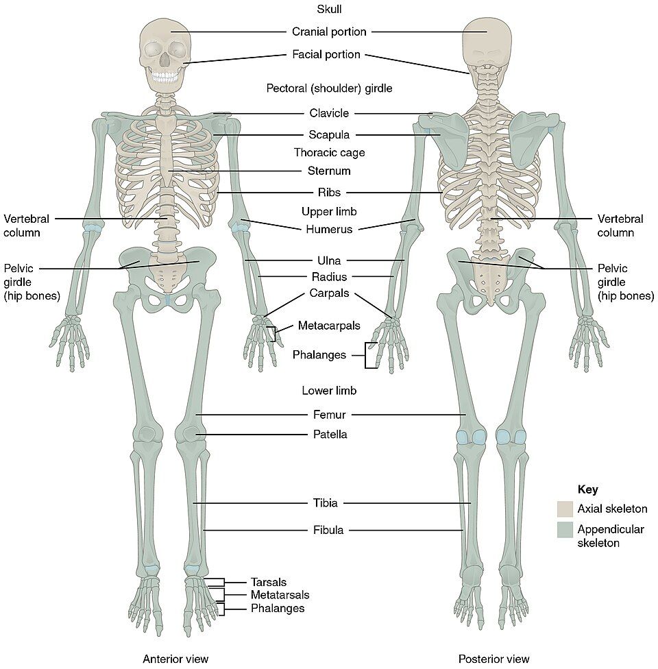
An adult skeleton has 206 bones, and they split into two named divisions. The axial skeleton (about 80 bones) runs down the body's central axis — the skull, the vertebral column, and the thoracic cage — and its main job is protection and support. The appendicular skeleton (about 126 bones) is everything that hangs off that axis: the arms and legs plus the two girdles (pectoral and pelvic) that anchor the limbs to the trunk. Its main job is movement. In the reference figure, the axial bones are tinted one color and the appendicular bones another — a useful way to keep the two straight.
Tap each of the skeleton's jobs in the interactive to see what carries it out.
- A bone is a living organ; the skeletal system is bones, cartilages, and joints together.
- Core jobs: support, protection, movement, mineral/fat storage, and blood-cell formation (hematopoiesis).
- The axial skeleton (~80 bones) protects and supports; the appendicular skeleton (~126 bones) enables movement; 206 bones total in an adult.
- About 99% of the body's calcium is stored in bone.
Section 2Classifying bones by shape
Shape follows the job
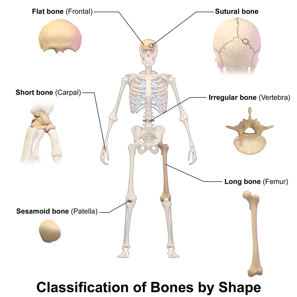
Bones come in a handful of recognizable shapes, and — just as with the rest of the body — the shape is a clue to the function. A bone built for leverage looks nothing like a bone built to shield a soft organ. Sorting the skeleton into five shape classes gives you a quick vocabulary for describing any bone you meet.
The five shapes
| Class | Shape & role | Examples |
|---|---|---|
| Long bone | Longer than wide, with a shaft and two ends; act as levers for movement | Femur, humerus, phalanges (fingers & toes) |
| Short bone | Roughly cube-shaped; provide stability with limited motion | Carpals (wrist), tarsals (ankle) |
| Flat bone | Thin, often curved plates; protect organs and give broad muscle attachment | Skull bones, sternum, ribs, scapula |
| Irregular bone | Complex shapes that fit no other class; varied protective and support roles | Vertebrae, hip bones, many facial bones |
| Sesamoid bone | Small bones embedded inside a tendon; protect the tendon and improve its pull | Patella (kneecap) |
Notice that the phalanges of your fingers count as long bones even though they are tiny — the label is about proportion (longer than wide, with a shaft) and about function (leverage), not about absolute size. The reference figure also shows a sutural (Wormian) bone, a small extra bone that can appear within the seams of the skull; think of it as a special case of a flat bone rather than a sixth class.
Tap each shape in the interactive to see the bone and where it lives in the body.
- Five shape classes: long, short, flat, irregular, sesamoid.
- Long bones are levers; short bones give stable support; flat bones protect; irregular bones are specialized; sesamoids sit inside tendons.
- The patella is the largest sesamoid; the phalanges are small long bones.
Section 3Inside a long bone
The shaft and the ends
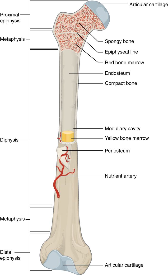
A long bone such as the femur has a clean architecture once you learn its parts. The long central tube is the diaphysis, or shaft. Its wall is a cylinder of dense compact bone, and its hollow center — the medullary cavity — is filled in adults with fatty yellow marrow. Each rounded end is an epiphysis, filled with lightweight spongy (trabecular) bone whose spaces hold red marrow, the site of blood-cell formation. Where the joint surface meets another bone, the epiphysis is capped with slick articular cartilage that cushions the joint and lets it glide. Between the shaft and each end lies the metaphysis, which in a growing child contains the epiphyseal (growth) plate — a disc of cartilage where the bone lengthens. When growth finishes, that plate ossifies into a thin epiphyseal line.
The living wrappers
Two thin membranes keep bone alive and connected. The periosteum is a tough sheath covering the outer surface everywhere except the joint caps; it carries the blood vessels and nerves, anchors tendons and ligaments, and holds bone-forming cells for growth and repair. Lining the inner surfaces — the medullary cavity and the spaces of spongy bone — is the delicate endosteum, also stocked with bone cells. A nutrient artery pierces the shaft through a small opening to supply the bone from within.
Zoom in: the osteon
Under the microscope, compact bone is not a solid block but a bundle of tiny weight-bearing pillars called osteons (Haversian systems). Each osteon is a set of concentric rings of bone matrix, the lamellae, wrapped around a central canal that carries a blood vessel and nerve. Trapped in small pockets called lacunae between the rings are the mature bone cells, the osteocytes, and they stay connected to one another and to the central vessel through hair-thin channels called canaliculi. This canal-and-channel plumbing is how a hard, mineralized tissue keeps its buried cells fed and alive.
| Part | What it is / holds |
|---|---|
| Diaphysis | The shaft; a tube of compact bone around the medullary cavity |
| Epiphysis | An expanded end filled with spongy bone and red marrow |
| Metaphysis | The region between shaft and end; site of the growth plate/line |
| Medullary cavity | The hollow center of the shaft; holds yellow (fatty) marrow in adults |
| Articular cartilage | Smooth cartilage capping the joint surface of each epiphysis |
| Periosteum | Outer membrane; vessels, nerves, attachment, bone-forming cells |
| Endosteum | Inner lining of the marrow cavity and spongy spaces |
Tap each labeled part in the interactive to find it on the bone and learn its role.
- The diaphysis (shaft) is compact bone around a marrow-filled medullary cavity; the epiphyses (ends) are spongy bone with red marrow.
- The metaphysis holds the epiphyseal plate (growth) that becomes the epiphyseal line when growth stops.
- The periosteum wraps the outside; the endosteum lines the inside.
- Compact bone is built of osteons — lamellae around a central canal, with osteocytes in lacunae linked by canaliculi.
Section 4Bone cells & remodeling

What bone is made of
Bone tissue is a masterpiece of engineering because it combines two opposite qualities. Its organic part — mostly collagen fibers — gives bone flexibility and tensile strength, so it bends a little under load instead of snapping. Its inorganic part — mineral salts of calcium and phosphate that crystallize as hydroxyapatite — gives bone its hardness and compressive strength. Take the mineral away and bone becomes rubbery; take the collagen away and it becomes brittle as chalk. Healthy bone needs both, in balance.
Four cells, one crew
Four cell types build and maintain that matrix, and the easiest way to remember them is by their jobs. Stem-like osteogenic (osteoprogenitor) cells divide to produce the builders. Osteoblasts lay down new bone matrix — they build. Once an osteoblast walls itself into the matrix it has made, it matures into an osteocyte, the caretaker cell that lives in a lacuna and keeps the tissue healthy. And the demolition crew are the osteoclasts, large multinucleated cells that dissolve old bone and release its minerals back into the blood.
| Cell | Job | Memory hook |
|---|---|---|
| Osteogenic cell | Stem cell that divides into osteoblasts | The source |
| Osteoblast | Builds new bone matrix | “b” for build |
| Osteocyte | Mature cell; maintains bone, senses stress | Retired builder, walled in |
| Osteoclast | Breaks down bone; frees calcium | “c” for consume/carve |
The tug-of-war called remodeling
Bone is never finished. Throughout life, osteoclasts dissolve old bone while osteoblasts lay down fresh bone right behind them — a continuous cycle called remodeling that replaces roughly 10% of the skeleton every year. When the two crews stay balanced, bone mass holds steady. Two things tune the balance. First, mechanical stress: bone thickens where it is loaded and thins where it is not — the principle known as Wolff's law, which is why weight-bearing exercise strengthens the skeleton and why bones weaken during bed rest or spaceflight. Second, hormones, especially the calcium-regulating pair you will meet next. When resorption outpaces formation for years — as often happens after menopause — the result is osteoporosis, porous bone that fractures easily.
Watch the crews trade places in the interactive, and meet the four cells one at a time.
- Bone matrix = collagen (flexibility) + hydroxyapatite mineral (hardness); it needs both.
- Four cells: osteogenic (source), osteoblast (build), osteocyte (maintain), osteoclast (break down).
- Remodeling balances resorption and deposition, replacing ~10% of bone yearly; it responds to load (Wolff's law) and hormones.
- Chronic imbalance toward resorption gives osteoporosis.
Section 5Calcium homeostasis
Why calcium is worth guarding
Blood calcium (Ca²⁺) must stay in a tight window, roughly 9–10 mg/dL, because so much depends on it: calcium triggers muscle contraction, lets nerves fire, is required for blood clotting, and acts as a messenger inside cells. Drift too low and nerves become jumpy, causing muscle spasms and cramps (tetany); drift too high and nerves and muscles go sluggish. This is a textbook example of the homeostasis idea from Chapter 1 — a value held near a set point by feedback — and the skeleton is the reservoir the body draws on to defend it.
PTH raises calcium
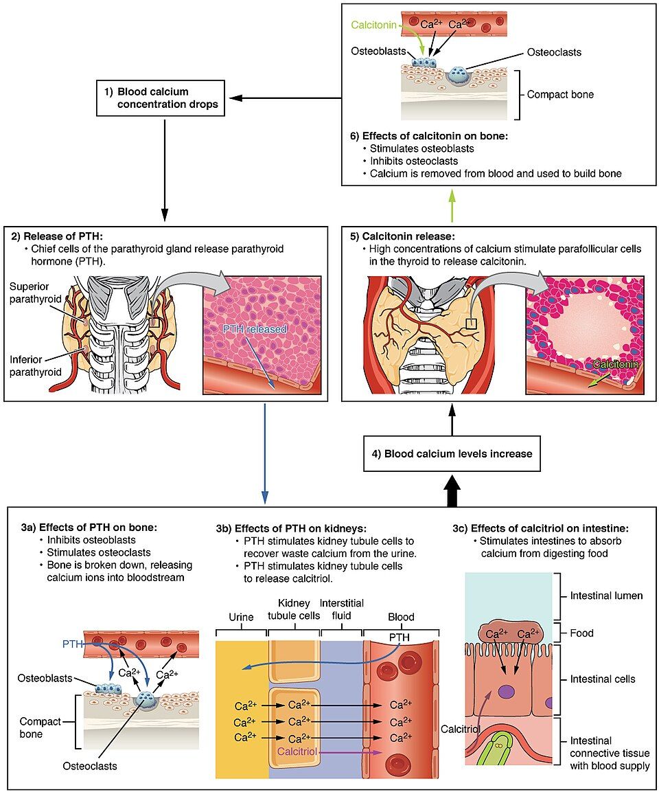
When blood calcium falls, the parathyroid glands (four small glands on the back of the thyroid) release parathyroid hormone (PTH). PTH pushes calcium up through three routes at once. In bone, it stimulates osteoclasts to dissolve matrix and release calcium into the blood. In the kidneys, it tells the tubules to reclaim calcium that would otherwise be lost in urine, and it activates vitamin D into calcitriol. And through calcitriol, it drives the intestines to absorb more calcium from food. Bone, kidney, and gut all pour calcium into the blood, and the level climbs back toward normal.
Calcitonin lowers calcium
When blood calcium runs high, the parafollicular (C) cells of the thyroid gland release calcitonin, which does the opposite of PTH. Calcitonin inhibits osteoclasts and favors osteoblasts, so calcium is pulled out of the blood and deposited back into bone, and the level settles down. The two hormones form a push–pull pair — PTH the accelerator, calcitonin the brake — that keeps blood calcium remarkably steady.
| Feature | Parathyroid hormone (PTH) | Calcitonin |
|---|---|---|
| Released when | Blood calcium is low | Blood calcium is high |
| Source | Parathyroid glands | Thyroid C (parafollicular) cells |
| Effect on calcium | Raises it | Lowers it |
| Bone | Stimulates osteoclasts (breakdown) | Inhibits osteoclasts (favors buildup) |
| Kidney / gut | Reabsorbs Ca; activates vitamin D → more gut absorption | Minor role |
Knock blood calcium off its set point in the interactive and watch the right hormone respond.
- Blood calcium is held near 9–10 mg/dL because it runs nerves, muscle, and clotting.
- PTH (from the parathyroids) raises calcium via bone, kidney, and gut (through calcitriol/vitamin D).
- Calcitonin (from thyroid C cells) lowers calcium by inhibiting osteoclasts.
- The body will withdraw calcium from bone to protect the blood level.
Section 6Fractures & repair
How bones break
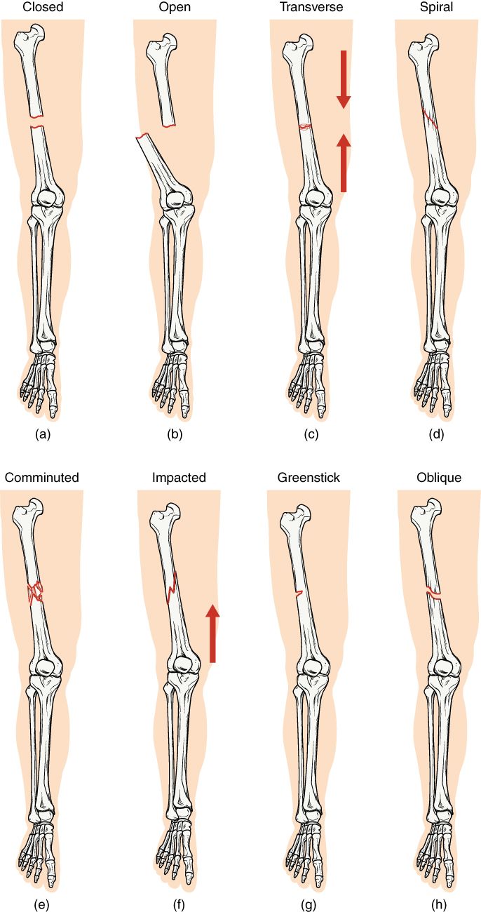
A fracture is simply a broken bone, but clinicians name the pattern of the break because the pattern hints at what caused it and how it should be treated. The first and most important distinction is whether the skin stayed intact. A closed (simple) fracture leaves the skin unbroken; an open (compound) fracture breaks through the skin, which is far more serious because it opens a door for infection. Beyond that, breaks are named for their line and severity.
| Type | What it looks like |
|---|---|
| Closed vs open | Skin intact (closed) vs bone piercing the skin (open) — open carries infection risk |
| Transverse | Break runs straight across the shaft |
| Oblique | Break runs at an angle across the bone |
| Spiral | Twisting break that winds around the shaft; suggests a rotational force |
| Comminuted | Bone shatters into three or more fragments |
| Impacted | One broken end is driven into the other |
| Greenstick | Incomplete break; bone bends and cracks on one side (mostly in children) |
How a bone heals: four stages
A broken bone repairs itself in a predictable sequence that can take months to finish. First, torn vessels bleed and clot, forming a fracture hematoma that seals the gap and sets the stage. Next, cells build a soft fibrocartilaginous (soft) callus of collagen and cartilage that splints the ends together. Osteoblasts then replace that soft bridge with a bony (hard) callus of spongy bone, roughly six weeks after the injury. Finally, over months, the bone remodels — osteoclasts and osteoblasts reshape the callus back toward the bone's original form, guided by the same stresses that shape every bone.
| Stage | What happens |
|---|---|
| 1. Fracture hematoma | Bleeding forms a clot that fills and seals the break |
| 2. Soft (fibrocartilaginous) callus | Collagen and cartilage bridge the gap, splinting the ends |
| 3. Hard (bony) callus | Osteoblasts replace the soft callus with spongy bone (~6 weeks) |
| 4. Bone remodeling | The callus is reshaped over months toward the original contour |
To help this happen, a displaced fracture is first reduced — the ends are lined back up, either by manipulation from outside (closed reduction) or by surgery (open reduction) — and then immobilized in a cast or with hardware so the delicate callus is not disturbed while it matures.
Explore each fracture pattern in the interactive, then step through the four stages of repair.
- Closed = skin intact; open (compound) = bone breaks skin and risks infection.
- Patterns include transverse, oblique, spiral, comminuted, impacted, greenstick.
- Healing runs in four stages: hematoma → soft callus → hard callus → remodeling.
- Treatment is reduction (realigning) then immobilization to protect the callus.
The chapter in one breath
- Bone is living tissue, and the skeletal system does five jobs: support, protection, movement, mineral/fat storage, and blood-cell formation.
- The skeleton splits into the axial (~80 bones) and appendicular (~126 bones) divisions, 206 in all.
- Bones are classed by shape as long, short, flat, irregular, or sesamoid, and the shape follows the function.
- A long bone has a diaphysis of compact bone around a marrow cavity and spongy epiphyses; compact bone is built of osteons.
- Four cells — osteogenic, osteoblast, osteocyte, osteoclast — drive remodeling, the lifelong balance of building and dissolving bone.
- The matrix combines collagen (flexibility) with hydroxyapatite mineral (hardness).
- PTH raises and calcitonin lowers blood calcium, keeping it near 9–10 mg/dL — the skeleton is the calcium bank.
- Fractures come in named patterns and heal in four stages: hematoma → soft callus → hard callus → remodeling.
ReferenceWords worth knowing
A quick reference for the terms in this chapter — skim it now, or circle back whenever a word trips you up.
- Appendicular skeleton
- The bones of the limbs plus the pectoral and pelvic girdles that attach them to the axis (~126 bones); specialized for movement.
- Axial skeleton
- The bones along the body's central axis — skull, vertebral column, and thoracic cage (~80 bones); specialized for support and protection.
- Calcitonin
- A hormone from the thyroid's parafollicular (C) cells that lowers blood calcium by inhibiting osteoclasts.
- Calcitriol
- The active form of vitamin D; increases calcium absorption from the intestine and is activated with the help of PTH.
- Callus
- The repair bridge that forms across a fracture — first a soft (fibrocartilaginous) callus, later a hard (bony) callus.
- Compact bone
- Dense, strong bone tissue built of osteons; forms the outer wall of bones and the shaft of long bones.
- Diaphysis
- The tubular shaft of a long bone; compact bone surrounding the medullary cavity.
- Endosteum
- The thin membrane lining the internal surfaces of bone — the medullary cavity and spongy-bone spaces.
- Epiphyseal plate
- The cartilage growth plate in the metaphysis where a long bone lengthens; becomes the epiphyseal line when growth ends.
- Epiphysis
- The expanded end of a long bone, made of spongy bone with red marrow and capped by articular cartilage.
- Hematopoiesis
- The formation of blood cells (red cells, white cells, platelets) in the red marrow of bone.
- Hydroxyapatite
- The crystalline calcium-phosphate mineral of bone matrix that gives bone its hardness.
- Medullary cavity
- The hollow center of a long bone's shaft; stores fatty yellow marrow in adults.
- Osteoblast
- The bone-building cell that secretes new matrix; matures into an osteocyte once surrounded by bone.
- Osteoclast
- The large, multinucleated cell that dissolves bone matrix and releases its minerals into the blood.
- Osteocyte
- A mature bone cell housed in a lacuna that maintains the matrix and senses mechanical stress.
- Osteon
- The structural unit of compact bone — concentric lamellae of matrix around a central (Haversian) canal.
- Parathyroid hormone (PTH)
- The hormone from the parathyroid glands that raises blood calcium via bone, kidney, and intestine.
- Periosteum
- The tough outer membrane of a bone; carries vessels and nerves, anchors tendons, and holds bone-forming cells.
- Remodeling
- The lifelong cycle of bone resorption by osteoclasts balanced against deposition by osteoblasts.
- Sesamoid bone
- A small bone embedded within a tendon (such as the patella) that protects the tendon and improves its leverage.
- Spongy bone
- Lightweight, porous bone tissue of struts (trabeculae) found in the epiphyses; its spaces hold red marrow.